ABCA4 Antibodies
Background
The ABCA4 gene encodes a transmembrane transport protein mainly expressed on the outer segment disc membrane of rod cells in retinal photoreceptor cells. This protein utilizes the energy generated by ATP hydrolysis to transport metabolic products with potential phototoxic effects in the retina, such as all-trans retinaldehyde, to the cytoplasm, thereby maintaining the normal operation of the visual cycle and the stability of photoreceptor cells. The pathogenic mutation of this gene can lead to impaired transport function, causing toxic substances to accumulate within the retinal pigment epithelial cells, and ultimately triggering typical pathological changes in hereditary retinal degenerative diseases such as Stargardt's disease. Since its successful cloning in 1997, the ABCA4 gene has become an important model for studying the molecular mechanisms of hereditary eye diseases. Its complex allelic heterogeneity and phenotypic diversity provide a key scientific basis for understanding the pathogenic mechanisms of autosomal recessive genetic diseases.
Structure of ABCA4
ABCA4 is a large transmembrane protein with a molecular weight of approximately 250 kDa. The molecular weight of this protein remains highly conserved across different mammals, mainly due to the strict evolutionary constraints of its functional domain.
| Species | Human | Mouse | Bovine | Macaque |
| Molecular Weight (kDa) | 250.2 | 249.8 | 250.3 | 250.1 |
| Primary Structural Differences | Benchmark sequence, containing 50 exons | Sequence similarity of transmembrane regions > 95% | The C-end domain is highly conservative | The ATP binding domain is exactly the same as that of the human source |
The ABCA4 protein contains approximately 2,273 amino acid residues, which form a typical modular architecture of alternating "transmembrane domains - nucleotide binding domains" through its primary structure. The three-dimensional structure of this protein presents hydrophobic channels composed of multiple transmembrane α -helices, among which two nucleotide binding domains provide transport impetus by hydrolyzing ATP. The Walker A motif located on the cytoplasmic side is responsible for binding ATP molecules, while the LSGGQ motif regulates the substrate transport process through allosteric effects, jointly completing the efficient transport of n-retina-phosphatidylethanolamine.
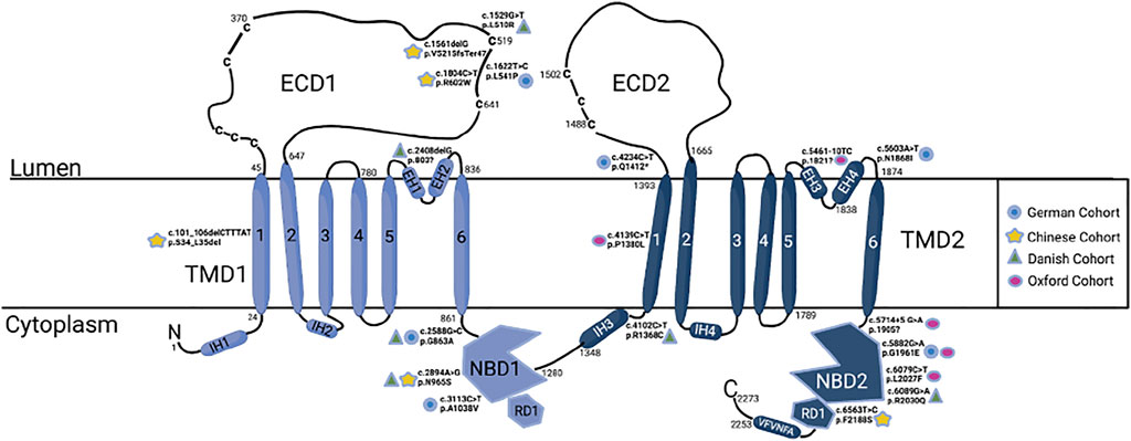 Fig. 1 ABCA4 molecular structure with the five most mutations from each cohort.1
Key structural properties of ABCA4:
- Multi-domain transmembrane topological configuration
- Substrate transport channels mediated by hydrophobic chambers
- Mechanism of ATP hydrolysis driven by nucleotide binding domains
Functions of ABCA4
The core function of the ABCA4 protein is to mediate the transport of visual circulation products in the retinal pigment epithelium. However, this protein is also involved in a variety of cellular physiological processes, including maintaining lipid homeostasis and protecting against photo-oxidative damage.
| Function | Description |
| Visual circulatory metabolism | Transport the all-trans retinaldehyde-phospholipid complex from the outer segment of photoreceptor cells to the cytoplasm to complete the recycling and utilization of vitamin A derivatives. |
| Removal of toxic substances | Prevent the abnormal accumulation of N-ascoryloxyl-phosphatidylethanolamine in the retina through an ATP-dependent transport mechanism. |
| Relief of oxidative stress | Reduce the formation of lipofuscin precursors and lower the risk of photo-oxidative damage to photoreceptor cells. |
| Membrane lipid homeostasis | Maintain the integrity of the phospholipid bilayer on the membrane of photoreceptor cells to ensure the efficiency of visual signal transmission. |
| Genetic phenotypic regulation | Gene mutations lead to phenotypes such as Stargardt disease, and their severity is related to residual transport activity. |
The substrate transport of ABCA4 follows a typical alternating access model. Unlike the synergistic effect of hemoglobin, its individual transport units exhibit Michelson-type kinetic characteristics, which explains the protein's high affinity for specific retinal derivatives and its exclusive role in the visual cycle.
Applications of ABCA4 and ABCA4 Antibody in Literature
1. Cremers, Frans PM, et al. "Clinical spectrum, genetic complexity and therapeutic approaches for retinal disease caused by ABCA4 mutations." Progress in retinal and eye research 79 (2020): 100861. https://doi.org/10.1016/j.preteyeres.2020.100861
The article indicates that mutations in the ABCA4 gene can cause various retinopathy types, including not only typical Stargardt disease but also other phenotypes such as cone and rod cell dystrophy. There are over 1,200 pathogenic mutations in this gene, covering a wide range of types. Relevant research has deepened the understanding of the complexity of genetic diseases. This article reviews its clinical manifestations, pathological mechanisms and treatment progress.
2. Wang, Liang, et al. "Updates on emerging interventions for autosomal recessive ABCA4-associated Stargardt disease." Journal of Clinical Medicine 12.19 (2023): 6229. https://doi.org/10.3390/jcm12196229
The article indicates that mutations in the ABCA4 gene cause Stargardt disease, and there is currently no effective treatment. This article reviews the latest clinical trial progress in the treatment of this disease, including gene therapy, small molecule drugs (such as ALK-001), and stem cell therapy, etc. Although multiple schemes have shown potential to delay the disease, their safety and effectiveness still need to be further verified.
3. Al-Khuzaei, Saoud, et al. "An overview of the genetics of ABCA4 retinopathies, an evolving story." Genes 12.8 (2021): 1241. https://doi.org/10.3390/genes12081241
The article indicates that mutations in the ABCA4 gene cause retinopathy such as Stargardt disease, and its diagnosis is difficult due to diverse phenotypes and complex variations. More than 2,000 pathogenic variations of this gene have been discovered, but some patients still have difficulty being diagnosed. With the development of targeted therapy, precise genetic diagnosis is becoming increasingly important. This article aims to update the latest progress in ABCA4 genotyping and therapeutic research.
4. Piotter, Elena, Michelle E. McClements, and Robert E. MacLaren. "The scope of pathogenic ABCA4 mutations targetable by CRISPR DNA base editing systems—a systematic review." Frontiers in Genetics 12 (2022): 814131. https://doi.org/10.3389/fgene.2021.814131
The article indicates that mutations in the ABCA4 gene cause Stargardt disease, and there is currently no treatment available. Given that there are over 1,200 pathogenic mutations in this gene, the research explored the therapeutic potential of gene editing technologies such as CRISPR-Cas. Analysis reveals that over half of the pathogenic mutations can theoretically be corrected through base editing, providing a new direction for the development of gene therapies targeting ABCA4.
5. Kim, Bo Min, et al. "Functional characterization of ABCA4 genetic variants related to Stargardt disease." Scientific Reports 12.1 (2022): 22282. https://doi.org/10.1038/s41598-022-26912-6
This study focuses on how variations in the ABCA4 gene affect protein expression and thereby trigger Stargardt disease. Studies have found that four missense variants can lead to protein degradation, while specific haplotypes in the promoter region can significantly alter transcriptional activity, a process regulated by GATA-2 and HLF transcription factors.
Creative Biolabs: ABCA4 Antibodies for Research
Creative Biolabs specializes in the production of high-quality ABCA4 antibodies for research and industrial applications. Our portfolio includes monoclonal antibodies tailored for ELISA, Flow Cytometry, Western blot, immunohistochemistry, and other diagnostic methodologies.
- Custom ABCA4 Antibody Development: Tailor-made solutions to meet specific research requirements.
- Bulk Production: Large-scale antibody manufacturing for industry partners.
- Technical Support: Expert consultation for protocol optimization and troubleshooting.
- Aliquoting Services: Conveniently sized aliquots for long-term storage and consistent experimental outcomes.
For more details on our ABCA4 antibodies, custom preparations, or technical support, contact us at email.
Reference
- Piotter, Elena, Michelle E. McClements, and Robert E. MacLaren. "The scope of pathogenic ABCA4 mutations targetable by CRISPR DNA base editing systems—a systematic review." Frontiers in Genetics 12 (2022): 814131. https://doi.org/10.3389/fgene.2021.814131
Anti-ABCA4 antibodies
 Loading...


Hot products 
-
Mouse Anti-CASQ1 Recombinant Antibody (CBFYC-0863) (CBMAB-C0918-FY)

-
Mouse Anti-BIRC7 Recombinant Antibody (88C570) (CBMAB-L0261-YJ)

-
Mouse Anti-AMIGO2 Recombinant Antibody (CBYY-C0756) (CBMAB-C2192-YY)

-
Mouse Anti-ADGRL2 Recombinant Antibody (V2-58519) (CBMAB-L0166-YJ)

-
Mouse Anti-ARHGDIA Recombinant Antibody (CBCNA-009) (CBMAB-R0415-CN)

-
Mouse Anti-CAPZB Recombinant Antibody (CBYY-C0944) (CBMAB-C2381-YY)

-
Mouse Anti-CD24 Recombinant Antibody (HIS50) (CBMAB-C10123-LY)

-
Mouse Anti-GFAP Recombinant Antibody (24) (CBMAB-G2927-LY)

-
Rat Anti-CD63 Recombinant Antibody (7G4.2E8) (CBMAB-C8725-LY)

-
Human Anti-SARS-CoV-2 S1 Monoclonal Antibody (CBFYR-0120) (CBMAB-R0120-FY)

-
Mouse Anti-CARTPT Recombinant Antibody (113612) (CBMAB-C2450-LY)

-
Mouse Anti-ABCA3 Recombinant Antibody (V2-178911) (CBMAB-A0145-YC)

-
Mouse Anti-CRYAB Recombinant Antibody (A4345) (CBMAB-A4345-YC)

-
Mouse Anti-CD83 Recombinant Antibody (HB15) (CBMAB-C1765-CQ)

-
Mouse Anti-BAX Recombinant Antibody (CBYY-0216) (CBMAB-0217-YY)

-
Mouse Anti-ASTN1 Recombinant Antibody (H-9) (CBMAB-1154-CN)

-
Rabbit Anti-B2M Recombinant Antibody (CBYY-0059) (CBMAB-0059-YY)

-
Mouse Anti-CTCF Recombinant Antibody (CBFYC-2371) (CBMAB-C2443-FY)

-
Mouse Anti-ENO2 Recombinant Antibody (85F11) (CBMAB-0276CQ)

-
Mouse Anti-ACO2 Recombinant Antibody (V2-179329) (CBMAB-A0627-YC)

- AActivation
- AGAgonist
- APApoptosis
- BBlocking
- BABioassay
- BIBioimaging
- CImmunohistochemistry-Frozen Sections
- CIChromatin Immunoprecipitation
- CTCytotoxicity
- CSCostimulation
- DDepletion
- DBDot Blot
- EELISA
- ECELISA(Cap)
- EDELISA(Det)
- ESELISpot
- EMElectron Microscopy
- FFlow Cytometry
- FNFunction Assay
- GSGel Supershift
- IInhibition
- IAEnzyme Immunoassay
- ICImmunocytochemistry
- IDImmunodiffusion
- IEImmunoelectrophoresis
- IFImmunofluorescence
- IGImmunochromatography
- IHImmunohistochemistry
- IMImmunomicroscopy
- IOImmunoassay
- IPImmunoprecipitation
- ISIntracellular Staining for Flow Cytometry
- LALuminex Assay
- LFLateral Flow Immunoassay
- MMicroarray
- MCMass Cytometry/CyTOF
- MDMeDIP
- MSElectrophoretic Mobility Shift Assay
- NNeutralization
- PImmunohistologyp-Paraffin Sections
- PAPeptide Array
- PEPeptide ELISA
- PLProximity Ligation Assay
- RRadioimmunoassay
- SStimulation
- SESandwich ELISA
- SHIn situ hybridization
- TCTissue Culture
- WBWestern Blot
