AATK Antibodies
Background
The AATK (apoptosis-associated tyrosine kinase) gene encodes a protein kinase that is mainly expressed during the development of the nervous system. This gene plays a crucial role in the formation and maintenance of the central nervous system by regulating processes such as cell apoptosis, cell cycle, and neuronal differentiation. AATK was initially identified in 1994, and its abnormal expression is associated with the occurrence and development of various neurodegenerative diseases and tumors. As an important model molecule in cell signal transduction research, the functional study of AATK has deepened our understanding of the mechanism of programmed cell death and the principles of neurodevelopmental biology, providing a theoretical basis for targeted treatment of related diseases.
Structure of AATK
The protein encoded by the AATK (apoptosis-associated tyrosine kinase) gene has a molecular weight of approximately 140 kDa. This molecular weight may vary among different splicing variants. This protein belongs to the tyrosine kinase family. Its primary structure contains a typical kinase domain and is regulated by post-translational modifications such as autophosphorylation. Its advanced structure is composed of multiple functional domains, collectively forming the molecular interface for signal transduction. Key structural features include an N-terminal region that regulates cell localization and a C-terminal kinase domain responsible for catalytic activity. The two work together to determine its specific function in the process of neuronal apoptosis and differentiation. This structural framework provides a foundation for understanding how it responds to intracellular and extracellular signals and regulates downstream pathways.
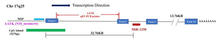 Fig. 1 Diagram showing the AATK gene, CpG island, miR-1250, and associated PCR targets.1
Key structural properties of AATK:
- Typical tyrosine kinase catalytic domain
- The N-terminal domain that regulates the localization and activity of proteins
- The phosphorylation-dependent conformational changes of the self-inhibitory loop
- The signal transduction interface formed by multiple functional modules
Functions of AATK
The main function of the AATK gene is to regulate the programmed death and differentiation of neurons, and it also participates in key processes such as cell cycle regulation.
| Function | Description |
| Regulate neuronal apoptosis | During the development of the nervous system, induce programmed cell death of excessive or damaged neurons to maintain the balance of cell numbers. |
| Promote Neuronal Differentiation | It participates in signal transduction and influences the maturation processes such as the establishment of neuronal polarity, axon guidance, and synapse formation. |
| Affects Cell Cycle | By regulating cyclins through kinase activity, it coordinates the cell proliferation and differentiation processes. |
| Involvement in Tumor Suppression | The absence or abnormal function of this expression is associated with the occurrence and development of various neuroblastomas and gliomas. |
| Maintaining Intracellular Homeostasis | As a crucial signaling node, it integrates internal and external environmental signals to determine the survival or death fate of the cell. |
The function of AATK relies on its kinase activity and dynamic interactions with other signaling proteins (such as p53, members of the MAPK pathway), forming a sophisticated regulatory network rather than a single linear effect.
Applications of AATK and AATK Antibody in Literature
1. Zhang, Min Yue, Lu Qian Wang, and Chor Sang Chim. "miR-1250-5p is a novel tumor suppressive intronic miRNA hypermethylated in non-Hodgkin’s lymphoma: novel targets with impact on ERK signaling and cell migration." Cell Communication and Signaling 19.1 (2021): 62. https://doi.org/10.1186/s12964-021-00707-0
This study has discovered that miR-1250-5p, located in the intron of the AATK gene, is a novel tumor suppressor miRNA. In non-Hodgkin's lymphoma, it is jointly silenced by promoter methylation with the host gene AATK. The study has confirmed that miR-1250-5p directly targets MAPK1 and WDR1, thereby inhibiting the MAPK/ERK signaling pathway and cell migration.
2. Vrabec, Katarina, et al. "Differential expression of several miRNAs and the host genes AATK and DNM2 in leukocytes of sporadic ALS patients." Frontiers in molecular neuroscience 11 (2018): 106. https://doi.org/10.3389/fnmol.2018.00106
In this study, significant abnormal expressions of miR-124a, miR-206, miR-9, let-7b and miR-638 were observed in 84 patients with sporadic ALS. Additionally, for the first time, the upregulation of the AATK gene and the downregulation of the DNM2 gene were observed. These findings reveal the potential shared pathogenic mechanisms between ALS and some neurodegenerative diseases.
3. Gao, Ling, et al. "CircPTK2 suppresses the progression of gastric cancer by targeting the MiR-196a-3p/AATK axis." Frontiers in oncology 11 (2021): 706415. https://doi.org/10.3389/fonc.2021.706415
Studies have shown that circPTK2 acts as a "sponge" for miR-196a-3p, promoting the expression of the tumor suppressor gene AATK, thereby significantly inhibiting the proliferation, migration and invasion of gastric cancer cells. It is expected to become a new therapeutic target.
4. Woods, Michelle L., et al. "Epigenetically silenced apoptosis-associated tyrosine kinase (AATK) facilitates a decreased expression of Cyclin D1 and WEE1, phosphorylates TP53 and reduces cell proliferation in a kinase-dependent manner." Cancer Gene Therapy 29.12 (2022): 1975-1987. https://doi.org/10.1038/s41417-022-00513-x
Studies have shown that the AATK gene is subject to epigenetic silencing in various cancers, and its inactivation is associated with poor prognosis in patients. As a serine/threonine kinase, AATK phosphorylates TP53 and downregulates key cell cycle factors to inhibit tumor growth, thereby exerting tumor suppressor functions.
5. Ding, Li-Yun, et al. "Epigenetic silencing of AATK in acinar to ductal metaplasia in murine model of pancreatic cancer." Clinical Epigenetics 12.1 (2020): 87. https://doi.org/10.1186/s13148-020-00878-6
The research has found that the epigenetic silencing of the AATK gene is closely related to the transformation of malignant subtypes of pancreatic cancer. Activation of AATK can initiate the differentiation of acinar cells into ducts, while its silencing will promote the proliferation of intraductal neoplasms, thereby regulating the cell fate transformation and progression of pancreatic cancer.
Creative Biolabs: AATK Antibodies for Research
Creative Biolabs specializes in the production of high-quality AATK antibodies for research and industrial applications. Our portfolio includes monoclonal antibodies tailored for ELISA, Flow Cytometry, Western blot, immunohistochemistry, and other diagnostic methodologies.
- Custom AATK Antibody Development: Tailor-made solutions to meet specific research requirements.
- Bulk Production: Large-scale antibody manufacturing for industry partners.
- Technical Support: Expert consultation for protocol optimization and troubleshooting.
- Aliquoting Services: Conveniently sized aliquots for long-term storage and consistent experimental outcomes.
For more details on our AATK antibodies, custom preparations, or technical support, contact us at email.
Reference
- Zhang, Min Yue, Lu Qian Wang, and Chor Sang Chim. "miR-1250-5p is a novel tumor suppressive intronic miRNA hypermethylated in non-Hodgkin’s lymphoma: novel targets with impact on ERK signaling and cell migration." Cell Communication and Signaling 19.1 (2021): 62. https://doi.org/10.1186/s12964-021-00707-0
Anti-AATK antibodies
 Loading...


Hot products 
-
Rat Anti-ADGRE4 Recombinant Antibody (V2-160163) (CBMAB-F0011-CQ)

-
Mouse Anti-CD83 Recombinant Antibody (HB15) (CBMAB-C1765-CQ)

-
Rat Anti-CD34 Recombinant Antibody (MEC 14.7) (CBMAB-C10196-LY)

-
Rabbit Anti-DLK1 Recombinant Antibody (9D8) (CBMAB-D1061-YC)

-
Mouse Anti-DMD Recombinant Antibody (D1190) (CBMAB-D1190-YC)

-
Rabbit Anti-ENO2 Recombinant Antibody (BA0013) (CBMAB-0272CQ)

-
Mouse Anti-BRCA2 Recombinant Antibody (CBYY-1728) (CBMAB-2077-YY)

-
Mouse Anti-CD24 Recombinant Antibody (SN3) (CBMAB-C1037-CQ)

-
Rabbit Anti-AKT2 (Phosphorylated S474) Recombinant Antibody (V2-556130) (PTM-CBMAB-0605LY)

-
Mouse Anti-APP Recombinant Antibody (5C2A1) (CBMAB-A3314-YC)

-
Rabbit Anti-Acetyl-Histone H3 (Lys36) Recombinant Antibody (V2-623395) (CBMAB-CP0994-LY)

-
Mouse Anti-AQP2 Recombinant Antibody (G-3) (CBMAB-A3359-YC)

-
Rabbit Anti-AKT3 Recombinant Antibody (V2-12567) (CBMAB-1057-CN)

-
Rabbit Anti-CBL Recombinant Antibody (D4E10) (CBMAB-CP0149-LY)

-
Mouse Anti-ACE2 Recombinant Antibody (V2-179293) (CBMAB-A0566-YC)

-
Mouse Anti-dsRNA Recombinant Antibody (2) (CBMAB-D1807-YC)

-
Mouse Anti-FeLV g27 Recombinant Antibody (1) (CBMAB-V208-1714-FY)

-
Mouse Anti-CD24 Recombinant Antibody (ALB9) (CBMAB-0176CQ)

-
Mouse Anti-ATP1A2 Recombinant Antibody (M7-PB-E9) (CBMAB-A4013-YC)

-
Mouse Anti-AHCYL1 Recombinant Antibody (V2-180270) (CBMAB-A1703-YC)

- AActivation
- AGAgonist
- APApoptosis
- BBlocking
- BABioassay
- BIBioimaging
- CImmunohistochemistry-Frozen Sections
- CIChromatin Immunoprecipitation
- CTCytotoxicity
- CSCostimulation
- DDepletion
- DBDot Blot
- EELISA
- ECELISA(Cap)
- EDELISA(Det)
- ESELISpot
- EMElectron Microscopy
- FFlow Cytometry
- FNFunction Assay
- GSGel Supershift
- IInhibition
- IAEnzyme Immunoassay
- ICImmunocytochemistry
- IDImmunodiffusion
- IEImmunoelectrophoresis
- IFImmunofluorescence
- IGImmunochromatography
- IHImmunohistochemistry
- IMImmunomicroscopy
- IOImmunoassay
- IPImmunoprecipitation
- ISIntracellular Staining for Flow Cytometry
- LALuminex Assay
- LFLateral Flow Immunoassay
- MMicroarray
- MCMass Cytometry/CyTOF
- MDMeDIP
- MSElectrophoretic Mobility Shift Assay
- NNeutralization
- PImmunohistologyp-Paraffin Sections
- PAPeptide Array
- PEPeptide ELISA
- PLProximity Ligation Assay
- RRadioimmunoassay
- SStimulation
- SESandwich ELISA
- SHIn situ hybridization
- TCTissue Culture
- WBWestern Blot
