ACTA2 Antibodies
Background
ACTA2 encodes a cytoskeletal protein called α-smooth muscle actin, which is found in vascular smooth muscle cells and myofibroblasts. This protein forms a microfilament structure through polymerization, which not only maintains the cell morphology and mechanical stability but also participates in key physiological processes such as cell contraction, migration and signal transduction. Abnormal function of ACTA2 is directly related to a variety of vascular diseases, especially its expression disorder has been observed in atherosclerosis and aortic aneurysms. This gene was first cloned and identified in 1986. Its promoter region, as an important tool in molecular biology research, has greatly promoted the study of the pathological mechanisms of smooth muscle cell differentiation and fibrosis. Due to its core role in vascular dynamics, ACTA2 has become a key molecular target in targeted therapy research for cardiovascular diseases.
Structure of ACTA2
The molecular weight of α -smooth muscle actin encoded by the ACTA2 gene is approximately 42 kDa, and its molecular weight is highly conserved among different species. This protein is composed of 375 amino acids and has a typical actin folding structure. Its core structure includes an ATP binding slot and four subdomains. The secondary structure of ACTA2 is mainly β -lamellar and α -helical, among which the mutation of arginine at position 73 is closely related to familial thoracic aortic aneurysms. This protein forms microfilaments through polymerization, and the binding ability of the N-terminal acidic amino acid residues to calmodulin determines the contractile characteristics of vascular smooth muscle. Cysteine at position 312 of ACTA2 is a key site for reversible oxidative modification and directly affects the regulation of vascular tension.
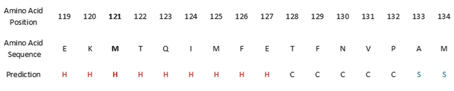 Fig. 1 The predicted secondary structure of ACTA2.1
Key structural properties of ACTA2:
- Conserved ATP-binding domains and divalent cation binding sites
- Actin polymerization interface mediated by clusters of N-terminal acidic amino acids
- Arginine mutation point 73rd (directly related to the onset of aortic aneurysm)
- Calmodulin-specific binding region composed of C-terminal phenylalanine residues
Functions of ACTA2
The ACTA2 gene encodes α -smooth muscle actin, whose core function is to maintain vascular structure and regulate contraction, and it is also involved in multiple pathophysiological processes:
| Function | Description |
| Regulation of vascular contraction | Through the calmodulin-dependent myosin light chain phosphorylation pathway, it regulates vascular tone and blood pressure. |
| Cytoskeleton construction | Aggregation forms a microfilament network, maintaining the mechanical stability and morphology of vascular smooth muscle cells. |
| Regulation of wound healing | After tissue damage to promote muscle fibroblast differentiation and reconstruction of extracellular matrix. |
| Atherosclerosis | Abnormal expression under pathological conditions leads to intimal proliferation of blood vessels and migration of smooth muscle cells. |
| Aortic aneurysm formation | Gene mutations (such as R179H) cause the disintegration of mesenchymal smooth muscle, increasing the risk of vascular rupture. |
Unlike the ACTB gene in skeletal muscle, the expression of ACTA2 is strictly limited by the smooth muscle lineage, and its promoter contains specific binding elements for serum response factor (SRF) and Myocardin. This regulatory characteristic makes ACTA2 a core molecular marker for vascular development and disease.
Applications of ACTA2 and ACTA2 Antibody in Literature
1. Yuan-Min, Shi. "[alpha]-Smooth Muscle Actin and ACTA2 Gene Expressions in Vasculopathies." BJCVS 30.6 (2015). https://doi.org/10.5935/1678-9741.20150081
The article indicates that the ACTA2 gene encodes α -smooth muscle actin, and its mutation can lead to various vascular lesions such as thoracic aortic aneurysm and coronary artery disease. Currently, the related pathogenic mechanism has not been fully elucidated. This article aims to explore the mechanism of action of ACTA2 mutations in vascular lesions.
2. Peng, Yi, aoxi Huang, and Hongmei Wang. "lncRNA ACTA2-AS1 predicts malignancy and poor prognosis of triple-negative breast cancer and regulates tumor progression via modulating miR-532-5p." BMC molecular and cell biology 23.1 (2022): 34. https://doi.org/10.1186/s12860-022-00432-7
This study found that in triple-negative breast cancer (TNBC), the low expression of ACTA2-AS1 was significantly negatively correlated with the high expression of miR-532-5p, and both were closely related to the poor prognosis of patients. ACTA2-AS1 inhibits tumor progression by regulating miR-532-5p and can serve as a potential biomarker for TNBC.
3. Bobba, Christopher M., et al. "A highly penetrant ACTA2 mutation of thoracic aortic disease." Journal of Cardiothoracic Surgery 18.1 (2023): 352. https://doi.org/10.1186/s13019-023-02420-0
This study indicates that the R118Q mutation of the ACTA2 gene is associated with familial aortic disease. The study found that the penetrant rate of this mutation in carriers over 50 years old is as high as 100%, with an average age of onset of 57.8 years. It is recommended that such patients undergo strict aortic monitoring and actively consider elective surgery when the diameter reaches 4.5 centimeters.
4. Cooper, Kylie, and Stephen Brown. "ACTA2 mutation and postpartum hemorrhage: a case report." BMC Medical Genetics 18.1 (2017): 143. https://doi.org/10.1186/s12881-017-0505-5
This study indicates that ACTA2 gene mutations are commonly seen in vascular diseases such as thoracic aortic aneurysms. This study reports for the first time a case of a pregnant female carrier experiencing postpartum uterine atony and severe bleeding due to the mutation, suggesting that this mutation may cause uterine smooth muscle dysfunction, which requires obstetric vigilance.
5. Lin, Chenxiao, et al. "Knockdown of lncRNA ACTA2-AS1 reverses cisplatin resistance of ovarian cancer cells via inhibition of miR-378a-3p-regulated Wnt5a." Bioengineered 13.4 (2022): 9829-9838. https://doi.org/10.1080/21655979.2022.2061181
This study indicates that in ovarian cancer, ACTA2-AS1 promotes cisplatin resistance by competitively binding to miR-378a-3p, upregulating the expression of Wnt5a. Inhibition of ACTA2-AS1 can enhance the activity of miR-378a-3p and reduce Wnt5a, thereby reversing drug resistance and providing a new target for treatment.
Creative Biolabs: ACTA2 Antibodies for Research
Creative Biolabs specializes in the production of high-quality ACTA2 antibodies for research and industrial applications. Our portfolio includes monoclonal antibodies tailored for ELISA, Flow Cytometry, Western blot, immunohistochemistry, and other diagnostic methodologies.
- Custom ACTA2 Antibody Development: Tailor-made solutions to meet specific research requirements.
- Bulk Production: Large-scale antibody manufacturing for industry partners.
- Technical Support: Expert consultation for protocol optimization and troubleshooting.
- Aliquoting Services: Conveniently sized aliquots for long-term storage and consistent experimental outcomes.
For more details on our ACTA2 antibodies, custom preparations, or technical support, contact us at email.
Reference
- Keravnou, Anna, et al. "Novel variants in the ACTA2 and MYH11 genes in a Cypriot family with thoracic aortic aneurysms: a case report." BMC Medical Genetics 19.1 (2018): 208. https://doi.org/10.1186/s12881-018-0728-0
Anti-ACTA2 antibodies
 Loading...


Hot products 
-
Mouse Anti-CCT6A/B Recombinant Antibody (CBXC-0168) (CBMAB-C5570-CQ)

-
Mouse Anti-ASH1L Monoclonal Antibody (ASH5H03) (CBMAB-1372-YC)

-
Mouse Anti-NSUN6 Recombinant Antibody (D-5) (CBMAB-N3674-WJ)

-
Mouse Anti-BPGM Recombinant Antibody (CBYY-1806) (CBMAB-2155-YY)

-
Mouse Anti-BAD (Phospho-Ser136) Recombinant Antibody (CBYY-0138) (CBMAB-0139-YY)

-
Mouse Anti-BANF1 Recombinant Antibody (3F10-4G12) (CBMAB-A0707-LY)

-
Mouse Anti-CGAS Recombinant Antibody (CBFYM-0995) (CBMAB-M1146-FY)

-
Mouse Anti-DLL4 Recombinant Antibody (D1090) (CBMAB-D1090-YC)

-
Mouse Anti-GFAP Recombinant Antibody (20) (CBMAB-G2914-LY)

-
Mouse Anti-BSN Recombinant Antibody (219E1) (CBMAB-1228-CN)

-
Mouse Anti-AMOT Recombinant Antibody (CBYC-A564) (CBMAB-A2552-YC)

-
Mouse Anti-FTH1 Recombinant Antibody (CBXF-1896) (CBMAB-F3426-CQ)

-
Rat Anti-EMCN Recombinant Antibody (28) (CBMAB-E0280-FY)

-
Mouse Anti-ALB Recombinant Antibody (V2-363290) (CBMAB-S0173-CQ)

-
Mouse Anti-Acetyl-α-Tubulin (Lys40) Recombinant Antibody (V2-623485) (CBMAB-CP2897-LY)

-
Mouse Anti-CCS Recombinant Antibody (CBFYC-1093) (CBMAB-C1150-FY)

-
Mouse Anti-CAT Recombinant Antibody (724810) (CBMAB-C8431-LY)

-
Mouse Anti-CASQ1 Recombinant Antibody (CBFYC-0863) (CBMAB-C0918-FY)

-
Mouse Anti-ADGRE2 Recombinant Antibody (V2-261270) (CBMAB-C0813-LY)

-
Mouse Anti-APC Recombinant Antibody (CBYC-A661) (CBMAB-A3036-YC)

- AActivation
- AGAgonist
- APApoptosis
- BBlocking
- BABioassay
- BIBioimaging
- CImmunohistochemistry-Frozen Sections
- CIChromatin Immunoprecipitation
- CTCytotoxicity
- CSCostimulation
- DDepletion
- DBDot Blot
- EELISA
- ECELISA(Cap)
- EDELISA(Det)
- ESELISpot
- EMElectron Microscopy
- FFlow Cytometry
- FNFunction Assay
- GSGel Supershift
- IInhibition
- IAEnzyme Immunoassay
- ICImmunocytochemistry
- IDImmunodiffusion
- IEImmunoelectrophoresis
- IFImmunofluorescence
- IGImmunochromatography
- IHImmunohistochemistry
- IMImmunomicroscopy
- IOImmunoassay
- IPImmunoprecipitation
- ISIntracellular Staining for Flow Cytometry
- LALuminex Assay
- LFLateral Flow Immunoassay
- MMicroarray
- MCMass Cytometry/CyTOF
- MDMeDIP
- MSElectrophoretic Mobility Shift Assay
- NNeutralization
- PImmunohistologyp-Paraffin Sections
- PAPeptide Array
- PEPeptide ELISA
- PLProximity Ligation Assay
- RRadioimmunoassay
- SStimulation
- SESandwich ELISA
- SHIn situ hybridization
- TCTissue Culture
- WBWestern Blot
