ARG1 Antibodies
Background
The ARG1 gene encodes an argininease, which is mainly present in the liver cells of mammals and is widely expressed in various tissues and cell types. The protein expressed by this gene can catalyze the hydrolysis of arginine into ornithine and urea, and is one of the key enzymes in the urea cycle. It plays an important role in detoxifying ammonia in the body and maintaining nitrogen metabolism balance. The loss or mutation of ARG1's function is closely related to metabolic disorders such as hyperarginineemia, often leading to neurological damage and abnormal growth and development. Since its discovery in the mid-20th century, ARG1 has become an important model for metabolic diseases and gene function research. The study of its structure and mechanism has deepened our understanding of enzyme kinetics, metabolic regulation, and the pathology of hereditary metabolic diseases.
Structure of ARG1
The argininease encoded by the ARG1 gene is a homologous trimeric protein with a molecular weight of approximately 107 kDa. This molecular weight is relatively conserved among different species, but there are slight differences due to subtypes or post-translational modifications.
| Species | Human | Mouse | Rat | Bovine |
| Molecular Weight (kDa) | ~107 | ~105 | ~106 | ~107 |
| Primary Structural Differences | Contains 322 amino acids and has a conserved manganese ion binding site | Highly homologous, with conserved active center sequence | Has an extremely high similarity to the human ARG1 sequence | Highly conserved catalytic core structure |
This protein is composed of three identical subunits. Each subunit has a primary structure containing a dual-manganese ion cluster active center, which is the key to its hydrolytic function. Its secondary structure is mainly α/β folding, which together form the catalytic domain. On the tertiary structure, the three subunits are assembled through non-covalent interactions to form the central active channel. The key amino acid residues include His101 and Asp124, etc., which are responsible for coordinating the manganese ions and stabilizing the transition state, jointly mediating the hydrolysis reaction of arginine.
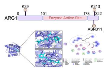 Fig. 1 Molecular Docking of ARG1 with BHB: A Binding Affinity Study.1
Key structural properties of ARG1:
- Presenting a conservative α/β folded trimeric structure
- Each sub containing dinuclear manganese ion (Mn squared ⁺) active center, as the catalytic core
- Active pockets contain highly conserved histidine and aspartic acid residues
- The flexible loop region in the structure is involved in the formation of substrate channels and conformational changes during catalysis
Functions of ARG1
The argininease encoded by the ARG1 gene mainly functions to catalyze the key steps of the urea cycle, regulating nitrogen metabolism and arginine homeostasis in the body. Additionally, it is also widely involved in various pathological and physiological processes.
| Function | Description |
| Urea Synthesis | In the liver, it catalyzes the hydrolysis of arginine into ornithine and urea, which is the main pathway for the body to eliminate toxic ammonia. |
| Regulation of Arginine Homeostasis | By consuming arginine and competing with nitric oxide synthase (NOS) for the substrate, it indirectly regulates the production of nitric oxide (NO). |
| Initiation of Polyamine Synthesis | The catalytic product, ornithine, is a precursor for polyamine biosynthesis and affects cell proliferation and tissue repair. |
| Immune Regulation | It is expressed in immune cells such as macrophages. Changes in its activity can affect the arginine metabolism in the local microenvironment and regulate the immune response. |
| Associated with diseases | Genetic ARG1 deficiency leads to hyperarginineemia; under pathological conditions such as tumors and cardiovascular diseases, abnormal expression of this gene is often associated with metabolic reprogramming. |
The catalytic kinetics of arginase conforms to the Michaelis-Menten equation. Its activity is highly dependent on the bimetallic manganese ion (Mn²⁺) center and is precisely regulated by substrate concentration, pH value, and various intracellular signaling pathways.
Applications of ARG1 and ARG1 Antibody in Literature
- Stratoulias, Vassilis, et al. "ARG1-expressing microglia show a distinct molecular signature and modulate postnatal development and function of the mouse brain." Nature Neuroscience 26.6 (2023): 1008-1020. https://doi.org/10.1038/s41593-023-01326-3
Research in the early postnatal mice basal forebrain and found expression in the ventral striatum arginase 1 unique subtype of microglial cells. This subtype has active phagocytic function, and its molecular markers include upregulation of genes such as Apoe and Clec7a. Specific knockdown of Arg1 by microglia can lead to impaired cholinergic neural innervation and synaptic development in the hippocampus of female mice, thereby causing cognitive dysfunction.
- Yadav, Preeti, et al. "Myeloid-mesenchymal crosstalk drives ARG1-dependent profibrotic metabolism via ornithine in lung fibrosis." The Journal of Clinical Investigation 135.21 (2025). https://doi.org/10.1172/JCI188734
Research has found that in pulmonary fibrosis, myeloid cells produce ornithine through arginase-1 (ARG1), which drives lung fibroblasts to synthesize collagen. Inhibition of ARG1 can reduce collagen expression. This pathway is regulated by the ATP-P2RX4-IL-6 signaling axis, revealing the immune-interstitial metabolic circuit of fibrosis.
- Lin, Chuman, et al. "Abnormal β‐Hydroxybutyrylation Modification of ARG1 Drives Reprogramming of Arginine Metabolism to Promote the Progression of Colorectal Cancer." Advanced Science 12.38 (2025): e02402. https://doi.org/10.1002/advs.202502402
Studies have found that the level of arginine is elevated in colorectal cancer, and the β -hydroxybutyrylation (Kbhb) of its metabolic reprogramming of fertilizinase-1 (ARG1) is regulated. Low levels of ARG1-Kbhb inhibit its binding to the transporter SLC3A2, reduce arginine efflux, and thereby promote tumorigenesis. The research found that inducing ARG1-Kbhb with P300/BHB could reverse this effect.
- Tran, Dinh Nam, et al. "ARG1 is a potential prognostic marker in metastatic and recurrent endometrial cancer." Research Square (2023): rs-3. https://doi.org/10.21203/rs.3.rs-2917380/v1
Studies have found that the expression of arginase-1 (ARG1) is elevated in endometrial cancer of mice. Compared with Pten mutations alone, mice with dual mutations of Pten and Mig-6 had significantly upregulated ARG1 in early cancer and precancerous lesions, and were more prone to distant metastasis, suggesting that high expression of ARG1 was associated with a poor prognosis.
- Fleischer, Amelie B., et al. "Differential Expression of ARG1 and MRC2 in Retinal Müller Glial Cells During Autoimmune Uveitis." Biomolecules 15.2 (2025): 288. https://doi.org/10.3390/biom15020288
Studies have found that in the spontaneous uveitis model, the expression of arginase-1 (ARG1) and MHC class II molecules in retinal Muller glial cells is significantly upregulated, while MRC2 and THBS1 are enriched in healthy cells. This reveals that ARG1 can serve as a potential biomarker for cells' participation in immune regulation under neuroinflammatory conditions.
Creative Biolabs: ARG1 Antibodies for Research
Creative Biolabs specializes in the production of high-quality ARG1 antibodies for research and industrial applications. Our portfolio includes monoclonal antibodies tailored for ELISA, Flow Cytometry, Western blot, immunohistochemistry, and other diagnostic methodologies.
- Custom ARG1 Antibody Development: Tailor-made solutions to meet specific research requirements.
- Bulk Production: Large-scale antibody manufacturing for industry partners.
- Technical Support: Expert consultation for protocol optimization and troubleshooting.
- Aliquoting Services: Conveniently sized aliquots for long-term storage and consistent experimental outcomes.
For more details on our ARG1 antibodies, custom preparations, or technical support, contact us at email.
Reference
- Lin, Chuman, et al. "Abnormal β‐Hydroxybutyrylation Modification of ARG1 Drives Reprogramming of Arginine Metabolism to Promote the Progression of Colorectal Cancer." Advanced Science 12.38 (2025): e02402. https://doi.org/10.1002/advs.202502402
Anti-ARG1 antibodies
 Loading...


Hot products 
-
Rabbit Anti-CCN1 Recombinant Antibody (CBWJC-3580) (CBMAB-C4816WJ)

-
Mouse Anti-ADAM29 Recombinant Antibody (V2-179787) (CBMAB-A1149-YC)

-
Mouse Anti-BrdU Recombinant Antibody (IIB5) (CBMAB-1038CQ)

-
Mouse Anti-BZLF1 Recombinant Antibody (BZ.1) (CBMAB-AP705LY)

-
Mouse Anti-ADAM12 Recombinant Antibody (V2-179752) (CBMAB-A1114-YC)

-
Mouse Anti-CD83 Recombinant Antibody (HB15) (CBMAB-C1765-CQ)

-
Mouse Anti-AOC3 Recombinant Antibody (CBYY-0014) (CBMAB-0014-YY)

-
Mouse Anti-CEMIP Recombinant Antibody (3C12) (CBMAB-K0296-LY)

-
Mouse Anti-ADGRL2 Recombinant Antibody (V2-58519) (CBMAB-L0166-YJ)

-
Mouse Anti-ATP1B1 Recombinant Antibody (E4) (CBMAB-0463-LY)

-
Mouse Anti-ARHGAP5 Recombinant Antibody (54/P190-B) (CBMAB-P0070-YC)

-
Rat Anti-(1-5)-α-L-Arabinan Recombinant Antibody (V2-501861) (CBMAB-XB0003-YC)

-
Mouse Anti-BHMT Recombinant Antibody (CBYY-0547) (CBMAB-0550-YY)

-
Mouse Anti-AQP2 Recombinant Antibody (G-3) (CBMAB-A3359-YC)

-
Mouse Anti-GFAP Recombinant Antibody (5) (CBMAB-G0346-LY)

-
Mouse Anti-ARID3A Antibody (A4) (CBMAB-0128-YC)

-
Mouse Anti-CASP8 Recombinant Antibody (CBYY-C0987) (CBMAB-C2424-YY)

-
Mouse Anti-AKT1 (Phosphorylated S473) Recombinant Antibody (V2-505430) (PTM-CBMAB-0067LY)

-
Mouse Anti-EGR1 Recombinant Antibody (CBWJZ-100) (CBMAB-Z0289-WJ)

-
Mouse Anti-FTH1 Recombinant Antibody (CBXF-1896) (CBMAB-F3426-CQ)

- AActivation
- AGAgonist
- APApoptosis
- BBlocking
- BABioassay
- BIBioimaging
- CImmunohistochemistry-Frozen Sections
- CIChromatin Immunoprecipitation
- CTCytotoxicity
- CSCostimulation
- DDepletion
- DBDot Blot
- EELISA
- ECELISA(Cap)
- EDELISA(Det)
- ESELISpot
- EMElectron Microscopy
- FFlow Cytometry
- FNFunction Assay
- GSGel Supershift
- IInhibition
- IAEnzyme Immunoassay
- ICImmunocytochemistry
- IDImmunodiffusion
- IEImmunoelectrophoresis
- IFImmunofluorescence
- IGImmunochromatography
- IHImmunohistochemistry
- IMImmunomicroscopy
- IOImmunoassay
- IPImmunoprecipitation
- ISIntracellular Staining for Flow Cytometry
- LALuminex Assay
- LFLateral Flow Immunoassay
- MMicroarray
- MCMass Cytometry/CyTOF
- MDMeDIP
- MSElectrophoretic Mobility Shift Assay
- NNeutralization
- PImmunohistologyp-Paraffin Sections
- PAPeptide Array
- PEPeptide ELISA
- PLProximity Ligation Assay
- RRadioimmunoassay
- SStimulation
- SESandwich ELISA
- SHIn situ hybridization
- TCTissue Culture
- WBWestern Blot
