ART1 Antibodies
Background
ART1 encodes an ADP-ribosyltransferase protein that is mainly expressed on the surface of human T lymphocytes and some tumor cells. This protein participates in immunomodulatory signal transduction by mediating ADP-ribonylation modification. It can not only regulate the activation function of T cells but also affect the apoptotic pathway. Due to its dual role in autoimmune diseases and cancer immune escape, ART1 has become an important target in immunotherapy research. This gene was first identified by Guse et al. in 1999. Its unique glycosylation mechanism provides a molecular basis for the development of novel immune checkpoint inhibitors. Scientists have resolved the complex structure of ART1 and its receptor through cryo-electron microscopy, revealing its "induction-alignment" catalytic mechanism. These findings not only advance the development of tumor immunotherapy strategies but also deepen our understanding of the regulatory network of post-translational modifications of proteins.
Structure of ART1
ART1 is a protein with a molecular weight of approximately 36 kDa. This molecular weight shows slight differences among different mammals, mainly due to interspecific variations in amino acid sequences.
| Species | Human | Mouse | Rat | Rhesus monkey |
| Molecular Weight (kDa) | 36.0 | 35.8 | 35.9 | 36.1 |
| Primary Structural Differences | Conserved domains with catalytic activity | Across the membrane area exists two amino acid replacement | The C-terminal glycosylation sites are slightly different | Highly homologous to human ART1 |
ART1 is composed of 316 amino acids and presents a typical topological structure of type I transmembrane proteins as a whole. Its extracellular domain contains multiple conserved ADP-ribosyltransferase active sites, which can catalyze the ADP-ribosylation modification of substrate proteins. The spatial conformation of this enzyme forms a deep hydrophobic active pocket, and the key residues arginine-188 and glutamine-193 jointly participate in the NAD+ hydrolysis and ADP-ribose transfer processes. The cysteine residues near the cell membrane can stabilize the protein structure through disulfide bonds, while the intracellular segment contains multiple phosphorylation sites and is involved in signal transduction regulation.
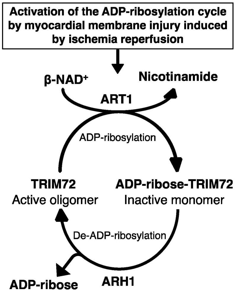 Fig. 1 The key role of ART1-mediated TRIM72 ADP-ribonylation in cell membrane repair.1
Key structural properties of ART1:
- Typical structure of glycosyl phosphatidylinositol (GPI) -anchored membrane proteins
- Extracellular domain contains a conservative ADP ribose base transferase catalytic core
- Have NAD + combined with pockets and specific substrate recognition area
Functions of ART1
The main function of the ART1 gene is to mediate the ADP-ribonylation modification of proteins and participate in immune regulation and cellular signal transduction. In addition, this gene also plays a role in a variety of pathophysiological processes, including inflammatory responses and tumor immune escape.
| Function | Description |
| Immune regulation | By modifying the surface proteins of T cells, it affects the activation of T cells and the intensity of immune responses, maintaining immune homeostasis. |
| Cell signal regulation | Involved in the ADP-ribosylation of G protein-coupled receptors and other signaling molecules, affecting downstream pathways such as apoptosis and proliferation. |
| Inflammatory response | In the high expression of immune cells such as macrophages, regulate inflammatory factors and inflammatory signaling pathways. |
| Tumor immune escape | Expressed abnormally in some cancer cells, it helps tumors evade host immune surveillance by modifying molecules on the surface of immune cells. |
| Protection against ischemia-reperfusion injury | Participate in regulating oxidative stress in the cardiac muscle and nerve tissue and cell survival signals, reduce tissue injury caused by ischemia. |
ART1 has a high affinity for NAD⁺, and its enzymatic activity depends on specific basic residues and conserved catalytic domains. It can rapidly catalyze single ADP-ribonylation in a local microenvironment. This characteristic is closely related to its precise functional regulation in immune synapses.
Applications of ART1 and ART1 Antibody in Literature
1. Lin, Ting, et al. "ART1 knockdown decreases the IL-6-induced proliferation of colorectal cancer cells." BMC cancer 24.1 (2024): 354. https://doi.org/10.1186/s12885-024-12120-0
The article indicates that ART1 affects the IL-6 signaling pathway by regulating gp130, promoting the proliferation of colorectal cancer cells. Inhibition of ART1 can reduce the phosphorylation of STAT3 and the expression of downstream target proteins, and inhibit tumor growth, indicating its potential as a therapeutic target.
2. Wu, Zhi, et al. "Pan-cancer analysis of ART1 and its potential value in gastric cancer." Journal of Cancer 15.12 (2024): 3684. https://doi.org/10.7150/jca.96033
The article indicates that the expression of ART1 is closely related to the prognosis of various cancers and the immune microenvironment, especially playing a significant role in digestive tract tumors. Studies have confirmed that ART1 promotes tumor development by regulating signaling pathways such as NF-κB and is a potential target for immunotherapy.
3. Friedrich, Maik, et al. "Identification of two regulatory binding sites which confer myotube specific expression of the mono-ADP-ribosyltransferase ART1 gene." BMC Molecular Biology 9.1 (2008): 91. https://doi.org/10.1186/1471-2199-9-91
The article indicates that the expression of the ART1 gene in skeletal muscle cells is regulated by myoietin and MEF-2. The two bind to the E box and A/T enrichment elements in the promoter region respectively, and synergically induce its transcription, thereby promoting the formation of myotubes and the adhesion of extracellular matrix.
4. Martínez-Márquez, Jorge Y., and Mara C. Duncan. "Investigation of Ldb19/Art1 localization and function at the late Golgi." PLoS One 13.11 (2018): e0206944. https://doi.org/10.1371/journal.pone.0206944
Studies have found that yeast protein Ldb19/Art1, in addition to mediating endocytosis, is also located in the late stage of reverse Golgi (TGN). Its function is related to the AP-1 adaptor protein complex but the mechanism of action is different, indicating that it has a unique regulatory function in TGN membrane transport.
5. Ishiwata-Endo, Hiroko, et al. "ARH1 in health and disease." Cancers 12.2 (2020): 479. https://doi.org/10.3390/cancers12020479
The article indicates that ARH1 is a demodifying enzyme that specifically hydrolyzes arginine ADP-ribonylation. Together with ART1, it forms a reversible modification cycle and participates in processes such as myocardial injury repair and tumorigenesis. Its absence will promote tumor formation in a gender-specific manner.
Creative Biolabs: ART1 Antibodies for Research
Creative Biolabs specializes in the production of high-quality ART1 antibodies for research and industrial applications. Our portfolio includes monoclonal antibodies tailored for ELISA, Flow Cytometry, Western blot, immunohistochemistry, and other diagnostic methodologies.
- Custom ART1 Antibody Development: Tailor-made solutions to meet specific research requirements.
- Bulk Production: Large-scale antibody manufacturing for industry partners.
- Technical Support: Expert consultation for protocol optimization and troubleshooting.
- Aliquoting Services: Conveniently sized aliquots for long-term storage and consistent experimental outcomes.
For more details on our ART1 antibodies, custom preparations, or technical support, contact us at email.
Reference
- Ishiwata-Endo, Hiroko, et al. "ARH1 in health and disease." Cancers 12.2 (2020): 479. https://doi.org/10.3390/cancers12020479
Anti-ART1 antibodies
Products List
 Loading...


Hot products 
-
Mouse Anti-AFM Recombinant Antibody (V2-634159) (CBMAB-AP185LY)

-
Mouse Anti-AAV-5 Recombinant Antibody (V2-503416) (CBMAB-V208-1402-FY)

-
Rabbit Anti-AKT3 Recombinant Antibody (V2-12567) (CBMAB-1057-CN)

-
Mouse Anti-CD33 Recombinant Antibody (6C5/2) (CBMAB-C8126-LY)

-
Mouse Anti-ENO1 Recombinant Antibody (CBYC-A950) (CBMAB-A4388-YC)

-
Mouse Anti-2C TCR Recombinant Antibody (V2-1556) (CBMAB-0951-LY)

-
Mouse Anti-EGR1 Recombinant Antibody (CBWJZ-100) (CBMAB-Z0289-WJ)

-
Mouse Anti-FTH1 Recombinant Antibody (CBXF-1896) (CBMAB-F3426-CQ)

-
Mouse Anti-ATP1A2 Recombinant Antibody (M7-PB-E9) (CBMAB-A4013-YC)

-
Mouse Anti-CSPG4 Recombinant Antibody (CBFYM-1050) (CBMAB-M1203-FY)

-
Mouse Anti-AFDN Recombinant Antibody (V2-58751) (CBMAB-L0408-YJ)

-
Mouse Anti-4-Hydroxynonenal Recombinant Antibody (V2-502280) (CBMAB-C1055-CN)

-
Rat Anti-FABP3 Recombinant Antibody (CBXF-2299) (CBMAB-F1612-CQ)

-
Mouse Anti-FLI1 Recombinant Antibody (CBXF-0733) (CBMAB-F0435-CQ)

-
Mouse Anti-CCN2 Recombinant Antibody (CBFYC-2383) (CBMAB-C2456-FY)

-
Mouse Anti-FOXA3 Recombinant Antibody (2A9) (CBMAB-0377-YC)

-
Mouse Anti-BIRC3 Recombinant Antibody (315304) (CBMAB-1214-CN)

-
Mouse Anti-ENO2 Recombinant Antibody (85F11) (CBMAB-0276CQ)

-
Mouse Anti-HTLV-1 gp46 Recombinant Antibody (CBMW-H1006) (CBMAB-V208-1154-FY)

-
Mouse Anti-ALB Recombinant Antibody (V2-55272) (CBMAB-H0819-FY)

- AActivation
- AGAgonist
- APApoptosis
- BBlocking
- BABioassay
- BIBioimaging
- CImmunohistochemistry-Frozen Sections
- CIChromatin Immunoprecipitation
- CTCytotoxicity
- CSCostimulation
- DDepletion
- DBDot Blot
- EELISA
- ECELISA(Cap)
- EDELISA(Det)
- ESELISpot
- EMElectron Microscopy
- FFlow Cytometry
- FNFunction Assay
- GSGel Supershift
- IInhibition
- IAEnzyme Immunoassay
- ICImmunocytochemistry
- IDImmunodiffusion
- IEImmunoelectrophoresis
- IFImmunofluorescence
- IGImmunochromatography
- IHImmunohistochemistry
- IMImmunomicroscopy
- IOImmunoassay
- IPImmunoprecipitation
- ISIntracellular Staining for Flow Cytometry
- LALuminex Assay
- LFLateral Flow Immunoassay
- MMicroarray
- MCMass Cytometry/CyTOF
- MDMeDIP
- MSElectrophoretic Mobility Shift Assay
- NNeutralization
- PImmunohistologyp-Paraffin Sections
- PAPeptide Array
- PEPeptide ELISA
- PLProximity Ligation Assay
- RRadioimmunoassay
- SStimulation
- SESandwich ELISA
- SHIn situ hybridization
- TCTissue Culture
- WBWestern Blot
