BNIP3 Antibodies
Background
BNIP3 is a pro-apoptotic protein located on the outer mitochondrial membrane and belongs to the Bcl-2 homology domain protein family. This protein binds to the mitochondrial membrane through its transmembrane domain and its expression significantly increases under hypoxic conditions. It can induce mitochondrial permeability transition and initiate the apoptotic program. As a key regulatory factor of cellular stress response, BNIP3 not only participates in the death regulation of cardiomyocytes in ischemia-reperfusion injury, but also plays an important role in the hypoxic microenvironment of various solid tumors. It maintains tumor cell survival or induces cell death through the dual functions of mediating mitochondrial autophagy. This gene was initially discovered and cloned in the 1990s and was named after its sequence homology with the interaction protein of adenovirus E1B 19kDa. Subsequent studies revealed that it interacts with Bcl-2 family members through the BH3 domain and established a molecular link between cell death and mitochondrial quality control. Currently, BNIP3 has been confirmed as a target gene of hypoxia-inducible factor HIF-1. The mechanism of its role in cardiovascular diseases, neurodegenerative disorders, and tumor occurrence and development is becoming a research hotspot.
Structure of BNIP3
BNIP3 is a mitochondrial membrane protein with a molecular weight of approximately 21.5 kDa. Its size varies slightly among different species due to differences in amino acid sequences.
| Species | Human | Mouse | Rat | Pig | Bovine |
| Molecular Weight (kDa) | 21.5 | 21.3 | 21.4 | 21.6 | 21.5 |
| Primary Structural Differences | The classic BH3 domain, with the most in-depth functional research | The C-terminal transmembrane region is highly conserved | Similarity of hypoxia response element sequences | High homology of transmembrane domains | Has a homology of over 85% with humans |
This protein is composed of 194 amino acids and possesses a characteristic mitochondrial targeting structure. BNIP3 contains an N-terminal PEST sequence that participates in the regulation of protein degradation, a conserved BH3 domain that mediates interaction with Bcl-2 family members, and a C-terminal transmembrane domain responsible for anchoring to the outer mitochondrial membrane. This transmembrane domain is also a key region for protein dimerization and can form ion channel-like pores. The pro-apoptotic activity of BNIP3 strictly depends on the integrity of its transmembrane domain, where the glycine residue at position 193 is crucial for dimer formation. This protein plays a central role in mitochondrial permeability transition by inserting its hydrophobic amino acids into the mitochondrial membrane through its transmembrane region.
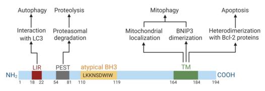 Fig. 1 Domain structure of BNIP3 and its domain-dependent functions.1
Key structural properties of BNIP3:
- The conserved BH3 domain mediates protein-protein interactions
- The C-terminal transmembrane domain is responsible for mitochondrial localization and dimerization
- The hydrophobic amino acid region required for membrane integration
- The PEST sequence that regulates protein stability
Functions of BNIP3
The core function of BNIP3 is to regulate mitochondrial homeostasis and cell death. However, it also plays a role in various cellular processes, including mitochondrial autophagy and hypoxia response.
| Function | Description |
| Induction of cell apoptosis | BNIP3 inserts into the mitochondrial membrane through its transmembrane domain, increasing membrane permeability and releasing pro-apoptotic factors. |
| Regulating Mitochondrial Autophagy | As a mitochondrial receptor, it directly interacts with LC3 and mediates the selective clearance of damaged mitochondria. |
| Hypoxia response | Activated by HIF-1 under hypoxic conditions, it helps cells adapt to the low-oxygen environment and balance survival and death signals. |
| Regulation of Mitochondrial Dynamics | BNIP3 affects the fusion and fission processes of mitochondria, and participates in the maintenance of mitochondrial network morphology and quality control. |
| Heart Protection and Injury | It plays a dual role in ischemia-reperfusion. Moderate induction can trigger protective autophagy, while excessive activation leads to cardiomyocyte death. |
The expression regulation of BNIP3 is closely related to its functional performance. Under normal circumstances, it remains at a low level. However, it rapidly increases in response to hypoxia or stress stimuli. Unlike other members of the Bcl-2 family, the mechanism by which BNIP3 induces cell death does not rely on the classic BH3 domain binding pattern, but rather more on the mitochondrial membrane integration and dimerization mediated by its transmembrane domain.
Applications of BNIP3 and BNIP3 Antibody in Literature
1. Gorbunova, Anna S., et al. "BNIP3 in lung cancer: to kill or rescue?." Cancers 12.11 (2020): 3390. https://doi.org/10.3390/cancers12113390
The research has found that BNIP3 is an apoptotic protein that participates in autophagy and tumor metabolism. Its role in lung cancer is still unclear, but mitochondrial function affects the progression of lung cancer. This article reviews the research on BNIP3 in various cancers, especially lung cancer, and explores its potential as a therapeutic target for lung cancer.
2. Fornelli, Claudia, et al. "BNIP3 Downregulation ameliorates muscle atrophy in cancer cachexia." Cancers 16.24 (2024): 4133. https://doi.org/10.3390/cancers16244133
The research has found that cancer cachexia leads to muscle atrophy and is related to mitochondrial autophagy. In this study, by silencing the elevated BNIP3 in the muscles, it was discovered that adenovirus-mediated knockdown could maintain mitochondrial quality and partially improve the size of muscle fibers, providing a potential strategy for alleviating muscle loss caused by cancer.
3. Li, Jialin, et al. "HIF1α-BNIP3-mediated mitophagy protects against renal fibrosis by decreasing ROS and inhibiting activation of the NLRP3 inflammasome." Cell death & disease 14.3 (2023): 200. https://doi.org/10.1038/s41419-023-05587-5
The research has found that BNIP3 mediates mitochondrial autophagy in renal fibrosis. By reducing mitochondrial reactive oxygen species and inhibiting the activation of NLRP3 inflammasome, it plays a protective role in hypoxic renal tubular epithelial cell damage, providing a new target for treatment.
4. Jin, Yuxin, et al. "Hypoxia‐preconditioned BMSC‐derived exosomes induce mitophagy via the BNIP3–ANAX2 axis to alleviate intervertebral disc degeneration." Advanced Science 11.34 (2024): 2404275. https://doi.org/10.1002/advs.202404275
The study found that hypoxia-induced BMSC exosomes deliver BNIP3, which activates the BNIP3/ANXA2/TFEB axis to improve mitochondrial autophagy, delay the aging of nucleus pulposus cells and repair intervertebral disc degeneration, providing a new target for treatment.
5. Zhang, Zeying, et al. "KPNB1-ATF4 induces BNIP3-dependent mitophagy to drive odontoblastic differentiation in dental pulp stem cells." Cellular & Molecular Biology Letters 29.1 (2024): 145. https://doi.org/10.1186/s11658-024-00664-9
The research has found that when dental pulp stem cells differentiate into odontoblasts, KPNB1 mediates the nuclear entry of ATF4, activating the transcription of BNIP3, enhancing mitochondrial autophagy, eliminating reactive oxygen species and promoting differentiation. This pathway provides a new target for dental pulp regeneration.
Creative Biolabs: BNIP3 Antibodies for Research
Creative Biolabs specializes in the production of high-quality BNIP3 antibodies for research and industrial applications. Our portfolio includes monoclonal and polyclonal antibodies tailored for ELISA, Flow Cytometry, Western blot, immunohistochemistry, and other diagnostic methodologies.
- Custom BNIP3 Antibody Development: Tailor-made solutions to meet specific research requirements.
- Bulk Production: Large-scale antibody manufacturing for industry partners.
- Technical Support: Expert consultation for protocol optimization and troubleshooting.
- Aliquoting Services: Conveniently sized aliquots for long-term storage and consistent experimental outcomes.
For more details on our BNIP3 antibodies, custom preparations, or technical support, contact us at email.
Reference
- Gorbunova, Anna S., et al. "BNIP3 in lung cancer: to kill or rescue?." Cancers 12.11 (2020): 3390. Distributed under Open Access license CC BY 4.0, without modification. https://doi.org/10.3390/cancers12113390
Anti-BNIP3 antibodies
 Loading...


Hot products 
-
Mouse Anti-C4B Recombinant Antibody (CBYY-C2996) (CBMAB-C4439-YY)

-
Mouse Anti-CAPZB Recombinant Antibody (CBYY-C0944) (CBMAB-C2381-YY)

-
Mouse Anti-FN1 Monoclonal Antibody (D6) (CBMAB-1240CQ)

-
Mouse Anti-DLG1 Monolconal Antibody (4F3) (CBMAB-0225-CN)

-
Human Anti-SARS-CoV-2 S1 Monoclonal Antibody (CBFYR-0120) (CBMAB-R0120-FY)

-
Mouse Anti-CCDC25 Recombinant Antibody (CBLC132-LY) (CBMAB-C9786-LY)

-
Mouse Anti-CD83 Recombinant Antibody (HB15) (CBMAB-C1765-CQ)

-
Rat Anti-FABP3 Recombinant Antibody (CBXF-2299) (CBMAB-F1612-CQ)

-
Mouse Anti-DMD Recombinant Antibody (D1190) (CBMAB-D1190-YC)

-
Mouse Anti-ARIH1 Recombinant Antibody (C-7) (CBMAB-A3563-YC)

-
Mouse Anti-C5AR1 Recombinant Antibody (R63) (CBMAB-C9553-LY)

-
Mouse Anti-CCN2 Recombinant Antibody (CBFYC-2383) (CBMAB-C2456-FY)

-
Mouse Anti-CTCF Recombinant Antibody (CBFYC-2371) (CBMAB-C2443-FY)

-
Mouse Anti-ARHGDIA Recombinant Antibody (CBCNA-009) (CBMAB-R0415-CN)

-
Mouse Anti-ALDOA Recombinant Antibody (A2) (CBMAB-A2316-YC)

-
Mouse Anti-AFM Recombinant Antibody (V2-634159) (CBMAB-AP185LY)

-
Mouse Anti-ATP1B1 Recombinant Antibody (E4) (CBMAB-0463-LY)

-
Mouse Anti-ACVR1C Recombinant Antibody (V2-179685) (CBMAB-A1041-YC)

-
Mouse Anti-APP Recombinant Antibody (DE2B4) (CBMAB-1122-CN)

-
Mouse Anti-BRCA2 Recombinant Antibody (CBYY-1728) (CBMAB-2077-YY)

- AActivation
- AGAgonist
- APApoptosis
- BBlocking
- BABioassay
- BIBioimaging
- CImmunohistochemistry-Frozen Sections
- CIChromatin Immunoprecipitation
- CTCytotoxicity
- CSCostimulation
- DDepletion
- DBDot Blot
- EELISA
- ECELISA(Cap)
- EDELISA(Det)
- ESELISpot
- EMElectron Microscopy
- FFlow Cytometry
- FNFunction Assay
- GSGel Supershift
- IInhibition
- IAEnzyme Immunoassay
- ICImmunocytochemistry
- IDImmunodiffusion
- IEImmunoelectrophoresis
- IFImmunofluorescence
- IGImmunochromatography
- IHImmunohistochemistry
- IMImmunomicroscopy
- IOImmunoassay
- IPImmunoprecipitation
- ISIntracellular Staining for Flow Cytometry
- LALuminex Assay
- LFLateral Flow Immunoassay
- MMicroarray
- MCMass Cytometry/CyTOF
- MDMeDIP
- MSElectrophoretic Mobility Shift Assay
- NNeutralization
- PImmunohistologyp-Paraffin Sections
- PAPeptide Array
- PEPeptide ELISA
- PLProximity Ligation Assay
- RRadioimmunoassay
- SStimulation
- SESandwich ELISA
- SHIn situ hybridization
- TCTissue Culture
- WBWestern Blot
