CCR4 Antibodies
Background
The CCR4 gene encodes a cell membrane protein belonging to the G protein-coupled receptor family and is mainly expressed on the surface of lymphocytes. This protein participates in the directed migration of immune cells by specifically binding to chemokines (such as CCL17 and CCL22), playing a key role in inflammatory responses and immune regulation. In 2001, the team led by Japanese scholar Yoshie first clarified its immune function. The research found that the deletion of this gene would lead to abnormal T cell migration, triggering autoimmune diseases or allergic reactions. Its unique seven-time transmembrane domain has become an important target for the development of immunotherapy drugs, and related research has greatly promoted the understanding of the mechanisms of Th2-related diseases such as asthma and atopic dermatitis.
Structure of CCR4
CCR4 is a G protein-coupled receptor with a molecular weight of approximately 41 kDa, and its molecular weight fluctuates slightly among different mammals due to differences in the degree of glycosylation.
| Species | Human | Mouse | Rat | Rhesus monkey |
| Molecular Weight (kDa) | 41 | 40.5 | 40.8 | 41.2 |
| Primary Structural Differences | Containing 355 amino acids, the N-terminal domain mediates ligand recognition | 78% homology with human | Intracellular area structure highly conservative | 95% homology with human sequence |
This protein is composed of 355 amino acids and forms a typical seven-transmembrane topological structure. Its N-terminal extracellular domain contains multiple glycosylation sites, which are responsible for specific binding to chemokines such as CCL17 and CCL22. The three intracellular loops and the C-terminal together form the G protein-coupled region, which mediates downstream signal transduction through Gai proteins. The acidic amino acid cluster on the second extracellular ring is the key site for ligand binding, while the conserved DRY motif in the third transmembrane region maintains the basic activity state of the receptor.
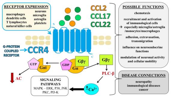 Fig. 1 CCR4—mechanisms of action, possible roles, cellular expression, and disease connections.1
Key structural properties of CCR4:
- Typical configuration of the seven-fold transmembrane helix topology
- Extracellular N-terminal domains mediate ligand-specific recognition
- Conserved DRY motifs maintain receptor signal transduction functions
Functions of CCR4
The main function of CCR4 is to mediate the migration of immune cells and inflammatory responses, and it also participates in multiple immune regulatory processes.
| Function | Description |
| Chemotaxis of immune cells | By recognizing chemokines such as CCL17 and CCL22, Th2 cells and Treg cells are guided to migrate directively to the inflammatory site. |
| Regulation of immune homeostasis | Participate in T cells in the thymus of the selection and development, to maintain their own immune tolerance. |
| Amplification of inflammatory response | Promote the aggregation of inflammatory cells such as eosinophils at the lesion site and aggravate allergic reactions. |
| Shaping of the tumor microenvironment | Assist tumor-associated T cells in infiltrating tumor tissues and affect the efficacy of tumor immunotherapy. |
| Pathogen defense | Guide immune cells to the infection site and participate in the immune response against parasites and viruses. |
The signal transduction of CCR4 exhibits rapid desensitization characteristics. After binding with ligands, it is quickly internalized. This mechanism not only ensures the timeliness of chemotactic responses but also prevents excessive inflammatory damage.
Applications of CCR4 and CCR4 Antibody in Literature
1. Yoshie, Osamu. "CCR4 as a therapeutic target for cancer immunotherapy." Cancers 13.21 (2021): 5542. https://doi.org/10.3390/cancers13215542
The article indicates that CCR4 is a receptor expressed in T cells such as Th2 and Treg, as well as in T-cell malignancies like ATLL and CTCL. Moglializumab, as a humanized monoclonal antibody targeting CCR4, can effectively eliminate malignant T cells and Tregs, showing potential in the treatment of T-cell lymphoma and solid tumors, but it may cause adverse skin reactions and graft-versus-host disease.
2. Chalabi Hagkarim, Nafiseh, and Roger J. Grand. "The regulatory properties of the Ccr4–Not complex." Cells 9.11 (2020): 2379. https://doi.org/10.3390/cells9112379
The article indicates that Ccr4-Not is a structurally conserved multifunctional complex that coordinates gene expression in the nucleus and cytoplasm in both yeast and mammals by regulating mechanisms such as transcription and mRNA degradation.
3. Bogacka, Joanna, et al. "CC chemokine receptor 4 (CCR4) as a possible new target for therapy." International Journal of Molecular Sciences 23.24 (2022): 15638. https://doi.org/10.3390/ijms232415638
The article indicates that CCR4 is a chemokine receptor, and its ligands are involved in various diseases such as immunity, tumors, and neuropathic pain. Its pharmacological blocking not only treats immune inflammation but also has analgesic effects, making it a potential new therapeutic target.
4. Khabipov, Aydar, et al. "CCR4 blockade diminishes intratumoral macrophage recruitment and augments survival of syngeneic pancreatic cancer-bearing mice." Biomedicines 11.6 (2023): 1517. https://doi.org/10.3390/biomedicines11061517
The article indicates that CCR4 plays a key role in regulating the infiltration of tumor-associated macrophages (Tams) and tumor progression in pancreatic cancer. Studies have confirmed that blocking CCR4 through gene knockout or antagonists can effectively reduce TAM recruitment, inhibit tumor growth and prolong the survival period of mice, suggesting its potential as a new target for pancreatic cancer treatment.
5. Deng, Shuixiang, et al. "Recombinant CCL17-dependent CCR4 activation alleviates neuroinflammation and neuronal apoptosis through the PI3K/AKT/Foxo1 signaling pathway after ICH in mice." Journal of Neuroinflammation 18.1 (2021): 62. https://doi.org/10.1186/s12974-021-02112-3
This study confirmed that activating the CCR4 receptor after cerebral hemorrhage can alleviate neuroinflammation and neuronal apoptosis through the PI3K/AKT/Foxo1 signaling pathway, improve cerebral edema and neurological deficits, and provide a new strategy for the early treatment of cerebral hemorrhage.
Creative Biolabs: CCR4 Antibodies for Research
Creative Biolabs specializes in the production of high-quality CCR4 antibodies for research and industrial applications. Our portfolio includes monoclonal antibodies tailored for ELISA, Flow Cytometry, Western blot, immunohistochemistry, and other diagnostic methodologies.
- Custom CCR4 Antibody Development: Tailor-made solutions to meet specific research requirements.
- Bulk Production: Large-scale antibody manufacturing for industry partners.
- Technical Support: Expert consultation for protocol optimization and troubleshooting.
- Aliquoting Services: Conveniently sized aliquots for long-term storage and consistent experimental outcomes.
For more details on our CCR4 antibodies, custom preparations, or technical support, contact us at email.
Reference
- Bogacka, Joanna, et al. "CC chemokine receptor 4 (CCR4) as a possible new target for therapy." International Journal of Molecular Sciences 23.24 (2022): 15638. https://doi.org/10.3390/ijms232415638
Anti-CCR4 antibodies
 Loading...


Hot products 
-
Mouse Anti-ENO1 Recombinant Antibody (CBYC-A950) (CBMAB-A4388-YC)

-
Mouse Anti-AGK Recombinant Antibody (V2-258056) (CBMAB-M0989-FY)

-
Mouse Anti-CD24 Recombinant Antibody (2Q1282) (CBMAB-C1624-CN)

-
Mouse Anti-CD2AP Recombinant Antibody (BR083) (CBMAB-BR083LY)

-
Mouse Anti-ADRB2 Recombinant Antibody (V2-180026) (CBMAB-A1420-YC)

-
Mouse Anti-CORO1A Recombinant Antibody (4G10) (V2LY-1206-LY806)

-
Mouse Anti-AMH Recombinant Antibody (5/6) (CBMAB-A2527-YC)

-
Mouse Anti-FPR2 Recombinant Antibody (1D6) (CBMAB-F2628-CQ)

-
Mouse Anti-CD63 Recombinant Antibody (CBXC-1200) (CBMAB-C1467-CQ)

-
Mouse Anti-BAX Recombinant Antibody (CBYY-0216) (CBMAB-0217-YY)

-
Mouse Anti-ENPP1 Recombinant Antibody (CBFYE-0159) (CBMAB-E0375-FY)

-
Mouse Anti-BAD (Phospho-Ser136) Recombinant Antibody (CBYY-0138) (CBMAB-0139-YY)

-
Mouse Anti-CRYAB Recombinant Antibody (A4345) (CBMAB-A4345-YC)

-
Mouse Anti-APOA1 Monoclonal Antibody (CBFYR0637) (CBMAB-R0637-FY)

-
Rabbit Anti-Acetyl-Histone H3 (Lys36) Recombinant Antibody (V2-623395) (CBMAB-CP0994-LY)

-
Rabbit Anti-CCN1 Recombinant Antibody (CBWJC-3580) (CBMAB-C4816WJ)

-
Human Anti-SARS-CoV-2 S1 Monoclonal Antibody (CBFYR-0120) (CBMAB-R0120-FY)

-
Mouse Anti-B2M Recombinant Antibody (CBYY-0050) (CBMAB-0050-YY)

-
Mouse Anti-AK4 Recombinant Antibody (V2-180419) (CBMAB-A1891-YC)

-
Mouse Anti-ADIPOR2 Recombinant Antibody (V2-179983) (CBMAB-A1369-YC)

- AActivation
- AGAgonist
- APApoptosis
- BBlocking
- BABioassay
- BIBioimaging
- CImmunohistochemistry-Frozen Sections
- CIChromatin Immunoprecipitation
- CTCytotoxicity
- CSCostimulation
- DDepletion
- DBDot Blot
- EELISA
- ECELISA(Cap)
- EDELISA(Det)
- ESELISpot
- EMElectron Microscopy
- FFlow Cytometry
- FNFunction Assay
- GSGel Supershift
- IInhibition
- IAEnzyme Immunoassay
- ICImmunocytochemistry
- IDImmunodiffusion
- IEImmunoelectrophoresis
- IFImmunofluorescence
- IGImmunochromatography
- IHImmunohistochemistry
- IMImmunomicroscopy
- IOImmunoassay
- IPImmunoprecipitation
- ISIntracellular Staining for Flow Cytometry
- LALuminex Assay
- LFLateral Flow Immunoassay
- MMicroarray
- MCMass Cytometry/CyTOF
- MDMeDIP
- MSElectrophoretic Mobility Shift Assay
- NNeutralization
- PImmunohistologyp-Paraffin Sections
- PAPeptide Array
- PEPeptide ELISA
- PLProximity Ligation Assay
- RRadioimmunoassay
- SStimulation
- SESandwich ELISA
- SHIn situ hybridization
- TCTissue Culture
- WBWestern Blot
