CD247 Antibodies
Background
CD247 is a transmembrane protein mainly present on the surface of immune cells, especially T cells and natural killer cells. As a key signaling subunit of the T cell receptor complex, it participates in the immune activation process after antigen recognition. The protein encoded by this gene binds to the CD3 molecule to form a complete T-cell receptor-CD3 complex, thereby transmitting external antigenic stimulation signals into the interior of the cell and initiating an immune response. First identified in the 1980s, the discovery of CD247 and the study of its functions have greatly advanced people's understanding of the signal transduction mechanism of the adaptive immune system, especially its role in T cell development, activation and immune regulation. The in-depth analysis of its structure and function provides an important molecular basis for fields such as autoimmune diseases, immunodeficiency disorders, and cancer immunotherapy.
Structure of CD247
The protein encoded by the CD247 gene (also known as the CD3ζ chain or T-cell receptor ζ subunit) is a transmembrane signaling protein with a molecular weight of approximately 16 kDa. The molecular weight of this protein is highly conserved among different species, with the main differences lying in the number of phosphorylation sites in the intracellular domain and the arrangement of signaling motifs (ITAM).
| Species | Human | Mouse | Rat |
| Molecular Weight (kDa) | About 16 | About 16 | About 16 |
| Primary Structural Differences | Contains three ITAM signal motifs | Contains three ITAM signal motifs | Contains three ITAM signal motifs |
The CD247 protein contains a short extracellular domain, a transmembrane domain and a long intracellular tail region in its primary structure. Its secondary structure mainly consists of irregular curling and a small amount of α -helix, and does not form a typical spherical structure. Its core functional domain is the three tandem immune receptor tyrosine activating motifs (ITAM) in its intracellular region. When the T-cell receptor is activated, these tyrosine residues undergo phosphorylation, thereby recruiting and activating downstream kinases such as ZAP-70, which is the key first step in initiating the T-cell immune response cascade.
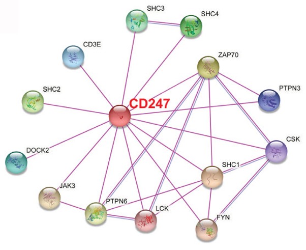 Fig. 1 CD247 Interaction Partners.1
Key structural properties of CD247:
- Intracellular region containing multiple phosphorylated tyrosine residues (ITAM motif)
- Across the membrane area contains positively charged arginine residues
- Extracellular region of simple structure is mainly responsible for complex assembly
Functions of CD247
The main function of the CD247 gene (encoding the CD3ζ chain) is to serve as a key signal transduction subunit of the T-cell receptor complex, but it is also involved in regulating multiple levels of the immune response.
| Function | Description |
| Signal activation | The phosphorylation of the intracellular ITAM motif is the initiating step of T cell activation, transmitting antigen recognition signals into the interior of the cell. |
| Receptor assembly and stability | It is a necessary component for the correct assembly of the TCR-CD3 complex and stable expression on the surface of the cell membrane. |
| Immune synaptic formation | Participate in the molecular organization of the immune synapse, help signal molecule in T cells and antigen presenting cells contact interface. |
| Signal amplification and regulation | The quantity and sequence of ITAM motifs affect signal strength and are involved in the fine balance of positive or negative regulatory signals. |
| Thymus selection | During the development of T cells, the signals they transmit are crucial for the positive and negative selection of thymocytes. |
Unlike hemoglobin, which has a synergistic effect, the signal transduction of CD247 is more like a "digital" switch. The activation of a single TCR complex can trigger early signaling events such as calcium ion influx, but its complete activation requires the accumulation of phosphorylation of multiple ITAM motifs to ensure the specificity and controllability of the immune response.
Applications of CD247 and CD247 Antibody in Literature
1. Briones, Alejandro C., et al. "Discordant restoration of TCR expression and function by CD247 somatic reversions." Journal of Clinical Immunology 45.1 (2025): 116. https://doi.org/10.1007/s10875-025-01908-9
This study found that somatic reverse mutations of the CD247 gene had different abilities to restore TCR expression and function in different models: although they could partially repair expression, they were all unable to effectively activate ZAP70 phosphorylation, revealing the inconsistency in the rescue of its expression and function.
2. Li, Yupeng, et al. "CD247, a potential T cell–derived disease severity and prognostic biomarker in patients with idiopathic pulmonary fibrosis." Frontiers in immunology 12 (2021): 762594. https://doi.org/10.3389/fimmu.2021.762594
Research has found that CD247 is lowly expressed in the blood and lung tissue of patients with idiopathic pulmonary fibrosis, and its low expression is associated with decreased lung function indicators, poor prognosis and changes in immune cell activity, suggesting that it can serve as a potential T-cell-derived biomarker for the severity and prognosis of IPF.
3. Danquah, Bright D., et al. "Mass Spectrometric analysis of antibody—Epitope peptide complex dissociation: Theoretical concept and practical procedure of binding strength characterization." Molecules 25.20 (2020): 4776. https://doi.org/10.1371/journal.pone.0068295
Research has found that the CD247 gene variation (rs864537) is significantly associated with the risk of rheumatoid arthritis in the European population across the entire genome, but has no independent correlation with anti-CCP antibodies. This is the first time that the GWAS-level association between this locus and RA has been confirmed.
4. Deng, Lin-Fang. "Identification of immune-related hub genes in thymoma: defects in CD247 and characteristics of paraneoplastic syndrome." Frontiers in Genetics 13 (2022): 895587. https://doi.org/10.3389/fgene.2022.895587
Research has found that in thymoma, the low expression of 14 immune hub genes such as CD247 is associated with defects in the T-cell receptor signaling pathway and a reduction in the number of mature T cells, which is a common feature of thymoma and some autoimmune diseases.
5. Holmberg, Dan, et al. "Association of CD247 (CD3ζ) gene polymorphisms with T1D and AITD in the population of northern Sweden." BMC medical genetics 17.1 (2016): 70. https://doi.org/10.1186/s12881-016-0333-z
This study found in the population of northern Sweden that the CD247 gene polymorphism is significantly associated with type 1 diabetes and autoimmune thyroid diseases, and it is a novel susceptibility gene locus.
Creative Biolabs: CD247 Antibodies for Research
Creative Biolabs specializes in the production of high-quality CD247 antibodies for research and industrial applications. Our portfolio includes monoclonal antibodies tailored for ELISA, Flow Cytometry, Western blot, immunohistochemistry, and other diagnostic methodologies.
- Custom CD247 Antibody Development: Tailor-made solutions to meet specific research requirements.
- Bulk Production: Large-scale antibody manufacturing for industry partners.
- Technical Support: Expert consultation for protocol optimization and troubleshooting.
- Aliquoting Services: Conveniently sized aliquots for long-term storage and consistent experimental outcomes.
For more details on our CD247 antibodies, custom preparations, or technical support, contact us at email.
Reference
- Li, Yupeng, et al. "CD247, a potential T cell–derived disease severity and prognostic biomarker in patients with idiopathic pulmonary fibrosis." Frontiers in immunology 12 (2021): 762594. https://doi.org/10.3389/fimmu.2021.762594
Anti-CD247 antibodies
 Loading...


Hot products 
-
Mouse Anti-BCL6 Recombinant Antibody (CBYY-0435) (CBMAB-0437-YY)

-
Mouse Anti-AKT1 Recombinant Antibody (V2-180546) (CBMAB-A2070-YC)

-
Mouse Anti-ACE2 Recombinant Antibody (V2-179293) (CBMAB-A0566-YC)

-
Rabbit Anti-CCL5 Recombinant Antibody (R0437) (CBMAB-R0437-CN)

-
Mouse Anti-Acetyl SMC3 (K105/K106) Recombinant Antibody (V2-634053) (CBMAB-AP052LY)

-
Mouse Anti-FTH1 Recombinant Antibody (CBXF-1896) (CBMAB-F3426-CQ)

-
Mouse Anti-AAV-5 Recombinant Antibody (V2-503416) (CBMAB-V208-1402-FY)

-
Mouse Anti-C1QC Recombinant Antibody (CBFYC-0600) (CBMAB-C0654-FY)

-
Mouse Anti-EMP3 Recombinant Antibody (CBFYE-0100) (CBMAB-E0207-FY)

-
Mouse Anti-CARD11 Recombinant Antibody (CBFYC-0811) (CBMAB-C0866-FY)

-
Mouse Anti-FN1 Monoclonal Antibody (71) (CBMAB-1241CQ)

-
Mouse Anti-AFDN Recombinant Antibody (V2-58751) (CBMAB-L0408-YJ)

-
Mouse Anti-AZGP1 Recombinant Antibody (CBWJZ-007) (CBMAB-Z0012-WJ)

-
Mouse Anti-ADRB2 Recombinant Antibody (V2-180026) (CBMAB-A1420-YC)

-
Mouse Anti-ADIPOR2 Recombinant Antibody (V2-179983) (CBMAB-A1369-YC)

-
Mouse Anti-ACTN4 Recombinant Antibody (V2-6075) (CBMAB-0020CQ)

-
Mouse Anti-CIITA Recombinant Antibody (CBLC160-LY) (CBMAB-C10987-LY)

-
Mouse Anti-AMACR Recombinant Antibody (CB34A) (CBMAB-CA034LY)

-
Mouse Anti-COL1A2 Recombinant Antibody (CF108) (V2LY-1206-LY626)

-
Mouse Anti-ADGRE2 Recombinant Antibody (V2-261270) (CBMAB-C0813-LY)

- AActivation
- AGAgonist
- APApoptosis
- BBlocking
- BABioassay
- BIBioimaging
- CImmunohistochemistry-Frozen Sections
- CIChromatin Immunoprecipitation
- CTCytotoxicity
- CSCostimulation
- DDepletion
- DBDot Blot
- EELISA
- ECELISA(Cap)
- EDELISA(Det)
- ESELISpot
- EMElectron Microscopy
- FFlow Cytometry
- FNFunction Assay
- GSGel Supershift
- IInhibition
- IAEnzyme Immunoassay
- ICImmunocytochemistry
- IDImmunodiffusion
- IEImmunoelectrophoresis
- IFImmunofluorescence
- IGImmunochromatography
- IHImmunohistochemistry
- IMImmunomicroscopy
- IOImmunoassay
- IPImmunoprecipitation
- ISIntracellular Staining for Flow Cytometry
- LALuminex Assay
- LFLateral Flow Immunoassay
- MMicroarray
- MCMass Cytometry/CyTOF
- MDMeDIP
- MSElectrophoretic Mobility Shift Assay
- NNeutralization
- PImmunohistologyp-Paraffin Sections
- PAPeptide Array
- PEPeptide ELISA
- PLProximity Ligation Assay
- RRadioimmunoassay
- SStimulation
- SESandwich ELISA
- SHIn situ hybridization
- TCTissue Culture
- WBWestern Blot
