CD37 Antibodies
Background
CD37 belongs to the tetrameric transmembrane protein superfamily and is mainly highly expressed on the surface of mature B cells. This protein plays a crucial role in B cell development, activation, and functional maintenance by participating in cell signal transduction and membrane organization regulation. Studies have found that abnormal expression of CD37 is closely related to the occurrence and development of B-cell lymphomas and leukemias, and has become an important target for immunotherapy. Currently, several monoclonal antibodies targeting CD37 and CAR-T therapies are in the clinical trial stage, showing promising results. As a typical representative of membrane protein structure and function, the research on CD37 provides an important model for understanding the role of the tetrameric transmembrane protein family in immune regulation and disease mechanisms.
Structure of CD37
CD37 is a tetrameric transmembrane protein with a molecular weight ranging from 40 to 52 kDa, which varies depending on the degree of glycosylation.
| Species | Human | Mouse | Rat | Dog | Monkey |
|---|---|---|---|---|---|
| Molecular Weight (kDa) | 40-52 | 40-50 | 40-50 | 42-50 | 40-52 |
| Primary Structural Differences | Contains 4 transmembrane domains | Conservative glycosylation sites | High homology with rat | High sequence conservation | Higher homology with human |
The CD37 protein consists of 281 amino acids and has a typical four-pass transmembrane structure. Its structure includes a short N-terminal and C-terminal within the cell, two extracellular loops (small loop and large loop), and four transmembrane domains. The large extracellular loop contains multiple cysteine residues, forming disulfide bonds to stabilize the conformation. CD37 interacts with other tetraspanin proteins and integrins through its transmembrane domains, forming microdomains on the cell membrane and participating in signal transduction. Its intracellular domain contains phosphorylation sites, which can recruit signaling molecules to regulate B cell activation, proliferation, and antigen presentation functions.
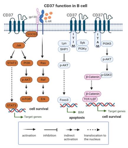 Fig. 1 CD37 function in B cell.1
The main structural features of CD37:
- Four transmembrane domains
- Glycosylated large extracellular loop
- Cysteines forming disulfide bonds
- The transmembrane region mediates protein interactions
- The intracellular segment contains phosphorylation sites that regulate signal transduction
Functions of CD37
The main function of CD37 is to participate in B-cell signal transduction and immune regulation. However, its role involves various physiological processes, including cell adhesion, activation regulation, and membrane structure organization.
| Function | Description |
| Signal transduction regulation | CD37 regulates the activity of MAPK and PI3K-AKT pathways downstream of the B cell receptor by recruiting signaling molecules. |
| Cell adhesion mediation | Interacts with integrins to promote the adhesion of B cells to stromal cells or antigen-presenting cells. |
| Membrane structure organization | Participates in the formation of microdomains enriched with transmembrane proteins, maintaining the stability of membrane protein complexes. |
| Immune response regulation | Affects the activation threshold of B cells, antibody production, and antigen presentation functions. |
| Tumor suppression | Downregulated in B-cell lymphomas. CD37 deficiency can lead to reprogramming of fatty acid metabolism, promoting tumor progression. |
CD37 forms a dynamic network with various membrane proteins through four transmembrane structures. Its function is dependent on the synergistic interaction with other four-transmembrane family members and integrins, and it plays a crucial regulatory role in the formation of immune synapses and the maintenance of B cell homeostasis.
Applications of CD37 and CD37 Antibody in Literature
1. Bobrowicz, Malgorzata, et al. "CD37 in B cell derived tumors—more than just a docking point for monoclonal antibodies." International Journal of Molecular Sciences 21.24 (2020): 9531.https://doi.org/10.3390/ijms21249531
The article indicates that CD37 is a key protein on the surface of B cells and has become an important target for immunotherapy in lymphoma and leukemia. This article reviews the mechanism of CD37 in B-cell malignancies, the latest progress in targeted therapy, and the future prospects, providing new ideas for patients with relapsed and refractory diseases.
2. Caulier, Benjamin, et al. "CD37 is a safe chimeric antigen receptor target to treat acute myeloid leukemia." Cell Reports Medicine 5.6 (2024). https://doi.org/10.1016/j.xcrm.2024.101572
The study found that CD37 is expressed in acute myeloid leukemia (AML) cells and is related to risk stratification. The modified CD37 CAR-T cells can specifically kill AML cells, control tumor progression, and are non-toxic to hematopoietic stem cells, providing a safe new target for AML treatment.
3. Peeters, Rens, et al. "Fatty acid metabolism in aggressive B-cell lymphoma is inhibited by tetraspanin CD37." Nature communications 13.1 (2022): 5371. https://doi.org/10.1038/s41467-022-33138-7
The study found that CD37 negatively regulates fatty acid metabolism by inhibiting FATP1. The absence of CD37 promotes the uptake and utilization of exogenous fatty acids by lymphoma cells, leading to lipid deposition and enhancing sensitivity to CPT1A inhibitors, thereby revealing the crucial role of CD37 in metabolic reprogramming.
4. Lu, Jinyuan, et al. "CD37 regulates the self-renewal of leukemic stem cells via integrin-mediated signaling in acute myeloid leukemia." Stem cell reports 20.5 (2025). https://doi.org/10.1016/j.stemcr.2025.102476
The study found that CD37 maintains the self-renewal of acute myeloid leukemia stem cells. The absence of CD37 inhibits the progression of leukemia by affecting the integrin signaling pathway, but has little impact on normal hematopoiesis. This provides a safe and effective new target for AML immunotherapy.
5. Zhang, Qi, et al. "CD37 high expression as a potential biomarker and association with poor outcome in acute myeloid leukemia." Bioscience reports 40.5 (2020): BSR20200008. https://doi.org/10.1042/BSR20200008
The study found that CD37 is highly expressed in acute myeloid leukemia, and is significantly associated with shortened overall survival and disease-free survival of patients. CD37 is involved in cell cycle, DNA replication and other pathways, and is a potential prognostic marker and a new therapeutic target.
Creative Biolabs: CD37 Antibodies for Research
Creative Biolabs specializes in the production of high-quality CD37 antibodies for research and industrial applications. Our portfolio includes monoclonal and polyclonal antibodies tailored for ELISA, Flow Cytometry, Western blot, immunohistochemistry, and other diagnostic methodologies.
- Custom CD37 Antibody Development: Tailor-made solutions to meet specific research requirements.
- Bulk Production: Large-scale antibody manufacturing for industry partners.
- Technical Support: Expert consultation for protocol optimization and troubleshooting.
- Aliquoting Services: Conveniently sized aliquots for long-term storage and consistent experimental outcomes.
For more details on our CD37 antibodies, custom preparations, or technical support, contact us at email.
Reference
- Bobrowicz, Malgorzata, et al. "CD37 in B cell derived tumors—more than just a docking point for monoclonal antibodies." International Journal of Molecular Sciences 21.24 (2020): 9531. Distributed under Open Access license CC BY 4.0, and cropped from the original figure. https://doi.org/10.3390/ijms21249531
Anti-CD37 antibodies
 Loading...


Hot products 
-
Mouse Anti-ATP5F1A Recombinant Antibody (51) (CBMAB-A4043-YC)

-
Mouse Anti-AGK Recombinant Antibody (V2-258056) (CBMAB-M0989-FY)

-
Mouse Anti-ACO2 Recombinant Antibody (V2-179329) (CBMAB-A0627-YC)

-
Mouse Anti-CCND2 Recombinant Antibody (DCS-3) (CBMAB-G1318-LY)

-
Mouse Anti-BAX Recombinant Antibody (CBYY-0216) (CBMAB-0217-YY)

-
Mouse Anti-CD33 Recombinant Antibody (P67.6) (CBMAB-C10189-LY)

-
Mouse Anti-ARIH1 Recombinant Antibody (C-7) (CBMAB-A3563-YC)

-
Mouse Anti-BACE1 Recombinant Antibody (CBLNB-121) (CBMAB-1180-CN)

-
Mouse Anti-CCDC6 Recombinant Antibody (CBXC-0106) (CBMAB-C5397-CQ)

-
Mouse Anti-ARG1 Recombinant Antibody (CBYCL-103) (CBMAB-L0004-YC)

-
Mouse Anti-ACTN4 Recombinant Antibody (V2-6075) (CBMAB-0020CQ)

-
Mouse Anti-ACVR1C Recombinant Antibody (V2-179685) (CBMAB-A1041-YC)

-
Mouse Anti-BCL6 Recombinant Antibody (CBYY-0435) (CBMAB-0437-YY)

-
Mouse Anti-EIF4G1 Recombinant Antibody (2A9) (CBMAB-A2544-LY)

-
Mouse Anti-GFAP Recombinant Antibody (24) (CBMAB-G2927-LY)

-
Mouse Anti-AKT1 Recombinant Antibody (V2-180546) (CBMAB-A2070-YC)

-
Mouse Anti-FN1 Monoclonal Antibody (D6) (CBMAB-1240CQ)

-
Human Anti-SARS-CoV-2 Spike Recombinant Antibody (CR3022) (CBMAB-CR014LY)

-
Mouse Anti-CTCF Recombinant Antibody (CBFYC-2371) (CBMAB-C2443-FY)

-
Mouse Anti-AMH Recombinant Antibody (5/6) (CBMAB-A2527-YC)

- AActivation
- AGAgonist
- APApoptosis
- BBlocking
- BABioassay
- BIBioimaging
- CImmunohistochemistry-Frozen Sections
- CIChromatin Immunoprecipitation
- CTCytotoxicity
- CSCostimulation
- DDepletion
- DBDot Blot
- EELISA
- ECELISA(Cap)
- EDELISA(Det)
- ESELISpot
- EMElectron Microscopy
- FFlow Cytometry
- FNFunction Assay
- GSGel Supershift
- IInhibition
- IAEnzyme Immunoassay
- ICImmunocytochemistry
- IDImmunodiffusion
- IEImmunoelectrophoresis
- IFImmunofluorescence
- IGImmunochromatography
- IHImmunohistochemistry
- IMImmunomicroscopy
- IOImmunoassay
- IPImmunoprecipitation
- ISIntracellular Staining for Flow Cytometry
- LALuminex Assay
- LFLateral Flow Immunoassay
- MMicroarray
- MCMass Cytometry/CyTOF
- MDMeDIP
- MSElectrophoretic Mobility Shift Assay
- NNeutralization
- PImmunohistologyp-Paraffin Sections
- PAPeptide Array
- PEPeptide ELISA
- PLProximity Ligation Assay
- RRadioimmunoassay
- SStimulation
- SESandwich ELISA
- SHIn situ hybridization
- TCTissue Culture
- WBWestern Blot
