DLAT Antibodies
Background
The DLAT gene encodes the E2 component of pyruvate dehydrogenase, a protein located in the mitochondrial matrix. It serves as the core scaffold of the multi-enzyme complex and catalyzes the key step of converting pyruvate to acetyl coenzyme A, thereby connecting glycolysis and the tricarboxylic acid cycle to maintain the homeostasis of cellular energy metabolism. Abnormalities in its function are associated with various metabolic diseases, especially in mitochondrial dysfunction and neurological disorders. This gene was first identified in mitochondrial enzymology research in the 1970s. The structure of its complex was deeply revealed through techniques such as low-temperature electron microscopy, highlighting the molecular mechanisms of multi-enzyme collaboration and metabolic regulation, providing an important foundation for understanding cellular energy conversion and the treatment of related diseases.
Structure of DLAT
The DLAT gene encodes the E2 subunit of pyruvate dehydrogenase (the DLAT protein), with a molecular weight of approximately 65 kDa (containing about 550 amino acids). This value may vary slightly among different species due to minor differences in their sequences.
| Species | Human | Mouse | Rat | Bovine |
| Molecular Weight (kDa) | 65 | 64.5 | 65 | 64.8 |
| Primary Structural Differences | Containing a fatty acyl domain, the catalytic active center is highly conserved | Catalytic core and anthropogenic highly homologous | The sequence similarity is extremely high. | Fat acylation loci conservative, individual amino acid replacement |
The DLAT protein self-assembles through its N-terminal lipid-acylated domain to form a icosahedral core scaffold, serving as the core support of the pyruvate dehydrogenase complex (PDC). Its C-terminal catalytic domain contains a lipoic acid binding site, which can swing between the E1 and E3 components to transfer the acetyl group, playing a key role in connecting glycolysis and the tricarboxylic acid cycle. The tertiary structure of this protein is composed of multiple globular domains, and its flexible linker region is the structural basis for realizing the "waving arm" function, while the conserved cysteine residues in the catalytic pocket are the active center for acetyl transfer.
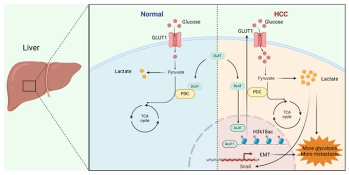 Fig. 1 DLAT Drives HCC Metastasis by Reprogramming Glycolysis and Inducing EMT.1
Key structural properties of DLAT:
- Multi-domain core
- Fatty acylation of the N-terminus
- Swing the C-end of the arm
- Catalytic active center
Functions of DLAT
The core function of the protein encoded by the DLAT gene is to serve as the catalytic and structural core of the pyruvate dehydrogenase complex (PDC). Additionally, it is involved in various cellular regulatory processes.
| Function | Description |
| Acetyl Transfer | As the core catalytic component of PDC, it transfers the acetyl group generated from the decarboxylation of pyruvate to coenzyme A, forming acetyl coenzyme A. This is a crucial step connecting glycolysis and the tricarboxylic acid cycle. |
| Complex scaffold | The N-terminal lipid-acylated domain self-assembles to form a icosahedral core framework, providing an ordered assembly platform for the E1 and E3 components in PDC, ensuring the efficient operation of the metabolic pathway. |
| Metabolic Regulatory Node | It regulates the overall activity of PDC through post-translational modifications such as phosphorylation, thereby responding to the cellular energy state (such as the ATP/ADP ratio) and controlling the flux of glucose oxidation. |
| Disease Association | Defects in the function of DLAT are associated with various diseases, such as primary lactic acidosis, certain neurodegenerative disorders, and the metabolic reprogramming of cancer. These are the focus of research on its pathological mechanism. |
The catalytic kinetic characteristics of DLAT follow the classic "ping-pong mechanism". The "pendulum arm" domain of DLAT efficiently swings between the E1 and E3 active sites, ensuring the high efficiency and specificity of acetyl transfer. Together with the "metabolite channel" effect of the multi-enzyme complex, this constitutes the structural basis for its efficient catalysis.
Applications of DLAT and DLAT Antibody in Literature
1. Li, Zhipeng, et al. "MELK promotes HCC carcinogenesis through modulating cuproptosis-related gene DLAT-mediated mitochondrial function." Cell Death & Disease 14.11 (2023): 733. https://doi.org/10.1038/s41419-023-06264-3
This study reveals that MELK enhances DLAT expression through the PI3K/mTOR pathway, stabilizes mitochondrial function, thereby promoting the progression of hepatocellular carcinoma and inducing resistance to the copper death inducer elesclomol.
2. Yin, Qian, et al. "DLAT activates EMT to promote HCC metastasis by regulating GLUT1-mediated aerobic glycolysis." Molecular Medicine 31.1 (2025): 71. https://doi.org/10.1186/s10020-025-01125-5
Studies have found that DLAT is highly expressed in hepatocellular carcinoma and indicates a poor prognosis. It promotes tumor metastasis by mediating H3K18 acetylation to upregulate GLUT1, driving aerobic glycolysis and epithelial-mesenchymal transition.
3. Liu, Diya, et al. "DLAT promotes triple-negative breast cancer progression via YAP1 activation." Cancer Biology & Therapy 25.1 (2024): 2421578. https://doi.org/10.1080/15384047.2024.2421578
Studies have found that DLAT is highly expressed in triple-negative breast cancer and has a poor prognosis. It directly binds to YAP1, promoting its dephosphorylation and nuclear translocation, thereby activating the transcription of downstream oncogenes and driving the malignant progression of tumors.
4. Jia, Yanhan, et al. "Mitochondrial KMT9 methylates DLAT to control pyruvate dehydrogenase activity and prostate cancer growth." Nature Communications 16.1 (2025): 1191. https://doi.org/10.1038/s41467-025-56492-8
This study reveals that KMT9 monomethylates DLAT (K596me1) in mitochondria, thereby regulating the activity of the pyruvate dehydrogenase complex and promoting lipid synthesis and proliferation in prostate cancer. This mechanism may provide new targets for treatment.
5. Xu, Haitao, et al. "Pan-cancer analysis of DLAT reveals it as a prognostic Biomarker involved in immune infiltration of liver hepatocellular carcinoma." Journal of Cancer 16.7 (2025): 2167. https://doi.org/10.7150/jca.102256
Studies have found that the expression of DLAT is significantly elevated in hepatocellular carcinoma and is associated with a poor prognosis. Its expression is closely related to m6A modification, immune cell infiltration and multiple key signaling pathways (such as PI3K-AKT), suggesting that it may become a new therapeutic target.
Creative Biolabs: DLAT Antibodies for Research
Creative Biolabs specializes in the production of high-quality DLAT antibodies for research and industrial applications. Our portfolio includes monoclonal antibodies tailored for ELISA, Flow Cytometry, Western blot, immunohistochemistry, and other diagnostic methodologies.
- Custom DLAT Antibody Development: Tailor-made solutions to meet specific research requirements.
- Bulk Production: Large-scale antibody manufacturing for industry partners.
- Technical Support: Expert consultation for protocol optimization and troubleshooting.
- Aliquoting Services: Conveniently sized aliquots for long-term storage and consistent experimental outcomes.
For more details on our DLAT antibodies, custom preparations, or technical support, contact us at email.
Reference
- Yin, Qian, et al. "DLAT activates EMT to promote HCC metastasis by regulating GLUT1-mediated aerobic glycolysis." Molecular Medicine 31.1 (2025): 71. https://doi.org/10.1186/s10020-025-01125-5
Anti-DLAT antibodies
 Loading...


Hot products 
-
Mouse Anti-A2M Recombinant Antibody (V2-178822) (CBMAB-A0036-YC)

-
Mouse Anti-BANF1 Recombinant Antibody (3F10-4G12) (CBMAB-A0707-LY)

-
Mouse Anti-2C TCR Recombinant Antibody (V2-1556) (CBMAB-0951-LY)

-
Mouse Anti-AGO2 Recombinant Antibody (V2-634169) (CBMAB-AP203LY)

-
Mouse Anti-ACTG1 Recombinant Antibody (V2-179597) (CBMAB-A0916-YC)

-
Mouse Anti-EIF4G1 Recombinant Antibody (2A9) (CBMAB-A2544-LY)

-
Mouse Anti-FOXA3 Recombinant Antibody (2A9) (CBMAB-0377-YC)

-
Mouse Anti-BRD3 Recombinant Antibody (CBYY-0801) (CBMAB-0804-YY)

-
Mouse Anti-CCL18 Recombinant Antibody (64507) (CBMAB-C7910-LY)

-
Mouse Anti-AK4 Recombinant Antibody (V2-180419) (CBMAB-A1891-YC)

-
Mouse Anti-BCL6 Recombinant Antibody (CBYY-0442) (CBMAB-0445-YY)

-
Mouse Anti-CCT6A/B Recombinant Antibody (CBXC-0168) (CBMAB-C5570-CQ)

-
Mouse Anti-ATP1A2 Recombinant Antibody (M7-PB-E9) (CBMAB-A4013-YC)

-
Mouse Anti-APP Recombinant Antibody (DE2B4) (CBMAB-1122-CN)

-
Mouse Anti-GIPC2 Recombinant Antibody (10) (CBMAB-G0476-LY)

-
Mouse Anti-CTCF Recombinant Antibody (CBFYC-2371) (CBMAB-C2443-FY)

-
Mouse Anti-CFL1 Recombinant Antibody (CBFYC-1771) (CBMAB-C1833-FY)

-
Mouse Anti-DES Monoclonal Antibody (440) (CBMAB-AP1857LY)

-
Mouse Anti-ACKR3 Recombinant Antibody (V2-261265) (CBMAB-C1023-LY)

-
Mouse Anti-GLP1R Recombinant Antibody (4F3) (CBMAB-G0521-LY)

- AActivation
- AGAgonist
- APApoptosis
- BBlocking
- BABioassay
- BIBioimaging
- CImmunohistochemistry-Frozen Sections
- CIChromatin Immunoprecipitation
- CTCytotoxicity
- CSCostimulation
- DDepletion
- DBDot Blot
- EELISA
- ECELISA(Cap)
- EDELISA(Det)
- ESELISpot
- EMElectron Microscopy
- FFlow Cytometry
- FNFunction Assay
- GSGel Supershift
- IInhibition
- IAEnzyme Immunoassay
- ICImmunocytochemistry
- IDImmunodiffusion
- IEImmunoelectrophoresis
- IFImmunofluorescence
- IGImmunochromatography
- IHImmunohistochemistry
- IMImmunomicroscopy
- IOImmunoassay
- IPImmunoprecipitation
- ISIntracellular Staining for Flow Cytometry
- LALuminex Assay
- LFLateral Flow Immunoassay
- MMicroarray
- MCMass Cytometry/CyTOF
- MDMeDIP
- MSElectrophoretic Mobility Shift Assay
- NNeutralization
- PImmunohistologyp-Paraffin Sections
- PAPeptide Array
- PEPeptide ELISA
- PLProximity Ligation Assay
- RRadioimmunoassay
- SStimulation
- SESandwich ELISA
- SHIn situ hybridization
- TCTissue Culture
- WBWestern Blot
