FLT1 Antibodies
Background
The FLT1 gene (also known as VEGFR1) belongs to the vascular endothelial growth factor receptor family. The protein it encodes exists as a transmembrane tyrosine kinase receptor on the surface of various cells. This gene mainly participates in regulating physiological processes such as angiogenesis, vascular permeability and cell migration by specifically binding to ligands like VEGF-A and PIGF. During embryonic development, FLT1 is crucial for the formation of the cardiovascular system. In adult individuals, it negatively regulates angiogenesis and maintains vascular homeostasis through the generation of soluble splicing variants. This gene was first identified in 1990. Its abnormal expression is closely related to a variety of diseases, including angiogenesis in solid tumors, the pathogenesis of preeclampsia, and the progression of retinopathy. The analysis of the FLT1 signaling pathway not only deepens the understanding of vascular biological mechanisms but also provides a key target for the development of anti-angiogenic therapies.
Structure of FLT1
Vascular endothelial growth factor receptor 1 encoded by the FLT1 gene is a type I transmembrane glycoprotein with a molecular weight of approximately 180 kDa. Its actual molecular weight fluctuates within the range of 150-180 kDa depending on the degree of glycosylation. The extracellular domain of this protein contains seven immunoglobulin-like domains (D1-D7), which are key regions for ligand binding. The D2 domain plays a core role in binding to VEGF-A, while the D3 domain is crucial for the binding of PIGF. Its intracellular part is composed of the perimembrane region, the split tyrosine kinase domain (separated by the kinase insertion region between TK1 and TK2), and the C-terminal tail. FLT1 exerts a unique negative regulatory function by generating a soluble splicing variant (sFLT1, approximately 100 kDa) lacking intracellular kinase domains, a characteristic that is of great significance in placental vascular physiology and the pathological mechanism of preeclampsia.
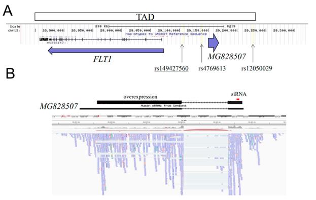 Fig. 1 Genomic structure of the FLT1 upstream region.1
Key structural properties of FLT1:
- Extracellular ligand binding domain
- Unique kinase regulation mechanism
- Transmembrane negative regulatory function
- Structure-function adaptability
Functions of FLT1
The core function of FLT1 (VEGFR1) is to regulate angiogenesis and homeostasis, and it also plays a complex role in various pathophysiological processes.
| Function | Description |
|---|---|
| Angiogenesis regulation | As a high-affinity receptor for VEGF and PIGF, its membrane-bound form (mFLT1) signaling can promote the migration and survival of vascular endothelial cells under specific conditions, which is particularly crucial for embryonic angiogenesis and monocyte/macrophage chemotaxis. |
| Negative regulation of angiogenesis | Its soluble variant (sFLT1) acts as a "bait receptor" to effectively isolate VEGF/PIGF in circulation and inhibit the VEGFR2 pathway, which is a key negative feedback mechanism for maintaining adult vascular homeostasis and normal remodeling of placental vessels. |
| Disease Association | Abnormally elevated sFLT1 levels are the core pathological marker of preeclampsia. In tumors, FLT1 signaling can promote tumor angiogenesis, the formation of an immunosuppressive microenvironment, and tumor cell invasion. |
| Non-vascular function | It is expressed in non-endothelial cells such as osteoclasts and monocytes, and participates in processes such as bone metabolism, inflammatory response and organ fibrosis. |
Compared with the potent pro-angiogenic signals mediated by VEGFR2, the signal output of FLT1 is usually weaker and more context-dependent, reflecting its unique role in vascular biology as a precise "regulatory valve" rather than a simple "switch".
Applications of FLT1 and FLT1 Antibody in Literature
1. Tai, Yifan, et al. "FLT1 activation in cancer cells promotes PARP-inhibitor resistance in breast cancer." EMBO Molecular Medicine 16.8 (2024): 1957-1980. https://doi.org/10.1038/s44321-024-00094-2
This study found that FLT1 drives the development of PARP inhibitor resistance in BRCA1/2 mutant breast cancer and protects tumor cells through non-classical pathways. Blocking FLT1 can inhibit AKT and enhance CD8+ T cell infiltration, thereby restoring sensitivity to the resistant tumors, making it a potential therapeutic target.
2. Ashar-Patel, Ami, et al. "FLT1 and transcriptome-wide polyadenylation site (PAS) analysis in preeclampsia." Scientific reports 7.1 (2017): 12139. https://doi.org/10.1038/s41598-017-11639-6
Studies have confirmed that the Flt1 gene is significantly upregulated in both early-onset and late-onset preeclampsia placental tissues. Its mRNA level is directly correlated with the increase in maternal blood pressure, suggesting that it could be a potential target for RNAi therapy.
3. Wujcicka, Wioletta Izabela, et al. "Single nucleotide polymorphisms from CSF2, FLT1, TFPI and TLR9 genes are associated with prelabor rupture of membranes." Genes 12.11 (2021): 1725. https://doi.org/10.3390/genes12111725
This study analyzed 360 pregnant women and found that the polymorphisms of genes related to hemostasis and angiogenesis (such as FLT1, etc.) acting together could affect the risk of premature rupture of membranes, but the single gene polymorphism of FLT1 had no independent association.
4. Raikwar, Nandita S., Kang Z. Liu, and Christie P. Thomas. "N-terminal cleavage and release of the ectodomain of Flt1 is mediated via ADAM10 and ADAM 17 and regulated by VEGFR2 and the Flt1 intracellular domain." PloS one 9.11 (2014): e112794. https://doi.org/10.1371/journal.pone.0112794
The study found that the Flt1 receptor can be cleaved by the ADAM10/17 proteases, generating fragments that can antagonize VEGF. PKC and the ubiquitination system regulate this cleavage process. The interaction between Flt1 and VEGFR2 affects the cleavage efficiency and the range of action of the fragments.
5. Morris, Brian J., et al. "Vascular endothelial growth factor receptor 1 gene (FLT1) longevity variant increases lifespan by reducing mortality risk posed by hypertension." Aging (Albany NY) 15.10 (2023): 3967. https://doi.org/10.18632/aging.204722
The research has found that a specific genotype (GG) of the longevity gene FLT1 can reduce the risk of death caused by hypertension, thereby prolonging lifespan. However, it has no significant protective effect against coronary heart disease, stroke or diabetes.
Creative Biolabs: FLT1 Antibodies for Research
Creative Biolabs specializes in the production of high-quality FLT1 antibodies for research and industrial applications. Our portfolio includes monoclonal antibodies tailored for ELISA, Flow Cytometry, Western blot, immunohistochemistry, and other diagnostic methodologies.
- Custom FLT1 Antibody Development: Tailor-made solutions to meet specific research requirements.
- Bulk Production: Large-scale antibody manufacturing for industry partners.
- Technical Support: Expert consultation for protocol optimization and troubleshooting.
- Aliquoting Services: Conveniently sized aliquots for long-term storage and consistent experimental outcomes.
For more details on our FLT1 antibodies, custom preparations, or technical support, contact us at email.
Reference
- Yoshizawa, Hikari, et al. "Characterization of the MG828507 lncRNA Located Upstream of the FLT1 Gene as an Etiology for Pre-Eclampsia." Journal of Clinical Medicine 11.15 (2022): 4603. https://doi.org/10.3390/jcm11154603
Anti-FLT1 antibodies
 Loading...


Hot products 
-
Mouse Anti-ARHGDIA Recombinant Antibody (CBCNA-009) (CBMAB-R0415-CN)

-
Mouse Anti-BRD3 Recombinant Antibody (CBYY-0801) (CBMAB-0804-YY)

-
Mouse Anti-ATP1A2 Recombinant Antibody (M7-PB-E9) (CBMAB-A4013-YC)

-
Mouse Anti-2C TCR Recombinant Antibody (V2-1556) (CBMAB-0951-LY)

-
Mouse Anti-BACE1 Recombinant Antibody (CBLNB-121) (CBMAB-1180-CN)

-
Mouse Anti-C1QC Recombinant Antibody (CBFYC-0600) (CBMAB-C0654-FY)

-
Mouse Anti-AKT1 (Phosphorylated S473) Recombinant Antibody (V2-505430) (PTM-CBMAB-0067LY)

-
Mouse Anti-AFDN Recombinant Antibody (V2-58751) (CBMAB-L0408-YJ)

-
Mouse Anti-DLL4 Recombinant Antibody (D1090) (CBMAB-D1090-YC)

-
Mouse Anti-ATP5F1A Recombinant Antibody (51) (CBMAB-A4043-YC)

-
Mouse Anti-ENO1 Recombinant Antibody (8G8) (CBMAB-E1329-FY)

-
Rat Anti-CCR2 Recombinant Antibody (475301) (CBMAB-C1338-LY)

-
Mouse Anti-DMD Recombinant Antibody (D1190) (CBMAB-D1190-YC)

-
Mouse Anti-Acetyl SMC3 (K105/K106) Recombinant Antibody (V2-634053) (CBMAB-AP052LY)

-
Mouse Anti-CD83 Recombinant Antibody (HB15) (CBMAB-C1765-CQ)

-
Human Anti-SARS-CoV-2 Spike Recombinant Antibody (CBC05) (CBMAB-CR005LY)

-
Rat Anti-EPO Recombinant Antibody (16) (CBMAB-E1578-FY)

-
Mouse Anti-FeLV g27 Recombinant Antibody (1) (CBMAB-V208-1714-FY)

-
Mouse Anti-FLT1 Recombinant Antibody (11) (CBMAB-V0154-LY)

-
Mouse Anti-ALB Recombinant Antibody (V2-55272) (CBMAB-H0819-FY)

- AActivation
- AGAgonist
- APApoptosis
- BBlocking
- BABioassay
- BIBioimaging
- CImmunohistochemistry-Frozen Sections
- CIChromatin Immunoprecipitation
- CTCytotoxicity
- CSCostimulation
- DDepletion
- DBDot Blot
- EELISA
- ECELISA(Cap)
- EDELISA(Det)
- ESELISpot
- EMElectron Microscopy
- FFlow Cytometry
- FNFunction Assay
- GSGel Supershift
- IInhibition
- IAEnzyme Immunoassay
- ICImmunocytochemistry
- IDImmunodiffusion
- IEImmunoelectrophoresis
- IFImmunofluorescence
- IGImmunochromatography
- IHImmunohistochemistry
- IMImmunomicroscopy
- IOImmunoassay
- IPImmunoprecipitation
- ISIntracellular Staining for Flow Cytometry
- LALuminex Assay
- LFLateral Flow Immunoassay
- MMicroarray
- MCMass Cytometry/CyTOF
- MDMeDIP
- MSElectrophoretic Mobility Shift Assay
- NNeutralization
- PImmunohistologyp-Paraffin Sections
- PAPeptide Array
- PEPeptide ELISA
- PLProximity Ligation Assay
- RRadioimmunoassay
- SStimulation
- SESandwich ELISA
- SHIn situ hybridization
- TCTissue Culture
- WBWestern Blot
