GLDC Antibodies
Background
The GLDC gene encodes glycine decarboxylase and is mainly distributed in the liver, kidneys, and brain tissues. This gene is responsible for catalyzing the key steps of glycine catabolism and participates in one-carbon unit metabolism and cellular energy supply, which is particularly important for maintaining normal neurological function. Mutations in the GLDC gene can lead to the autosomal recessive genetic disorder "non-ketotic hyperglycinemia", with clinical manifestations of severe brain disorders and developmental delays. Since its function was gradually clarified in the 1970s, this gene has become a key target in genetic metabolic disease research and newborn screening. Related studies not only revealed the regulatory mechanism of amino acid metabolism but also provided a molecular basis for targeted therapy.
Structure of GLDC
The GLDC gene encodes a glycine decarboxylase with a molecular weight of approximately 110 kDa. The exact value may vary slightly among different species due to differences in amino acid sequences.
| Species | Human | Mouse | Rat |
|---|---|---|---|
| Molecular Weight (kDa) | About 110 | About 109 | About 110 |
| Primary Structural Differences | Containing four P protein domains, forming a complete enzyme active center | The core catalytic domain is highly conserved. | With the human sequence homology is extremely high, function highly similar |
This protein is mainly composed of its pyridoxal phosphate (PLP) binding domain as the catalytic core. This region is covalently bound to the PLP coenzyme through conserved lysine residues and is crucial for completing the transcarbamylase reaction of glycine. The entire protein folds through multiple domains to form a compact tetramer spatial conformation, which is essential for maintaining its stability and catalytic efficiency in the mitochondrial matrix. The precise geometric arrangement of its active center directly determines the metabolic ability to convert glycine into a one-carbon unit.
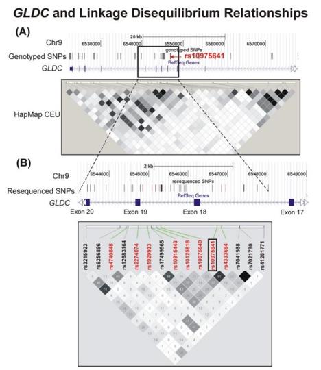 Fig. 1 The GLDC gene and its linkage disequilibrium relationships.1
Key structural properties of GLDC:
- Containing a conserved pyridoxal phosphate (PLP) binding domain
- Relying on the tight assembly of tetramers to maintain catalytic activity
- Active site contains the core of lysine residues, is responsible for the covalent binding PLP coenzyme
Functions of GLDC
The glycine decarboxylase encoded by the GLDC gene mainly functions in the catabolism of glycine, but it is also involved in a variety of crucial physiological and pathological processes.
| Function | Description |
|---|---|
| Glycine Degradation | In the mitochondrial matrix, it catalyzes the decarboxylation and deamination of glycine, generating carbon dioxide, ammonia and methylenetetrahydrofolate. This is the rate-limiting step of the glycine degradation metabolic pathway. |
| Supply of One-Carbon Units | Through the aforementioned reaction, "one-carbon units" essential for important life activities such as purine synthesis and methylation reactions are provided to the cells. |
| Neurotoxicity relief | By removing excessive glycine and preventing its excessive accumulation in the synaptic cleft, it avoids excitatory neurotoxicity. |
| Energy Metabolism Support | This reaction process is coupled with the mitochondrial respiratory chain, which helps maintain cellular energy metabolism, especially in organs with high metabolic demands (such as the liver and the brain). |
| Disease Association | Its functional defect directly leads to "non-ketotic hyperglycinemia", a severe congenital metabolic error, characterized by neurological damage and developmental delay. |
The catalytic reaction of this enzyme exhibits high substrate specificity, and its kinetic characteristics demonstrate a strong affinity for glycine, ensuring efficient and precise regulation of the homeostasis of this important but potentially neurotoxic amino acid.
Applications of GLDC and GLDC Antibody in Literature
1. Xie, Huaying, et al. "GLDC mitigated by miR-30e regulates cell proliferation and tumor immune infiltration in TNBC." Frontiers in Immunology 13 (2022): 1033367. https://doi.org/10.3389/fimmu.2022.1033367
The article indicates that GLDC is highly expressed in triple-negative breast cancer and has a poor prognosis, and is associated with specific immune cell infiltration. miR-30e regulates tumor cell proliferation by inhibiting GLDC, providing a basis for potential therapeutic targets.
2. Chen, Ming-kun, et al. "Glycine decarboxylase (GLDC) plays a crucial role in regulating energy metabolism, invasion, metastasis and immune escape for prostate cancer." International Journal of Biological Sciences 19.15 (2023): 4726. https://doi.org/10.7150/ijbs.85893
The article indicates that GLDC is regulated by HIF-1α in PCa. It promotes tumor invasion and metastasis by enhancing aerobic glycolysis and LDHA expression, and is associated with TP53 mutations, reduced CD8+ T cell infiltration, and immune escape. It is a potential therapeutic target.
3. Zhou, Jie, et al. "Identification and characterization of GLDC as host susceptibility gene to severe influenza." EMBO molecular medicine 11.1 (2019): e9528. https://doi.org/10.15252/emmm.201809528
The article indicates that GLDC has been identified as a susceptibility gene for severe human influenza. Inhibiting GLDC can enhance the antiviral response of type I interferons and downstream genes, effectively inhibiting the replication of H1N1 and H7N9 viruses. In vivo experiments have confirmed that the GLDC inhibitor has a significant protective effect on infected mice.
4. Cao, Yanyan, et al. "Novel GLDC compound heterozygous variant leading to nonketotic hyperglycinemia: case report and literature review." Frontiers in Pediatrics 9 (2021): 725930. https://doi.org/10.3389/fped.2021.725930
This case report describes a severely ill neonate with non-ketotic hyperglycinemia carrying a novel compound heterozygous mutation of GLDC. Genetic testing is crucial for diagnosis and prognosis, as its non-specific clinical manifestations increase the difficulty of making a definitive diagnosis.
5. Jäger, Katharina, et al. "Expression of neural crest markers GLDC and ERRFI1 is correlated with melanoma prognosis." Cancers 11.1 (2019): 76. https://doi.org/10.3390/cancers11010076
This study, using iPSC models, found that the neural crest marker GLDC was highly expressed in melanoma. The high expression of GLDC was significantly associated with early metastasis and poor prognosis in patients, and could serve as a potential biomarker for predicting prognosis.
Creative Biolabs: GLDC Antibodies for Research
Creative Biolabs specializes in the production of high-quality GLDC antibodies for research and industrial applications. Our portfolio includes monoclonal antibodies tailored for ELISA, Flow Cytometry, Western blot, immunohistochemistry, and other diagnostic methodologies.
- Custom GLDC Antibody Development: Tailor-made solutions to meet specific research requirements.
- Bulk Production: Large-scale antibody manufacturing for industry partners.
- Technical Support: Expert consultation for protocol optimization and troubleshooting.
- Aliquoting Services: Conveniently sized aliquots for long-term storage and consistent experimental outcomes.
For more details on our GLDC antibodies, custom preparations, or technical support, contact us at email.
Reference
- Ji, Yuan, et al. "Glycine and a glycine dehydrogenase (GLDC) SNP as citalopram/escitalopram response biomarkers in depression: pharmacometabolomics‐informed pharmacogenomics." Clinical Pharmacology & Therapeutics 89.1 (2011): 97-104. https://doi.org/10.1038/clpt.2010.250
Anti-GLDC antibodies
 Loading...


Hot products 
-
Mouse Anti-C5b-9 Recombinant Antibody (aE11) (CBMAB-AO138LY)

-
Mouse Anti-CARTPT Recombinant Antibody (113612) (CBMAB-C2450-LY)

-
Mouse Anti-FTH1 Recombinant Antibody (CBXF-1896) (CBMAB-F3426-CQ)

-
Mouse Anti-4-Hydroxynonenal Recombinant Antibody (V2-502280) (CBMAB-C1055-CN)

-
Mouse Anti-ATP1B1 Recombinant Antibody (E4) (CBMAB-0463-LY)

-
Mouse Anti-CEMIP Recombinant Antibody (3C12) (CBMAB-K0296-LY)

-
Rat Anti-ADGRE4 Recombinant Antibody (V2-160163) (CBMAB-F0011-CQ)

-
Mouse Anti-CD33 Recombinant Antibody (6C5/2) (CBMAB-C8126-LY)

-
Mouse Anti-DDC Recombinant Antibody (8E8) (CBMAB-0992-YC)

-
Mouse Anti-NSUN6 Recombinant Antibody (D-5) (CBMAB-N3674-WJ)

-
Mouse Anti-FLT1 Recombinant Antibody (11) (CBMAB-V0154-LY)

-
Mouse Anti-BCL2L1 Recombinant Antibody (H5) (CBMAB-1025CQ)

-
Mouse Anti-CHRNA9 Recombinant Antibody (8E4) (CBMAB-C9161-LY)

-
Mouse Anti-ALPL Antibody (B4-78) (CBMAB-1009CQ)

-
Mouse Anti-BPGM Recombinant Antibody (CBYY-1806) (CBMAB-2155-YY)

-
Mouse Anti-FYN Recombinant Antibody (10) (CBMAB-S6332-CQ)

-
Mouse Anti-CD24 Recombinant Antibody (HIS50) (CBMAB-C10123-LY)

-
Mouse Anti-BRD3 Recombinant Antibody (CBYY-0801) (CBMAB-0804-YY)

-
Mouse Anti-ATP1A2 Recombinant Antibody (M7-PB-E9) (CBMAB-A4013-YC)

-
Mouse Anti-ANXA7 Recombinant Antibody (A-1) (CBMAB-A2941-YC)

- AActivation
- AGAgonist
- APApoptosis
- BBlocking
- BABioassay
- BIBioimaging
- CImmunohistochemistry-Frozen Sections
- CIChromatin Immunoprecipitation
- CTCytotoxicity
- CSCostimulation
- DDepletion
- DBDot Blot
- EELISA
- ECELISA(Cap)
- EDELISA(Det)
- ESELISpot
- EMElectron Microscopy
- FFlow Cytometry
- FNFunction Assay
- GSGel Supershift
- IInhibition
- IAEnzyme Immunoassay
- ICImmunocytochemistry
- IDImmunodiffusion
- IEImmunoelectrophoresis
- IFImmunofluorescence
- IGImmunochromatography
- IHImmunohistochemistry
- IMImmunomicroscopy
- IOImmunoassay
- IPImmunoprecipitation
- ISIntracellular Staining for Flow Cytometry
- LALuminex Assay
- LFLateral Flow Immunoassay
- MMicroarray
- MCMass Cytometry/CyTOF
- MDMeDIP
- MSElectrophoretic Mobility Shift Assay
- NNeutralization
- PImmunohistologyp-Paraffin Sections
- PAPeptide Array
- PEPeptide ELISA
- PLProximity Ligation Assay
- RRadioimmunoassay
- SStimulation
- SESandwich ELISA
- SHIn situ hybridization
- TCTissue Culture
- WBWestern Blot
