IGF2 Antibodies
Background
IGF2 gene, as a paternally expressed imprinted gene, mainly exists in mammalian embryonic development stages. This gene encodes insulin-like growth factor 2 protein, which regulates fetal growth and development as well as nutrient metabolism by promoting cell proliferation and differentiation. Because the embryo is highly sensitive to the mother's nutritional supply, IGF2 maintains the energy balance of the fetus through a placenta-specific expression mechanism. This gene was first identified in 1978. Its unique genomic imprinting phenomenon provided a key model for epigenetic research, and the explanation of the related mechanism won the 2023 Nobel Prize in Physiology or Medicine. The cascade reaction of IGF2 gene expression regulation remains a research paradigm in developmental biology to this day, greatly promoting human understanding of embryo programming, imprinting regulatory networks, and intergenerational inheritance mechanisms.
Structure of IGF2
Insulin-like growth factor 2 encoded by the IGF2 gene is a protein polypeptide with a molecular weight of approximately 7.5 kDa. There are slight differences in this molecular weight among different species, mainly due to the post-translational processing of its precursor protein and the species specificity of the amino acid sequence.
| Species | Human | Mouse | Bovine | Rst |
| Molecular Weight (kDa) | 7.5 | 7.4 | 7.5 | 7.4 |
| Primary Structural Differences | Promote fetal growth and cell proliferation | Placental specific expression regulation | Affects muscle development in animal husbandry | Often used in neurodevelopmental studies |
This protein is processed by protease hydrolysis of its precursor protein and eventually forms a mature active polypeptide containing 67 amino acids. IGF2 binds to the IGF1 receptor and insulin receptor on the cell surface through its tertiary structure, thereby activating downstream signaling pathways. Its spatial conformation contains multiple key α -helical and β -folding regions, among which the B domain and C domain are directly involved in the specificity and affinity of receptor binding, which is crucial for maintaining normal embryonic development and metabolic regulation.
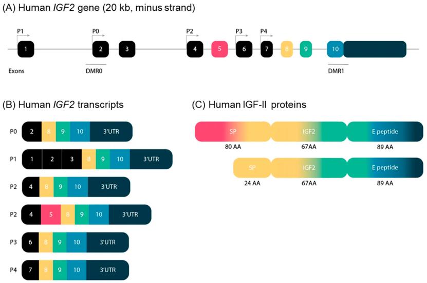 Fig. 1 Schematic representation of the structure of the IGF2 gene in humans.1
Key structural properties of IGF2:
- Complex imprinted regulatory regions for specific expression of paternity alleles
- Multiple promoters and differential methylation regions control spatiotemporal expression
- The non-translation area is rich in highly conserved cis-acting elements
Functions of IGF2
The main function of the IGF2 gene is to promote fetal growth and cell proliferation. However, it is also involved in various physiological and pathological processes, including metabolic regulation and tumorigenesis.
| Function | Description |
| Promote fetal growth | As a key embryonic growth factor, it strongly stimulates cell division and the growth and development of fetal tissues and organs. |
| Placental nutrient transport | Regulate the function of placental trophoblast cells and the efficiency of nutrient transport, directly connecting the maternal supply with the fetal demand. |
| Metabolic regulation | In adulthood, it works in synergy with insulin to regulate glucose uptake and the distribution of nutrients in muscles and fat. |
| Brain development support | Specifically expressed in the developing brain, it promotes nerve cell survival, synapse formation and cognitive function establishment. |
| Tumorigenesis association | Its abnormally high expression is closely related to the proliferation and invasion of various cancers, such as hepatoblastoma and colorectal cancer. |
The expression of this gene shows strict spatiotemporal specificity, and its activity significantly decreases after birth. Abnormal and persistent activation disrupts the growth balance and becomes a key factor driving tumor development.
Applications of IGF2 and IGF2 Antibody in Literature
1. Sélénou, Céline, et al. "IGF2: development, genetic and epigenetic abnormalities." Cells 11.12 (2022): 1886. https://doi.org/10.3390/cells11121886
The article indicates that 30 years after the first discovery of the Igf2 gene parental imprint, research has revealed the core role of IGF-II in growth and tumorigenesis. The abnormal expression of this gene is closely related to Silver-Russell and Beckwith-Wiedemann syndrome, confirming its profound impact on fetal growth, metabolism and tumor susceptibility.
2. Andersson, Mattias K., Pierre Åman, and Göran Stenman. "IGF2/IGF1R signaling as a therapeutic target in MYB-positive adenoid cystic carcinomas and other fusion gene-driven tumors." Cells 8.8 (2019): 913. https://doi.org/10.3390/cells8080913
The article indicates that oncogene fusions often drive tumors through the IGF signaling pathway. Research has found that multiple gene fusions of oncoproteins can activate IGF1R or be regulated by it. Therefore, therapies targeting IGF1R have become potential treatment strategies for this type of malignant tumor.
3. Pereira, Sofia S., et al. "IGF2 role in adrenocortical carcinoma biology." Endocrine 66.2 (2019): 326-337. https://doi.org/10.1007/s12020-019-02033-5
Studies have shown that IGF2 is highly expressed in adrenal cortical carcinoma and is a good biomarker for differentiating tumors. In cell experiments, IGF2 can promote the proliferation, survival of cancer cells and alter their metabolism. These effects are mainly achieved through the mTOR and MAPK pathways, revealing its key role in the development of ACC.
4. Kasprzak, Aldona, and Agnieszka Adamek. "Insulin-like growth factor 2 (IGF2) signaling in colorectal cancer—from basic research to potential clinical applications." International journal of molecular sciences 20.19 (2019): 4915. https://doi.org/10.3390/ijms20194915
Studies have shown that in colorectal cancer, overexpression of IGF2 and its imprinting loss are important risk factors. They drive the occurrence and development of cancer by promoting cell proliferation and are associated with liver metastasis. In addition, dysregulation of non-coding RNA within the IGF2 gene also has the potential to serve as a biomarker for diagnosis and prognosis.
5. Potalitsyn, Pavlo, et al. "Non-glycosylated IGF2 prohormones are more mitogenic than native IGF2." Communications Biology 6.1 (2023): 863. https://doi.org/10.1038/s42003-023-05239-6
Studies have shown that if the precursors of insulin-like growth factor-2 (IGF2) are processed abnormally, they can form various macromolecular forms. Research has found that these IGF2 precursor forms have stronger proliferative activity and receptor binding ability than mature IGF2, and are more difficult to be inhibited by binding proteins, which may play an important role in related diseases.
Creative Biolabs: IGF2 Antibodies for Research
Creative Biolabs specializes in the production of high-quality IGF2 antibodies for research and industrial applications. Our portfolio includes monoclonal antibodies tailored for ELISA, Flow Cytometry, Western blot, immunohistochemistry, and other diagnostic methodologies.
- Custom IGF2 Antibody Development: Tailor-made solutions to meet specific research requirements.
- Bulk Production: Large-scale antibody manufacturing for industry partners.
- Technical Support: Expert consultation for protocol optimization and troubleshooting.
- Aliquoting Services: Conveniently sized aliquots for long-term storage and consistent experimental outcomes.
For more details on our IGF2 antibodies, custom preparations, or technical support, contact us at email.
Reference
- Sélénou, Céline, et al. "IGF2: development, genetic and epigenetic abnormalities." Cells 11.12 (2022): 1886. https://doi.org/10.3390/cells11121886
Anti-IGF2 antibodies
 Loading...


Hot products 
-
Mouse Anti-CCND2 Recombinant Antibody (DCS-3) (CBMAB-G1318-LY)

-
Mouse Anti-ADIPOR2 Recombinant Antibody (V2-179983) (CBMAB-A1369-YC)

-
Mouse Anti-ADRB2 Recombinant Antibody (V2-180026) (CBMAB-A1420-YC)

-
Mouse Anti-AHCYL1 Recombinant Antibody (V2-180270) (CBMAB-A1703-YC)

-
Mouse Anti-CHRNA9 Recombinant Antibody (8E4) (CBMAB-C9161-LY)

-
Mouse Anti-8-oxoguanine Recombinant Antibody (V2-7697) (CBMAB-1869CQ)

-
Mouse Anti-CIITA Recombinant Antibody (CBLC160-LY) (CBMAB-C10987-LY)

-
Mouse Anti-DMD Recombinant Antibody (D1190) (CBMAB-D1190-YC)

-
Mouse Anti-DHFR Recombinant Antibody (D0821) (CBMAB-D0821-YC)

-
Mouse Anti-CECR2 Recombinant Antibody (CBWJC-2465) (CBMAB-C3533WJ)

-
Rat Anti-4-1BB Recombinant Antibody (V2-1558) (CBMAB-0953-LY)

-
Mouse Anti-CASQ1 Recombinant Antibody (CBFYC-0863) (CBMAB-C0918-FY)

-
Mouse Anti-BLNK Recombinant Antibody (CBYY-0623) (CBMAB-0626-YY)

-
Mouse Anti-FOXL1 Recombinant Antibody (CBXF-0845) (CBMAB-F0462-CQ)

-
Mouse Anti-ENPP1 Recombinant Antibody (CBFYE-0159) (CBMAB-E0375-FY)

-
Mouse Anti-BACE1 Recombinant Antibody (61-3E7) (CBMAB-1183-CN)

-
Mouse Anti-AMACR Recombinant Antibody (CB34A) (CBMAB-CA034LY)

-
Mouse Anti-CD33 Recombinant Antibody (P67.6) (CBMAB-C10189-LY)

-
Mouse Anti-ACTN4 Recombinant Antibody (V2-6075) (CBMAB-0020CQ)

-
Mouse Anti-BAD (Phospho-Ser136) Recombinant Antibody (CBYY-0138) (CBMAB-0139-YY)

- AActivation
- AGAgonist
- APApoptosis
- BBlocking
- BABioassay
- BIBioimaging
- CImmunohistochemistry-Frozen Sections
- CIChromatin Immunoprecipitation
- CTCytotoxicity
- CSCostimulation
- DDepletion
- DBDot Blot
- EELISA
- ECELISA(Cap)
- EDELISA(Det)
- ESELISpot
- EMElectron Microscopy
- FFlow Cytometry
- FNFunction Assay
- GSGel Supershift
- IInhibition
- IAEnzyme Immunoassay
- ICImmunocytochemistry
- IDImmunodiffusion
- IEImmunoelectrophoresis
- IFImmunofluorescence
- IGImmunochromatography
- IHImmunohistochemistry
- IMImmunomicroscopy
- IOImmunoassay
- IPImmunoprecipitation
- ISIntracellular Staining for Flow Cytometry
- LALuminex Assay
- LFLateral Flow Immunoassay
- MMicroarray
- MCMass Cytometry/CyTOF
- MDMeDIP
- MSElectrophoretic Mobility Shift Assay
- NNeutralization
- PImmunohistologyp-Paraffin Sections
- PAPeptide Array
- PEPeptide ELISA
- PLProximity Ligation Assay
- RRadioimmunoassay
- SStimulation
- SESandwich ELISA
- SHIn situ hybridization
- TCTissue Culture
- WBWestern Blot
