NOBOX Antibodies
Background
The NOBOX gene encodes a transcription factor specifically expressed in ovarian tissue, mainly involved in the formation of primordial follicles and the development of oocytes in mammals. This protein regulates the expression of multiple reproductive-related genes by binding to the promoter regions of target genes, playing a crucial role in maintaining ovarian function and female fertility. Research has found that NOBOX mutations are closely related to reproductive disorders such as primary ovarian insufficiency, and their abnormal expression can lead to premature depletion of follicular reserves. This gene was first identified as a germ cell-specific factor in 2000. Subsequent studies have gradually revealed its core position in physiological events such as oocyte meiosis arrest and granulosa cell differentiation. As an important regulatory factor in the field of reproductive biology, NOBOX provides key molecular targets for exploring the mechanisms of human infertility, the process of reproductive aging, and assisted reproductive technologies.
Structure of NOBOX
The molecular weight of the protein encoded by the NOBOX gene is approximately 68 kDa, and its size varies among different mammals.
| Species | Human | Mouse | Rat |
| Molecular Weight (kDa) | 68 | 67.5 | 68.2 |
| Primary Structural Differences | Contains the typical HOMEODOMAIN structure domain | High homology with human, functional conservation | Amino acid sequences have a high degree of similarity |
This protein belongs to the transcription factor category, and its primary structure contains a highly conserved Homeobox domain, which is responsible for specifically recognizing and binding to the DNA sequence of the target gene. The secondary structure of proteins is mainly composed of multiple α -helices, which jointly fold to form a specific three-dimensional conformation. Among them, the Homeobox domain forms a typical helical-angle-helical motif, which is crucial for its precise binding to the DNA groove.
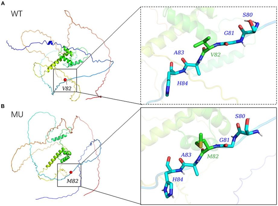 Fig. 1 Modelled tertiary structure of the protein encoded by NOBOX.1
Key structural properties of NOBOX:
- Contains typical Homeobox domains (transcription factor characteristics)
- Has approved a signal, to ensure that protein localization in the nucleus
- The C-terminal transcriptional activation domain is responsible for regulating gene expression
- Specific DNA binding helix-turn-helix motif
Functions of NOBOX
The core function of the NOBOX gene is to regulate oocyte development and maintain ovarian reserve. Its specific mechanism of action is as follows:
| Function | Description |
| Regulation of follicular development | Key gene networks are activated during primordial follicle activation and primary follicle formation to determine follicle fate. |
| Meiosis block | By regulating the signal transduction between granulosa cells and oocytes, the quiescent state of oocytes during the prophase of meiosis is maintained. |
| Specialization of germ cells | As a germline-specific transcription factor, it establishes oocyte identity and inhibits somatic cell differentiation pathways. |
| Transcriptional activation of target genes | By binding specific DNA sequences in the promoters of target genes, it regulates the expression of reproductive factors such as FIGLA and GDF9. |
| Association with ovarian aging | Its expression level is positively correlated with the volume of the primordial follicular pool. Functional deficiency will lead to premature ovarian failure and decreased fertility. |
The expression of this gene shows strict spatiotemporal specificity. Its transcriptional activity begins to rise in the ovaries during the fetal period and reaches its peak before puberty. This expression pattern is consistent with its core position in the establishment of germ cell lineages and the maintenance of early follicular pools.
Applications of NOBOX and NOBOX Antibody in Literature
1. Yao, Changfang, et al. "Hypo-hydroxymethylation of NOBOX is associated with ovarian dysfunction in rat offspring exposed to prenatal hypoxia." Reproductive Sciences 29.5 (2022): 1424-1436. https://doi.org/10.1007/s43032-022-00866-6
Research has found that hypoxia during pregnancy can inhibit the expression of the NOBOX gene in the ovaries of offspring rats. The mechanism is to reduce the hydroxymethylation level in specific regions of genes, ultimately leading to abnormal follicular development and decreased ovarian function.
2. Wu, Kun, et al. "Genetic evidence for differential functions of figla and nobox in zebrafish ovarian differentiation and folliculogenesis." Communications Biology 6.1 (2023): 1185. https://doi.org/10.1038/s42003-023-05551-1
Research has found that the absence of Nobox in zebrafish inhibits the expression of the aromatase gene cyp19a1a, reduces estrogen levels, and leads to the arrest of follicular development in the pre-oocyte formation stage, revealing that it drives follicular development by regulating estrogen signals.
3. Huang, Ruan Y, et al. "Association between polymorphisms in NOBOX and litter size traits in Xiangsu pigs." Frontiers in Veterinary Science 11 (2024): 1359312. https://doi.org/10.3389/fvets.2024.1359312
Research has found that NOBOX is dominant in the ovaries of Xiangsu pigs. Its missense mutation G.1858 G>A reduces protein stability. This mutation and the other two SNPS are significantly associated with litter size.
4. Tripurani, Swamy K., et al. "MicroRNA-196a regulates bovine newborn ovary homeobox gene (NOBOX) expression during early embryogenesis." BMC developmental biology 11.1 (2011): 25. https://doi.org/10.1186/1471-213X-11-25
This study confirmed that during the early embryonic development of cattle, microRNA miR-196a can directly target and bind to the 3'utr region of the NOBOX gene, achieving negative regulation of NOBOX by inhibiting its mRNA and protein expression. This discovery reveals that miR-196a is a key upstream regulatory factor of NOBOX, providing new insights into the mechanism by which maternal factors regulate embryonic development.
5. Bayne, Rosemary AL, et al. "GDF9 is transiently expressed in oocytes before follicle formation in the human fetal ovary and is regulated by a novel NOBOX transcript." PLoS One 10.3 (2015): e0119819. https://doi.org/10.1371/journal.pone.0119819
This study discovered a new NOBOX subtype in the ovaries of human fetuses, confirming that it can directly up-regulate the activity of the GDF9 promoter. During pregnancy, the mRNA levels of NOBOX, GDF9 and BMP15 increase significantly simultaneously, jointly regulating the formation of primordial follicles and the survival of germ cells. The expression differences of NOBOX may explain the differences in the age of onset among patients with primary ovarian insufficiency caused by its mutations.
Creative Biolabs: NOBOX Antibodies for Research
Creative Biolabs specializes in the production of high-quality NOBOX antibodies for research and industrial applications. Our portfolio includes monoclonal antibodies tailored for ELISA, Flow Cytometry, Western blot, immunohistochemistry, and other diagnostic methodologies.
- Custom NOBOX Antibody Development: Tailor-made solutions to meet specific research requirements.
- Bulk Production: Large-scale antibody manufacturing for industry partners.
- Technical Support: Expert consultation for protocol optimization and troubleshooting.
- Aliquoting Services: Conveniently sized aliquots for long-term storage and consistent experimental outcomes.
For more details on our NOBOX antibodies, custom preparations, or technical support, contact us at email.
Reference
- Huang, Ruan Y, et al. "Association between polymorphisms in NOBOX and litter size traits in Xiangsu pigs." Frontiers in Veterinary Science 11 (2024): 1359312. https://doi.org/10.3389/fvets.2024.1359312
Anti-NOBOX antibodies
 Loading...


Hot products 
-
Mouse Anti-DISP2 Monoclonal Antibody (F66A4B1) (CBMAB-1112CQ)

-
Mouse Anti-DMPK Recombinant Antibody (CBYCD-324) (CBMAB-D1200-YC)

-
Mouse Anti-BIRC3 Recombinant Antibody (16E63) (CBMAB-C3367-LY)

-
Mouse Anti-ATP5F1A Recombinant Antibody (51) (CBMAB-A4043-YC)

-
Mouse Anti-CCN2 Recombinant Antibody (CBFYC-2383) (CBMAB-C2456-FY)

-
Mouse Anti-ATM Recombinant Antibody (2C1) (CBMAB-A3970-YC)

-
Mouse Anti-C5b-9 Recombinant Antibody (aE11) (CBMAB-AO138LY)

-
Mouse Anti-ASTN1 Recombinant Antibody (H-9) (CBMAB-1154-CN)

-
Mouse Anti-AKT1 (Phosphorylated S473) Recombinant Antibody (V2-505430) (PTM-CBMAB-0067LY)

-
Mouse Anti-APP Recombinant Antibody (5C2A1) (CBMAB-A3314-YC)

-
Mouse Anti-CCT6A/B Recombinant Antibody (CBXC-0168) (CBMAB-C5570-CQ)

-
Mouse Anti-BLNK Recombinant Antibody (CBYY-0623) (CBMAB-0626-YY)

-
Mouse Anti-CARTPT Recombinant Antibody (113612) (CBMAB-C2450-LY)

-
Mouse Anti-BHMT Recombinant Antibody (CBYY-0547) (CBMAB-0550-YY)

-
Mouse Anti-CEMIP Recombinant Antibody (3C12) (CBMAB-K0296-LY)

-
Mouse Anti-ARHGAP5 Recombinant Antibody (54/P190-B) (CBMAB-P0070-YC)

-
Rat Anti-ADGRE4 Recombinant Antibody (V2-160163) (CBMAB-F0011-CQ)

-
Mouse Anti-BIRC5 Recombinant Antibody (6E4) (CBMAB-CP2646-LY)

-
Mouse Anti-CAT Recombinant Antibody (724810) (CBMAB-C8431-LY)

-
Mouse Anti-CAPZB Recombinant Antibody (CBYY-C0944) (CBMAB-C2381-YY)

- AActivation
- AGAgonist
- APApoptosis
- BBlocking
- BABioassay
- BIBioimaging
- CImmunohistochemistry-Frozen Sections
- CIChromatin Immunoprecipitation
- CTCytotoxicity
- CSCostimulation
- DDepletion
- DBDot Blot
- EELISA
- ECELISA(Cap)
- EDELISA(Det)
- ESELISpot
- EMElectron Microscopy
- FFlow Cytometry
- FNFunction Assay
- GSGel Supershift
- IInhibition
- IAEnzyme Immunoassay
- ICImmunocytochemistry
- IDImmunodiffusion
- IEImmunoelectrophoresis
- IFImmunofluorescence
- IGImmunochromatography
- IHImmunohistochemistry
- IMImmunomicroscopy
- IOImmunoassay
- IPImmunoprecipitation
- ISIntracellular Staining for Flow Cytometry
- LALuminex Assay
- LFLateral Flow Immunoassay
- MMicroarray
- MCMass Cytometry/CyTOF
- MDMeDIP
- MSElectrophoretic Mobility Shift Assay
- NNeutralization
- PImmunohistologyp-Paraffin Sections
- PAPeptide Array
- PEPeptide ELISA
- PLProximity Ligation Assay
- RRadioimmunoassay
- SStimulation
- SESandwich ELISA
- SHIn situ hybridization
- TCTissue Culture
- WBWestern Blot
