Phosphoserine Antibodies
Background
Phosphoserine is a phosphorylated and modified serine residue that is widely present in the proteins of eukaryotes and bacteria. It is formed through kinase-mediated post-translational modification and plays a core role in key pathways such as cell signal transduction, metabolic regulation, and DNA damage repair. Its phosphorylation state can dynamically regulate protein activity and interactions, directly affecting gene expression and the progress of the cell cycle. Since its discovery in the middle of the 20th century, phosphoserine, as a classic model for protein phosphorylation research, has promoted the analysis of the mechanism of kinase/phosphatase action and epigenetic regulatory mechanisms. In-depth research on this modification site continuously reveals its molecular regulatory logic in physiological and pathological processes such as neural development and cancer occurrence.
Structure of Phosphoserine
Phosphoserine is not an independent gene but a key post-translational modification. Its molecular weight is approximately 167.08 Da, and this value remains constant across different protein backgrounds because the modification itself does not alter the length of the core peptide chain.
| Characteristics | Signal transduction protein | Metabolic enzymes | Transcription factor | Cytoskeletal protein |
| Molecular Weight (kDa) | ≈167.08 | ≈167.08 | ≈167.08 | ≈167.08 |
| Primary Structural Differences | Activate the signal pathway | Regulate enzyme activity | Regulate gene expression | Affect protein aggregation |
This modification is attached to the hydroxyl group of serine residues through ester bonds, and the introduced phosphate group carries a strong negative charge, significantly altering the three-dimensional conformation and electrostatic properties of the protein. Its chemical structure consists of a phosphate group covalently bound to the hydroxyl group of serine through a phosphoester bond, forming a reversible modification switch. Protein kinases catalyze the transfer of phosphate groups from ATP to serine hydroxyl groups, while phosphatases are responsible for their removal, jointly constituting a dynamic regulatory mechanism. The modified negatively charged environment creates a recognition platform that promotes protein interactions and regulates substrate activity, playing a core role in the cellular signaling network.
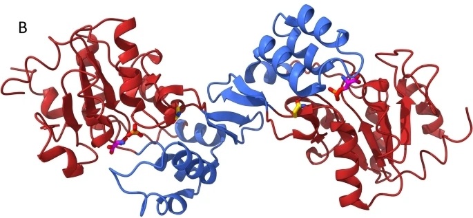 Fig. 1 The structure of phosphoserine phosphatase with its catalytic products.1
Key structural properties of phosphoserine:
- Phosphate groups through ester linkage covalent connection in the side chain amino acid serine
- Introduced by the strong negative significant changes in protein local static environment
- Dynamically reversible modification states constitute molecular switching functions
Functions of Phosphoserine
The main function of phosphoserine is to act as a molecular switch, regulating the activity of proteins and cellular signal transduction. However, it is also deeply involved in a variety of key cellular physiological processes, including cell cycle regulation and DNA damage repair.
| Function | Description |
| Signal transduction regulation | As a kinase substrate, its phosphorylation state directly activates or inhibits signaling pathways (such as the MAPK pathway), regulating cell growth, proliferation and apoptosis. |
| Regulation of metabolic enzyme activity | By reversibly phosphorylating and modifying key metabolic enzymes, their active states can be rapidly switched, thereby precisely regulating the metabolic flow and energy balance within cells. |
| Regulation of gene expression | Modify transcription factors and chromatin associated proteins, affect their DNA binding ability and transcriptional activity, and directly regulate the expression level of specific genes. |
| Cell cycle advancement | As a key regulatory point of the cell cycle engine, the phosphoserine modification of specific proteins is necessary to drive cells to complete the transitions at each stage of the cycle. |
| Stress response and repair | Under stress conditions such as DNA damage, a phosphoserine signaling network is rapidly formed to coordinate the initiation of repair mechanisms and cell fate decisions. |
The dynamic characteristics of this modification - catalyzed by kinases and removed by phosphatases - constitute an efficient binary switch, whose regulatory speed and reversibility are much faster than those of gene expression, making it the core mechanism of real-time signal processing within cells.
Applications of Phosphoserine and Phosphoserine Antibody in Literature
1. Wulfert, Sabine, and Stephan Krueger. "Phosphoserine aminotransferase1 is part of the phosphorylated pathways for serine biosynthesis and essential for light and sugar-dependent growth promotion." Frontiers in plant science 9 (2018): 1712. https://doi.org/10.3389/fpls.2018.01712
The article indicates that due to the lack of T-DNA insertion mutants, the physiological function of phosphoserine aminotransferase (PSAT1) remains unclear. In this study, by specifically silencing the PSAT1 gene, it was found that its absence would severely inhibit the growth of plant roots and stems, leading to amino acid accumulation. It was also demonstrated that the phosphorylated serine biosynthesis pathway is crucial for growth promoted by light and sucrose.
2. Collet, Jean-François, Vincent Stroobant, and Emile Van Schaftingen. "Mechanistic studies of phosphoserine phosphatase, an enzyme related to P-type ATPases." Journal of Biological Chemistry 274.48 (1999): 33985-33990. https://doi.org/10.1074/jbc.274.48.33985
The article indicates that phosphoserine phosphatase shares a catalytic mechanism with P-type ATPase, and the Asp-20 in its active center forms a key acyl phosphate intermediate in the catalysis. The study confirmed through mass spectrometry and site-directed mutagenesis that conserved residues such as Asp-179 and Lys-158 are crucial for substrate binding, magnesium ion coordination and enzyme activity.
3. Appel, Lisa-Marie, et al. "The SPOC domain is a phosphoserine binding module that bridges transcription machinery with co-and post-transcriptional regulators." Nature communications 14.1 (2023): 166. https://doi.org/10.1038/s41467-023-35853-1
This study reveals that the SPOC domain is a key reader for recognizing phosphorylated serine (such as Ser-2/Ser-5) on the carboxyl-terminal domain (CTD) of RNA polymerase II. This domain serves as a bridge, closely linking the transcriptional machine with key co-transcriptional regulatory processes such as m6A modification and X chromosome inactivation.
4. Miskiewicz, E. I., et al. "Phosphoserine-86-HSPB1 (pS86-HSPB1) is cytoplasmic and highly induced in rat myometrium at labour." Histochemistry and Cell Biology 159.2 (2023): 149-162. https://doi.org/10.1007/s00418-022-02158-1
This study focuses on the phosphorylation of heat shock protein HSPB1 at serine sites (such as S86/S15). Studies have found that during the third trimester of pregnancy and delivery, the level of pS86-HSPB1 in the myometrium of rats significantly increases, and this process is regulated by uterine dilation stress signals. The cellular localization (cytoplasm or adhesion plaque) of phosphorylated HSPB1 varies, suggesting that it plays an important role in labor preparation.
5. Kasuga, Masato, et al. "Insulin stimulation of phosphorylation of the beta subunit of the insulin receptor. Formation of both phosphoserine and phosphotyrosine." Journal of Biological Chemistry 257.17 (1982): 9891-9894. https://doi.org/10.1016/S0021-9258(18)33955-3
This study is the first to confirm that insulin can activate the self-phosphorylation of its receptor. Under insulin stimulation, the phosphorylation level of the insulin receptor β subunit in rat hepatoma cells was significantly enhanced. Among them, the increase in phosphoserine content was a newly discovered key event, suggesting that multi-site phosphorylation is an early mechanism of insulin action.
Creative Biolabs: Phosphoserine Antibodies for Research
Creative Biolabs specializes in the production of high-quality Phosphoserine antibodies for research and industrial applications. Our portfolio includes monoclonal antibodies tailored for ELISA, Flow Cytometry, Western blot, immunohistochemistry, and other diagnostic methodologies.
- Custom Phosphoserine Antibody Development: Tailor-made solutions to meet specific research requirements.
- Bulk Production: Large-scale antibody manufacturing for industry partners.
- Technical Support: Expert consultation for protocol optimization and troubleshooting.
- Aliquoting Services: Conveniently sized aliquots for long-term storage and consistent experimental outcomes.
For more details on our Phosphoserine antibodies, custom preparations, or technical support, contact us at email.
Reference
- Pollegioni, Loredano, et al. "L-serine deficiency: on the properties of the Asn133Ser variant of human phosphoserine phosphatase." Scientific Reports 14.1 (2024): 12463. https://doi.org/10.1038/s41598-024-63164-y
Anti-Phosphoserine antibodies
 Loading...


Hot products 
-
Mouse Anti-BRD3 Recombinant Antibody (CBYY-0801) (CBMAB-0804-YY)

-
Rat Anti-CD63 Recombinant Antibody (7G4.2E8) (CBMAB-C8725-LY)

-
Rat Anti-ADAM10 Recombinant Antibody (V2-179741) (CBMAB-A1103-YC)

-
Mouse Anti-BSN Recombinant Antibody (219E1) (CBMAB-1228-CN)

-
Mouse Anti-GFAP Recombinant Antibody (5) (CBMAB-G0346-LY)

-
Mouse Anti-ENO1 Recombinant Antibody (8G8) (CBMAB-E1329-FY)

-
Mouse Anti-AGO2 Recombinant Antibody (V2-634169) (CBMAB-AP203LY)

-
Mouse Anti-ENO2 Recombinant Antibody (85F11) (CBMAB-0276CQ)

-
Mouse Anti-ASB9 Recombinant Antibody (1D8) (CBMAB-A0529-LY)

-
Rabbit Anti-AP2M1 (Phosphorylated T156) Recombinant Antibody (D4F3) (PTM-CBMAB-0610LY)

-
Rabbit Anti-AKT2 (Phosphorylated S474) Recombinant Antibody (V2-556130) (PTM-CBMAB-0605LY)

-
Human Anti-SARS-CoV-2 S1 Monoclonal Antibody (CBFYR-0120) (CBMAB-R0120-FY)

-
Mouse Anti-FN1 Monoclonal Antibody (71) (CBMAB-1241CQ)

-
Rat Anti-CD300A Recombinant Antibody (172224) (CBMAB-C0423-LY)

-
Mouse Anti-C5B-9 Recombinant Antibody (CBFYA-0216) (CBMAB-X0304-FY)

-
Mouse Anti-AGK Recombinant Antibody (V2-258056) (CBMAB-M0989-FY)

-
Mouse Anti-C5b-9 Recombinant Antibody (aE11) (CBMAB-AO138LY)

-
Mouse Anti-C4B Recombinant Antibody (CBYY-C2996) (CBMAB-C4439-YY)

-
Rabbit Anti-ABL1 (Phosphorylated Y245) Recombinant Antibody (V2-505716) (PTM-CBMAB-0465LY)

-
Mouse Anti-FOSB Recombinant Antibody (CBXF-3593) (CBMAB-F2522-CQ)

- AActivation
- AGAgonist
- APApoptosis
- BBlocking
- BABioassay
- BIBioimaging
- CImmunohistochemistry-Frozen Sections
- CIChromatin Immunoprecipitation
- CTCytotoxicity
- CSCostimulation
- DDepletion
- DBDot Blot
- EELISA
- ECELISA(Cap)
- EDELISA(Det)
- ESELISpot
- EMElectron Microscopy
- FFlow Cytometry
- FNFunction Assay
- GSGel Supershift
- IInhibition
- IAEnzyme Immunoassay
- ICImmunocytochemistry
- IDImmunodiffusion
- IEImmunoelectrophoresis
- IFImmunofluorescence
- IGImmunochromatography
- IHImmunohistochemistry
- IMImmunomicroscopy
- IOImmunoassay
- IPImmunoprecipitation
- ISIntracellular Staining for Flow Cytometry
- LALuminex Assay
- LFLateral Flow Immunoassay
- MMicroarray
- MCMass Cytometry/CyTOF
- MDMeDIP
- MSElectrophoretic Mobility Shift Assay
- NNeutralization
- PImmunohistologyp-Paraffin Sections
- PAPeptide Array
- PEPeptide ELISA
- PLProximity Ligation Assay
- RRadioimmunoassay
- SStimulation
- SESandwich ELISA
- SHIn situ hybridization
- TCTissue Culture
- WBWestern Blot
