RAC2 Antibodies
Background
The RAC2 gene encodes a protein belonging to the Rho GTPase family, which is mainly expressed in hematopoietic cells such as neutrophils and phagocytes. This protein plays a central role in immune defense processes such as cell movement, phagocytosis, and reactive oxygen species production by regulating cytoskeleton reorganization. RAC2 was first identified in 1993, and its dysfunction is closely related to various immune deficiencies and hematological diseases. Notably, RAC2 is the first Rho family GTPase to be discovered to be directly associated with human primary immunodeficiency diseases. Its specific mutations can lead to leukocyte adhesion deficiency syndrome. The molecular mechanism study of this gene has deepened our understanding of immune cell signal transduction, host defense, and inflammatory regulation, providing a key theoretical basis for targeted immunoregulatory therapy.
Structure of RAC2
RAC2 is a small GTP-binding protein with a molecular weight of approximately 21.4 kDa. There are slight variations in its molecular weight among different species, mainly due to sequence changes outside the conserved domains.
| Species | Human | Mouse | Rat | Zebrafish |
| Molecular Weight (kDa) | 21.4 | 21.4 | 21.5 | 21.0 |
| Primary Structural Differences | Containing a highly conserved GTP-binding domain | High homologous to human RAC2 and similar functions | The core functional areas are highly conserved | Vertebrate homologs exist, with functions conserved in evolution |
This protein is composed of 191 amino acids and its structure belongs to the typical fold of the Rho GTPase family. The core of RAC2 is its GTPase domain, which contains highly conserved Switch I and Switch II regions. These conformational switches play a crucial role in the transition between its active (GTP-bound) and inactive (GDP-bound) states. Its C-terminal contains a multi-polyisoprenylation modification site (CAAX box), which is essential for anchoring the protein to the cell membrane and enabling its signaling function. Additionally, RAC2 possesses an insertion helix structure, which is one of the characteristics that distinguishes it from paralogous homologs such as RAC1 and RAC3, and may mediate its specific interactions with specific effector proteins.
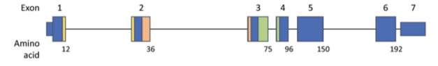 Fig. 1 Genomic schematic for RAC2.1
Key structural properties of RAC2:
- Gtpase core domain, containing the conserved Switch I/II conformational switch
- The C-terminal polyisoprene modification site (CAAX box) mediates membrane localization
- Different from the characteristic insertion helical structure of RAC1/RAC3
- Specific effector protein binding interface determines its immune cell function specificity
Functions of RAC2
The core function of the RAC2 gene is to act as a molecular switch to regulate the signal transduction and cytoskeletal dynamics of immune cells. Its main physiological functions are as follows:
| Function | Description |
| Immune Cell Movement and Chemotaxis | By activating downstream effectors (such as the WAVE complex), it regulates the reorganization of the actin cytoskeleton, driving the directional migration of neutrophils, macrophages, etc. to the infection site. |
| Regulation of Phagocytosis | It mediates the formation and closure of phagocytic cups, and is a crucial signaling molecule necessary for immune cells to engulf pathogens and apoptotic cells. |
| Production of Reactive Oxygen Species (ROS) | Activating the NADPH oxidase complex to catalyze the production of reactive oxygen species, which is the core mechanism for killing ingested microorganisms. |
| Cell Adhesion and Inflammation | Integrins, as important regulators of cell adhesion and inflammatory responses, influence the rolling and exudation of immune cells on the vascular endothelium. |
| Immune Synapse Formation | At the contact interface between T cells and antigen-presenting cells, it regulates the polarization of the cytoskeleton to ensure effective immune recognition and signal transmission. |
The activity of RAC2 is strictly regulated by the GDP/GTP binding state. Its activation depends on guanine nucleotide exchange factors (GEFs, such as DOCK2), while its inactivation is promoted by GTPase activating proteins (GAPs). Unlike the widely expressed RAC1, the function of RAC2 is mainly confined to the hematopoietic system, and its mutations are closely related to primary immune diseases such as leukocyte adhesion deficiency.
Applications of RAC2 and RAC2 Antibody in Literature
1. Hsu, Amy P. "Not too little, not too much: the impact of mutation types in Wiskott-Aldrich syndrome and RAC2 patients." Clinical and Experimental Immunology 212.2 (2023): 137-146. https://doi.org/10.1093/cei/uxad001
This article reviews the different clinical phenotypes caused by mutations in the WAS and RAC2 genes, and explores how variations in the protein functional domains affect the interaction of immune proteins, ultimately leading to a wide range of disease manifestations from severe syndromes to single symptoms.
2. Rakoczy, Katarzyna, et al. "The role of RAC2 and PTTG1 in cancer biology." Cells 14.5 (2025): 330. https://doi.org/10.3390/cells14050330
This article reviews the core roles of RAC2 and PTTG1 proteins in the regulation of cancer stem cells, revealing the molecular mechanisms by which they promote tumor proliferation, metastasis and drug resistance through signaling pathways such as PI3K/AKT and TGF-β. It also explores the potential value of these proteins as prognostic markers and therapeutic targets.
3. Liu, Ranran, et al. "Pancancer analysis revealed the value of RAC2 in immunotherapy and cancer stem cell." Stem Cells International 2023.1 (2023): 8485726. https://doi.org/10.1155/2023/8485726
Through multi-database analysis in this study, it was found that the RAC2 gene is highly expressed in various tumors and is significantly correlated with prognosis, immune cell infiltration, and tumor stem cell characteristics, suggesting that it can serve as a potential immunotherapy and prognostic marker.
4. Tanner, Christopher D., and Emily E. Rosowski. "Macrophages inhibit extracellular hyphal growth of A. fumigatus through Rac2 GTPase signaling." Infection and Immunity 92.2 (2024): e00380-23. https://doi.org/10.1128/iai.00380-23
This study, using the zebrafish model, found that although macrophages can normally migrate and engulf Aspergillus fumigatus spores, they rely on the Rac2 signaling pathway to effectively inhibit spore germination and control hyphal invasion. This function is crucial for the host's resistance to fungal infections.
5. Padmanabhan, Jagannath, et al. "Allometrically scaling tissue forces drive pathological foreign-body responses to implants via Rac2-activated myeloid cells." Nature Biomedical Engineering 7.11 (2023): 1419-1436. https://doi.org/10.1038/s41551-023-01091-5
This study reveals that the foreign body response (FBR) of the human body to implants is driven by surface forces and triggered by mechanical signals from immune cells mediated by RAC2. In a mouse model subjected to human-scale external forces, inhibition of Rac2 significantly alleviated this pathological response.
Creative Biolabs: RAC2 Antibodies for Research
Creative Biolabs specializes in the production of high-quality RAC2 antibodies for research and industrial applications. Our portfolio includes monoclonal antibodies tailored for ELISA, Flow Cytometry, Western blot, immunohistochemistry, and other diagnostic methodologies.
- Custom RAC2 Antibody Development: Tailor-made solutions to meet specific research requirements.
- Bulk Production: Large-scale antibody manufacturing for industry partners.
- Technical Support: Expert consultation for protocol optimization and troubleshooting.
- Aliquoting Services: Conveniently sized aliquots for long-term storage and consistent experimental outcomes.
For more details on our RAC2 antibodies, custom preparations, or technical support, contact us at email.
Reference
- Hsu, Amy P. "Not too little, not too much: the impact of mutation types in Wiskott-Aldrich syndrome and RAC2 patients." Clinical and Experimental Immunology 212.2 (2023): 137-146. https://doi.org/10.1093/cei/uxad001
Anti-RAC2 antibodies
 Loading...


Hot products 
-
Mouse Anti-FYN Recombinant Antibody (10) (CBMAB-S6332-CQ)

-
Mouse Anti-BCL2L1 Recombinant Antibody (H5) (CBMAB-1025CQ)

-
Rat Anti-ADAM10 Recombinant Antibody (V2-179741) (CBMAB-A1103-YC)

-
Mouse Anti-FN1 Monoclonal Antibody (D6) (CBMAB-1240CQ)

-
Mouse Anti-ACTN4 Recombinant Antibody (V2-6075) (CBMAB-0020CQ)

-
Rabbit Anti-ABL1 (Phosphorylated Y245) Recombinant Antibody (V2-505716) (PTM-CBMAB-0465LY)

-
Mouse Anti-DES Monoclonal Antibody (440) (CBMAB-AP1857LY)

-
Mouse Anti-CTCF Recombinant Antibody (CBFYC-2371) (CBMAB-C2443-FY)

-
Mouse Anti-AMIGO2 Recombinant Antibody (CBYY-C0756) (CBMAB-C2192-YY)

-
Mouse Anti-CD33 Recombinant Antibody (6C5/2) (CBMAB-C8126-LY)

-
Mouse Anti-CA9 Recombinant Antibody (CBXC-2079) (CBMAB-C0131-CQ)

-
Mouse Anti-AAV9 Recombinant Antibody (V2-634029) (CBMAB-AP023LY)

-
Mouse Anti-ENO2 Recombinant Antibody (85F11) (CBMAB-0276CQ)

-
Mouse Anti-CAPZB Recombinant Antibody (CBYY-C0944) (CBMAB-C2381-YY)

-
Mouse Anti-G6PD Recombinant Antibody (13B331) (CBMAB-G1553-LY)

-
Mouse Anti-CD59 Recombinant Antibody (CBXC-2097) (CBMAB-C4421-CQ)

-
Mouse Anti-CD83 Recombinant Antibody (HB15) (CBMAB-C1765-CQ)

-
Mouse Anti-FPR2 Recombinant Antibody (1D6) (CBMAB-F2628-CQ)

-
Mouse Anti-CD247 Recombinant Antibody (6B10.2) (CBMAB-C1583-YY)

-
Mouse Anti-AKT1 (Phosphorylated S473) Recombinant Antibody (V2-505430) (PTM-CBMAB-0067LY)

- AActivation
- AGAgonist
- APApoptosis
- BBlocking
- BABioassay
- BIBioimaging
- CImmunohistochemistry-Frozen Sections
- CIChromatin Immunoprecipitation
- CTCytotoxicity
- CSCostimulation
- DDepletion
- DBDot Blot
- EELISA
- ECELISA(Cap)
- EDELISA(Det)
- ESELISpot
- EMElectron Microscopy
- FFlow Cytometry
- FNFunction Assay
- GSGel Supershift
- IInhibition
- IAEnzyme Immunoassay
- ICImmunocytochemistry
- IDImmunodiffusion
- IEImmunoelectrophoresis
- IFImmunofluorescence
- IGImmunochromatography
- IHImmunohistochemistry
- IMImmunomicroscopy
- IOImmunoassay
- IPImmunoprecipitation
- ISIntracellular Staining for Flow Cytometry
- LALuminex Assay
- LFLateral Flow Immunoassay
- MMicroarray
- MCMass Cytometry/CyTOF
- MDMeDIP
- MSElectrophoretic Mobility Shift Assay
- NNeutralization
- PImmunohistologyp-Paraffin Sections
- PAPeptide Array
- PEPeptide ELISA
- PLProximity Ligation Assay
- RRadioimmunoassay
- SStimulation
- SESandwich ELISA
- SHIn situ hybridization
- TCTissue Culture
- WBWestern Blot
