RGMA Antibodies
Background
The RGMA gene encodes a glycosylated phosphatidylinositol-anchored membrane protein, which plays a crucial role in the development of the central nervous system. This protein acts as a repulsive guiding molecule, guiding axon growth paths and regulating neuronal migration by binding to the Neogenin receptors on the surface of nerve cells, thereby participating in the regulation of neural tube formation and the establishment of neural networks. Studies have shown that the expression of the RGMA protein is upregulated after spinal cord injury, and it may affect the repair process by inhibiting nerve regeneration. Since its first identification in 2002, this gene has become an important model for neurodevelopment and regeneration research. The repulsive signaling mechanism mediated by this gene provides a molecular basis for understanding neurological developmental disorders and has become a potential research direction for neurorepair treatment.
Structure of RGMA
The protein molecule size encoded by the RGMA gene is approximately 55 kDa. The exact value may vary slightly depending on different splicing variants or post-translational modifications (such as glycosylation).
| Species | Human | Mouse | Rat | Chicken |
|---|---|---|---|---|
| Molecular Weight (kDa) | ~55 | ~54 | ~55 | ~53 |
| Primary Structural Differences | Contains the RGM domain and binds to the Neogenin receptor | Highly conservative, functionally similar | High amino acid sequence homology | Consistent role in neural development |
This protein is composed of approximately 550 amino acids. Its primary structure is characterized by the presence of a conserved RGM (Repulsive Guidance Molecule) domain, which is the core for its biological function. The RGMA protein folds into a specific spatial conformation through its secondary structure (mainly α-helix and β-sheet), enabling it to specifically bind to the Neogenin receptor on the cell membrane as a ligand, thereby transmitting signals in the rejection-oriented guidance and cell migration of the nervous system.
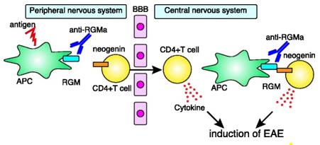 Fig. 1 RGMa-neogenin signaling mediates autoimmune encephalomyelitis.1
Key structural properties of RGMA:
- Contains a conservative RGM (rejection-oriented molecule) domain
- Attached to the cell membrane through glycosylated phosphatidylinositol (GPI)
- Having a specific region for recognizing and binding to the Neogenin receptor
- The protein folding conformation is very important to maintain the axon guidance function
Functions of RGMA
The main function of the protein encoded by the RGMA gene is to act as an exclusive guiding molecule, regulating the development and regeneration of the nervous system. Additionally, it is also involved in various pathological physiological processes.
| Function | Description |
|---|---|
| Axonal Guidance | As an inhibitory signaling molecule, it guides the growth cone path and participates in the precise wiring of neural networks. |
| Neuron Migration | By interacting with its receptor Neogenin, it regulates the positioning and layered distribution of developing neurons. |
| Neural tube closure | It plays a role in the early stages of embryonic development and influences the formation of the basic structure of the central nervous system. |
| Inhibition of nerve regeneration | It is upregulated after adult central nervous system injury, creating an inhibitory microenvironment that restricts axonal regeneration. |
| Apoptosis Regulation | In certain circumstances, it can participate in mediating programmed cell death and affect the neural progenitor cell population. |
The binding of RGMA protein to the receptor Neogenin exhibits a high-affinity characteristic, which is different from the co-binding of hemoglobin. This indicates its role as a precise and stable repulsive signaling molecule, and it strictly regulates the spatial and temporal growth path of nerve fibers during development.
Applications of RGMA and RGMA Antibody in Literature
1. Yamamoto, Masaya, et al. "Anti-RGMa neutralizing antibody ameliorates vascular cognitive impairment in mice." Neurotherapeutics 22.2 (2025): e00500. https://doi.org/10.1016/j.neurot.2024.e00500
This study explores the role of RGMa in a mouse model of vascular dementia. The elevated expression of RGMa in the hippocampal region of the model is accompanied by reduced neurogenesis and cognitive impairment. Anti-rgma neutralizing antibodies can reverse the above pathological changes, suggesting its potential as a new treatment strategy for vascular dementia.
2. Araya, Natsumi, et al. "Virus-induced RGMa expression drives neurodegeneration in HTLV-1–associated myelopathy." JCI insight 10.11 (2025): e184530. https://doi.org/10.1172/jci.insight.184530
This study reveals the neuronal injury mechanism of HTLV-1-related myelopathy: The viral Tax protein and Sp1 jointly upregulate the expression of RGMa, which remains highly expressed in infected cells due to reduced H3K27me3 methylation. The neutralizing antibody MT-3921 can effectively block this damage, suggesting that targeting RGMa is a potential therapy.
3. Yang, Xiaoyi, et al. "RGMA gene polymorphisms as predictive biomarkers for early relapse in neuromyelitis optica spectrum disorders." Neurobiology of Disease (2025): 107063. https://doi.org/10.1016/j.nbd.2025.107063
Research has found that the polymorphisms of rs725458-CC and rs4778099-AA in the RGMA gene are key markers for predicting early recurrence (early onset but low recurrence rate in the later stage) in patients with NMOSD. Combined with clinical features, the risk assessment of recurrence and individualized treatment can be optimized.
4. Cheng, Ruiqi, et al. "Artificial Microglia Nanoplatform Loaded With Anti‐RGMa in Acoustic/Magnetic Feld for Recanalization and Neuroprotection in Acute Ischemic Stroke." Advanced Science 11.48 (2024): 2410529. https://doi.org/10.1002/advs.202410529
This study constructed a bionic nanoplatform for targeted treatment of ischemic stroke. This platform delivers anti-RGMA antibodies and Fe₃O₄ through microglial cell membranes, achieving thrombolysis and neuroprotection under the influence of ultrasound and magnetic fields, and has demonstrated good safety in animal experiments.
5. Sempert, Kai, et al. "RGMa and Neogenin control dendritic spine morphogenesis via WAVE Regulatory Complex-mediated actin remodeling." Frontiers in Molecular Neuroscience 16 (2023): 1253801. https://doi.org/10.3389/fnmol.2023.1253801
Research has found that RGMa and its receptor Neogenin drive the polymerization of branched actin by activating the WRC complex, thereby regulating the structural plasticity and maturation of dendritic spines, providing new insights into the mechanism of RGMa in the induction of LTP related to learning and memory.
Creative Biolabs: RGMA Antibodies for Research
Creative Biolabs specializes in the production of high-quality RGMA antibodies for research and industrial applications. Our portfolio includes monoclonal antibodies tailored for ELISA, Flow Cytometry, Western blot, immunohistochemistry, and other diagnostic methodologies.
- Custom RGMA Antibody Development: Tailor-made solutions to meet specific research requirements.
- Bulk Production: Large-scale antibody manufacturing for industry partners.
- Technical Support: Expert consultation for protocol optimization and troubleshooting.
- Aliquoting Services: Conveniently sized aliquots for long-term storage and consistent experimental outcomes.
For more details on our RGMA antibodies, custom preparations, or technical support, contact us at email.
Reference
- Fujita, Yuki, and Toshihide Yamashita. "The roles of RGMa-neogenin signaling in inflammation and angiogenesis." Inflammation and regeneration 37.1 (2017): 6. https://doi.org/10.1186/s41232-017-0037-6
Anti-RGMA antibodies
 Loading...


Hot products 
-
Mouse Anti-AMIGO2 Recombinant Antibody (CBYY-C0756) (CBMAB-C2192-YY)

-
Rat Anti-CD34 Recombinant Antibody (MEC 14.7) (CBMAB-C10196-LY)

-
Rabbit Anti-AKT3 Recombinant Antibody (V2-12567) (CBMAB-1057-CN)

-
Mouse Anti-CFL1 (Phospho-Ser3) Recombinant Antibody (CBFYC-1770) (CBMAB-C1832-FY)

-
Mouse Anti-ALB Recombinant Antibody (V2-363290) (CBMAB-S0173-CQ)

-
Mouse Anti-BCL2L1 Recombinant Antibody (H5) (CBMAB-1025CQ)

-
Mouse Anti-BIRC7 Recombinant Antibody (88C570) (CBMAB-L0261-YJ)

-
Mouse Anti-BIRC3 Recombinant Antibody (16E63) (CBMAB-C3367-LY)

-
Mouse Anti-CCNH Recombinant Antibody (CBFYC-1054) (CBMAB-C1111-FY)

-
Mouse Anti-ARID3A Antibody (A4) (CBMAB-0128-YC)

-
Mouse Anti-CTCF Recombinant Antibody (CBFYC-2371) (CBMAB-C2443-FY)

-
Mouse Anti-AGO2 Recombinant Antibody (V2-634169) (CBMAB-AP203LY)

-
Mouse Anti-DLC1 Recombinant Antibody (D1009) (CBMAB-D1009-YC)

-
Mouse Anti-HTLV-1 gp46 Recombinant Antibody (CBMW-H1006) (CBMAB-V208-1154-FY)

-
Mouse Anti-GFP Recombinant Antibody (28) (CBMAB-G3038-LY)

-
Mouse Anti-CIITA Recombinant Antibody (CBLC160-LY) (CBMAB-C10987-LY)

-
Rabbit Anti-AP2M1 (Phosphorylated T156) Recombinant Antibody (D4F3) (PTM-CBMAB-0610LY)

-
Mouse Anti-ENO2 Recombinant Antibody (85F11) (CBMAB-0276CQ)

-
Mouse Anti-BAD (Phospho-Ser136) Recombinant Antibody (CBYY-0138) (CBMAB-0139-YY)

-
Rabbit Anti-CBL Recombinant Antibody (D4E10) (CBMAB-CP0149-LY)

- AActivation
- AGAgonist
- APApoptosis
- BBlocking
- BABioassay
- BIBioimaging
- CImmunohistochemistry-Frozen Sections
- CIChromatin Immunoprecipitation
- CTCytotoxicity
- CSCostimulation
- DDepletion
- DBDot Blot
- EELISA
- ECELISA(Cap)
- EDELISA(Det)
- ESELISpot
- EMElectron Microscopy
- FFlow Cytometry
- FNFunction Assay
- GSGel Supershift
- IInhibition
- IAEnzyme Immunoassay
- ICImmunocytochemistry
- IDImmunodiffusion
- IEImmunoelectrophoresis
- IFImmunofluorescence
- IGImmunochromatography
- IHImmunohistochemistry
- IMImmunomicroscopy
- IOImmunoassay
- IPImmunoprecipitation
- ISIntracellular Staining for Flow Cytometry
- LALuminex Assay
- LFLateral Flow Immunoassay
- MMicroarray
- MCMass Cytometry/CyTOF
- MDMeDIP
- MSElectrophoretic Mobility Shift Assay
- NNeutralization
- PImmunohistologyp-Paraffin Sections
- PAPeptide Array
- PEPeptide ELISA
- PLProximity Ligation Assay
- RRadioimmunoassay
- SStimulation
- SESandwich ELISA
- SHIn situ hybridization
- TCTissue Culture
- WBWestern Blot
