RICTOR Antibodies
Background
RICTOR is a highly conserved protein and is widely present in eukaryotic cells as a core component of the mTORC2 complex. The protein encoded by this gene regulates cell survival, metabolism and cytoskeletal recombination by integrating growth factors and nutritional signals, which is crucial for maintaining intracellular homeostasis. Research has found that RICTOR plays a key role in cancer occurrence because its abnormal expression can disrupt the balance of the mTOR signaling pathway. This gene was simultaneously identified by multiple research teams in 2004. The analysis of its three-dimensional structure revealed the precise assembly mechanism of the mTORC2 complex. As the core hub of the cell signal transduction network, the functional research of RICTOR continuously drives in-depth exploration of tumor metabolism and targeted therapy strategies.
Structure of RICTOR
RICTOR is a large-scale protein with a molecular weight of approximately 200 kDa. Its precise molecular weight varies among different species, mainly depending on the degree of splicing variants and post-translational modifications.
| Species | Human | Mouse | Rat | African clawed toad | Fruit fly |
| Molecular Weight (kDa) | ≈200 | ≈199 | ≈201 | ≈195 | ≈180 |
| Primary Structural Differences | Contains the HEAT repeat domain and the RBD domain | High homology with humans, highly conserved structure | Species-specific variation exists in the carboxy terminal sequence | Retain the core domain with a shorter amino terminal | Only the core conserved region is included, and the structure is relatively simplified |
The RICTOR protein contains over 1,700 amino acid residues and interacts with proteins such as mTOR and mLST8 through multiple domains (such as the amino-terminal RBD domain and the central CRIM domain), jointly forming the structural framework of the mTORC2 complex. The secondary structure of this protein is dominated by α-helices, forming typical HEAT repeat sequences. These repeat units are arranged in a bow shape, constituting the binding interface for protein-protein interactions. The conserved region of its carboxyl terminal is crucial for the assembly and stability of the complex, while multiple variable regions are involved in regulating the subcellular localization and substrate-specific recognition of the complex.
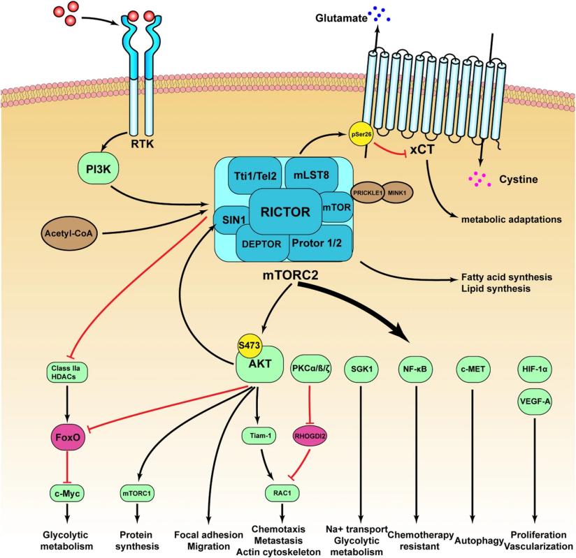 Fig. 1 The mechanisms by which RICTOR participating in tumor growth, invasion and drug resistance.1
Key structural properties of RICTOR:
- Extended conformation composed of multiple domains
- Contains amino end mediated RBD structure of protein interaction domain
- The central CRIM domain is involved in the assembly of the mTORC2 complex
- The carboxyl terminus contains multiple phosphorylation sites and is involved in signal regulation
Functions of RICTOR
The primary function of RICTOR is to serve as the core scaffold and regulatory protein of the mTORC2 complex, participating in cell survival, metabolism, proliferation, and signal integration of cytoskeletal tissue. Its specific functions are as follows:
| Function | Description |
| Assembly of mTORC2 complex | As an indispensable scaffold protein, it binds to mTOR, mLST8, etc., stabilizing and defining the structure and function of the mTORC2 complex. |
| Complete activation of the Akt signaling pathway | mTORC2 phosphorylates the Ser473 site of the Akt protein, which is a crucial step necessary for the complete activation of the Akt kinase and its function of promoting cell survival. |
| Regulation of cytoskeleton dynamics | By phosphorylating and activating proteins such as PKCα, it regulates the organization and arrangement of the actin cytoskeleton, thereby influencing cell morphology, migration and adhesion. |
| Cell growth and metabolic regulation | Integrate growth factors and nutritional signals, regulate anabolic processes such as glucose metabolism and lipid synthesis, and support cell growth and proliferation. |
| Coping with cellular stress | Under conditions such as DNA damage or survival stress, the mTORC2 signaling pathway is activated, promoting cell survival and inhibiting the apoptotic process. |
Unlike the mTORC1 pathway, which mainly senses nutritional and energy status and regulates anabolic metabolism, the signal response of mTORC2 is more complex. Its function is mainly reflected in the response to growth factor signals and the phosphorylation modification of downstream kinases (such as Akt and PKC), and it often shows abnormal activation in cancer occurrence.
Applications of RICTOR and RICTOR Antibody in Literature
1. Wang, Xinyue, et al. "A novel rabbit anti-myoglobin monoclonal antibody's potential application in rhabdomyolysis associated acute kidney injury." International Journal of Molecular Sciences 24.9 (2023): 7822. https://doi.org/10.1002/ctm2.1686
This study explored the role of the Rictor/mTORC2 signaling pathway in interstitial fibrosis after kidney transplantation. Research has found that Rictor degrades BNIP3 through K48 ubiquitination, inhibits mitochondrial autophagy, and promotes endothelial-mesenchymal transition and fibrosis. Inhibiting Rictor can enhance BNIP3-mediated mitochondrial autophagy, thereby improving fibrosis, indicating its potential as a therapeutic target.
2. Szalai, Fatime, et al. "Rictor—a mediator of progression and metastasis in lung cancer." Cancers 16.3 (2024): 543. https://doi.org/10.3390/cancers16030543
This study indicates that RICTOR gene amplification and protein overexpression can activate mTORC2, promoting the migration and metastasis of lung cancer cells. Studies have shown that inhibiting Rictor/mTORC2 can effectively reduce tumor metastasis, and it has significant potential as a therapeutic target and predictive marker.
3. Ponzone, Luca, et al. "RICTOR/mTORC2 downregulation in BRAFV600E melanoma cells promotes resistance to BRAF/MEK inhibition." Molecular Cancer 23.1 (2024): 105. https://doi.org/10.1186/s12943-024-02010-1
This study indicates that in BRAF-mutated melanoma, low RICTOR levels predict poor efficacy and prognosis of targeted therapy. Studies have found that the absence of RICTOR leads to resistance to BRAF/MEK inhibitors by activating the NAMPT-mitochondrial electron transport chain axis. Inhibiting this pathway can restore drug sensitivity, suggesting that RICTor levels have prognostic and therapeutic guiding value.
4. Wang, Chun, et al. "Rictor mediates p53 deactivation to facilitate the malignant transformation of hepatocytes and promote hepatocarcinogenesis." Journal of Translational Medicine 21.1 (2023): 919. https://doi.org/10.1186/s12967-023-04799-9
This study indicates that in hepatocellular carcinoma (HCC), Rictor exerts a promoting effect by binding to and inhibiting the activity of wild-type p53, and its dynamic nucleoplasmic translocation is associated with malignant progression. miR-192 targets and inhibits Rictor, restores p53 function and slows tumor growth. High expression of Rictor indicates a poor prognosis.
5. Jebali, Ahlem, and Nicolas Dumaz. "The role of RICTOR downstream of receptor tyrosine kinase in cancers." Molecular cancer 17.1 (2018): 39. https://doi.org/10.1186/s12943-018-0794-0
This study indicates that RICTOR is a key component of the mTORC2 complex and acts as an important signaling node in RTK-abnormal tumors to mediate carcinogenesis. Recent studies have found that RICTor variations are widely present in various cancers and have become a promising therapeutic target for RTK in tumor modification.
Creative Biolabs: RICTOR Antibodies for Research
Creative Biolabs specializes in the production of high-quality RICTOR antibodies for research and industrial applications. Our portfolio includes monoclonal antibodies tailored for ELISA, Flow Cytometry, Western blot, immunohistochemistry, and other diagnostic methodologies.
- Custom RICTOR Antibody Development: Tailor-made solutions to meet specific research requirements.
- Bulk Production: Large-scale antibody manufacturing for industry partners.
- Technical Support: Expert consultation for protocol optimization and troubleshooting.
- Aliquoting Services: Conveniently sized aliquots for long-term storage and consistent experimental outcomes.
For more details on our RICTOR antibodies, custom preparations, or technical support, contact us at email.
Reference
- Zhao, Deze, et al. "The role of RICTOR amplification in targeted therapy and drug resistance." Molecular Medicine 26.1 (2020): 20. https://doi.org/10.1186/s10020-020-0146-6
Anti-RICTOR antibodies
 Loading...


Hot products 
-
Mouse Anti-GFAP Recombinant Antibody (20) (CBMAB-G2914-LY)

-
Mouse Anti-C5B-9 Recombinant Antibody (CBFYA-0216) (CBMAB-X0304-FY)

-
Mouse Anti-4-Hydroxynonenal Recombinant Antibody (V2-502280) (CBMAB-C1055-CN)

-
Mouse Anti-AMOT Recombinant Antibody (CBYC-A564) (CBMAB-A2552-YC)

-
Mouse Anti-APOE Recombinant Antibody (A1) (CBMAB-0078CQ)

-
Mouse Anti-AMIGO2 Recombinant Antibody (CBYY-C0756) (CBMAB-C2192-YY)

-
Mouse Anti-GFAP Recombinant Antibody (5) (CBMAB-G0346-LY)

-
Mouse Anti-8-oxoguanine Recombinant Antibody (V2-7697) (CBMAB-1869CQ)

-
Mouse Anti-CSPG4 Recombinant Antibody (CBFYM-1050) (CBMAB-M1203-FY)

-
Rat Anti-CD34 Recombinant Antibody (MEC 14.7) (CBMAB-C10196-LY)

-
Mouse Anti-CD247 Recombinant Antibody (6B10.2) (CBMAB-C1583-YY)

-
Rabbit Anti-CAMK2A Recombinant Antibody (BA0032) (CBMAB-0137CQ)

-
Mouse Anti-FTH1 Recombinant Antibody (CBXF-1896) (CBMAB-F3426-CQ)

-
Mouse Anti-CCS Recombinant Antibody (CBFYC-1093) (CBMAB-C1150-FY)

-
Rabbit Anti-ABL1 (Phosphorylated Y185) Recombinant Antibody (V2-443434) (PTM-CBMAB-0001YC)

-
Mouse Anti-C5AR1 Recombinant Antibody (R63) (CBMAB-C9553-LY)

-
Mouse Anti-DHFR Recombinant Antibody (D0821) (CBMAB-D0821-YC)

-
Mouse Anti-CD24 Recombinant Antibody (ALB9) (CBMAB-0176CQ)

-
Mouse Anti-BACE1 Recombinant Antibody (CBLNB-121) (CBMAB-1180-CN)

-
Mouse Anti-CD24 Recombinant Antibody (SN3) (CBMAB-C1037-CQ)

- AActivation
- AGAgonist
- APApoptosis
- BBlocking
- BABioassay
- BIBioimaging
- CImmunohistochemistry-Frozen Sections
- CIChromatin Immunoprecipitation
- CTCytotoxicity
- CSCostimulation
- DDepletion
- DBDot Blot
- EELISA
- ECELISA(Cap)
- EDELISA(Det)
- ESELISpot
- EMElectron Microscopy
- FFlow Cytometry
- FNFunction Assay
- GSGel Supershift
- IInhibition
- IAEnzyme Immunoassay
- ICImmunocytochemistry
- IDImmunodiffusion
- IEImmunoelectrophoresis
- IFImmunofluorescence
- IGImmunochromatography
- IHImmunohistochemistry
- IMImmunomicroscopy
- IOImmunoassay
- IPImmunoprecipitation
- ISIntracellular Staining for Flow Cytometry
- LALuminex Assay
- LFLateral Flow Immunoassay
- MMicroarray
- MCMass Cytometry/CyTOF
- MDMeDIP
- MSElectrophoretic Mobility Shift Assay
- NNeutralization
- PImmunohistologyp-Paraffin Sections
- PAPeptide Array
- PEPeptide ELISA
- PLProximity Ligation Assay
- RRadioimmunoassay
- SStimulation
- SESandwich ELISA
- SHIn situ hybridization
- TCTissue Culture
- WBWestern Blot
