SLC17A7 Antibodies
Background
The SLC17A7 gene encodes a protein called vesicular glutamate transporter 1 (VGLUT1), which is mainly located on the synaptic vesicle membranes of neurons and is responsible for transporting glutamate from the cytoplasm into the synaptic vesicles, preparing for the release of neurotransmitters. This transport process relies on the electrochemical gradient on both sides of the vesicle membrane, ensuring that glutamate can be stored in the vesicles at high concentrations. VGLUT1 is highly expressed in excitatory neurons in regions such as the cerebral cortex, hippocampus, and cerebellum, and is one of the key markers of glutamate-mediated neural transmission. This gene was first identified through molecular cloning techniques in the late 1990s, and its discovery provided important clues for understanding the release mechanism of glutamate as the main excitatory neurotransmitter. Subsequent studies on SLC17A7 revealed its important role in synaptic plasticity, learning and memory, and various neuro-psychiatric diseases, further consolidating its core position in the field of neuroscience.
Structure of SLC17A7
The VGLUT1 protein encoded by the SLC17A7 gene has a molecular weight of approximately 60 kDa. There are certain differences among different species, which mainly result from minor variations in the amino acid sequence.
| Species | Human | Mouse | Rat | Pig | Macaque |
| Molecular Weight (kDa) | 60.2 | 59.8 | 59.9 | 60.0 | 60.1 |
| Primary Structural Differences | The key domain for glutamic acid transport is highly conserved | Species-specific modifications exist at the C-end | N-terminal sequence is highly homologous to rat | Structural stability in the transmembrane region | The highest sequence similarity to human |
The VGLUT1 protein encoded by the SLC17A7 gene consists of approximately 560 amino acids. Its secondary structure is mainly composed of 12 transmembrane α helices, forming a highly conserved hydrophobic core region. The active center of this protein contains key phosphate-binding sites and multiple conserved aspartic acid/glutamic acid residues. These amino acids drive the transmembrane transport of glutamate through forming charge interactions. The topology of VGLUT1 presents a cytoplasmic N-terminal and C-terminal region. There is a key glycosylation site near the fourth transmembrane helix, and the serine phosphorylation site located in the intracellular loop participates in regulating vesicle targeting and transport activity. The red fluorescence signal of the protein originates from its binding with specific antibodies, which makes it widely used in immunolabeling studies.
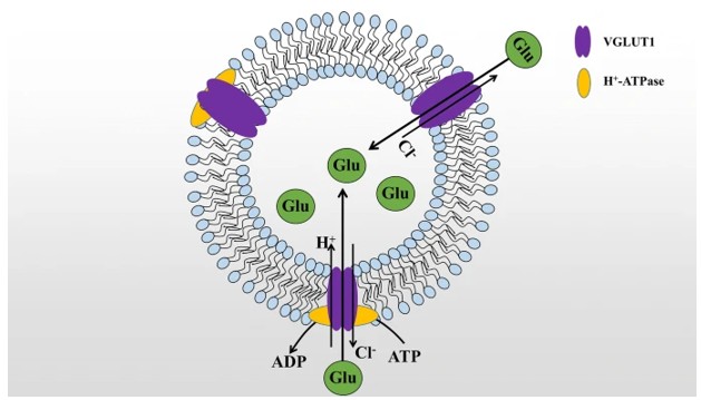 Fig. 1 SLC17A7(VGLUT1) transport glutamate (Glu) mechanism diagram.1
Key structural properties of SLC17A7:
- Topology of 12-fold transmembrane α-helix formation
- Highly conserved hydrophilic substrate binding cavity
- Phosphate recognition sites mediate glutamate transport
- Glycosylation modifications that regulate protein stability and vesicle localization
Functions of SLC17A7
The VGLUT1 protein encoded by the SLC17A7 gene mainly functions in packaging glutamate into synaptic vesicles. However, it is also involved in various physiological processes in the nervous system, including the regulation of synaptic plasticity and the balance of energy metabolism.
| Function | Description |
| Glutamate Loading | VGLUT1 utilizes the proton gradient of the vesicle membrane to transport cytosolic glutamate into synaptic vesicles, preparing for the release of neurotransmitters. |
| Synaptic Transmission Regulation | By controlling the glutamate filling volume of individual vesicles, it directly affects the intensity of excitatory synaptic transmission. |
| Plasticity Regulation | In the hippocampal long play a key role in the enhancement, its expression level affect learning and memory ability. |
| Metabolic Coupling | It interacts with the vesicle ATPase complex to coordinate the process of neurotransmitter loading and vesicle acidification. |
| Neuroprotection | Maintain the vesicle storage of glutamic acid and prevent the excitotoxicity caused by the accumulation of cytoplasmic glutamic acid. |
The VGLUT1 protein encoded by the SLC17A7 gene exhibits a typical saturation curve in the transport kinetics of glutamate, which is different from the linear kinetics of general metabolic transporters. This indicates its reserve capacity as a synaptic vesicle filling system. When the glutamate concentration inside the vesicle reaches 20-30 mM, the transport rate tends to plateau. This high-capacity and low-affinity characteristic enables it to maintain a stable neurotransmitter supply during high-frequency neuronal discharges.
Applications of SLC17A7 and SLC17A7 Antibody in Literature
1. Du, Xianchao, et al. "Research progress on the role of type I vesicular glutamate transporter (VGLUT1) in nervous system diseases." Cell & bioscience 10.1 (2020): 26. https://doi.org/10.1186/s13578-020-00393-4
The article indicates that SLC17A7(VGLUT1) is the most important vesicular glutamate transporter in the central nervous system, responsible for storing and releasing the excitatory neurotransmitter glutamate. Its expression is a specific marker of glutamatergic neurons. Abnormal function of this protein is closely related to various diseases such as learning and memory disorders, Alzheimer's disease, and pain pathologies.
2. Farris, Sean P., Robert A. Harris, and Igor Ponomarev. "Epigenetic modulation of brain gene networks for cocaine and alcohol abuse." Frontiers in neuroscience 9 (2015): 176. https://doi.org/10.3389/fnins.2015.00176
One study found that the expression of the gene SLC17A7/VGLUT1 in the hippocampus of cocaine and alcohol abusers coincides with changes in its histone modification H3K4me3. This gene encodes a glutamate transporter and is a functional network node related to addiction. Integrating epigenetic and transcriptomic analyses helps to reveal the molecular mechanism of drug addiction.
3. Lin, Biaoyang, et al. "Global analysis of H3K4me3 and H3K27me3 profiles in glioblastoma stem cells and identification of SLC17A7 as a bivalent tumor suppressor gene." Oncotarget 6.7 (2015): 5369. https://doi.org/10.18632/oncotarget.3030
The study found that SLC17A7 acts as a dual tumor suppressor gene in glioblastoma, with its expression being downregulated due to the simultaneous presence of H3K4me3 and H3K27me3 modifications. Restoring its expression can inhibit tumor cell proliferation and invasion, providing a new target for inducing differentiation therapy.
4. Poltavskaya, Evgeniya G., et al. "The role of glutamatergic gene polymorphisms in the clinical phenotypes of schizophrenia." Genes 14.3 (2023): 575. https://doi.org/10.3390/genes14030575
The study found that the rs62126236 locus of the glutamate system gene SLC17A7 was associated with the clinical phenotype of schizophrenia. The T allele at this locus provided significant protection against the negative symptoms of patients, providing a genetic basis for understanding the clinical heterogeneity of the disease.
5. Murao, Naoya, et al. "Essential roles of aspartate aminotransferase 1 and vesicular glutamate transporters in β-cell glutamate signaling for incretin-induced insulin secretion." PLoS One 12.11 (2017): e0187213. https://doi.org/10.1371/journal.pone.0187213
The research has found that the secretion of insulin by pancreatic β cells requires the participation of VGLUTs (including SLC17A7) in glutamate signal transduction. The knockout of SLC17A7 alone does not affect secretion due to compensation by other subtypes, but when all three are knocked out simultaneously, the function is lost, confirming its crucial role in insulin release.
Creative Biolabs: SLC17A7 Antibodies for Research
Creative Biolabs specializes in the production of high-quality SLC17A7 antibodies for research and industrial applications. Our portfolio includes monoclonal and polyclonal antibodies tailored for ELISA, Flow Cytometry, Western blot, immunohistochemistry, and other diagnostic methodologies.
- Custom SLC17A7 Antibody Development: Tailor-made solutions to meet specific research requirements.
- Bulk Production: Large-scale antibody manufacturing for industry partners.
- Technical Support: Expert consultation for protocol optimization and troubleshooting.
- Aliquoting Services: Conveniently sized aliquots for long-term storage and consistent experimental outcomes.
For more details on our SLC17A7 antibodies, custom preparations, or technical support, contact us at email.
Reference
- Du, Xianchao, et al. "Research progress on the role of type I vesicular glutamate transporter (VGLUT1) in nervous system diseases." Cell & bioscience 10.1 (2020): 26. Distributed under Open Access license CC BY 4.0, without modification. https://doi.org/10.1186/s13578-020-00393-4
Anti-SLC17A7 antibodies
 Loading...


Hot products 
-
Mouse Anti-CFL1 (Phospho-Ser3) Recombinant Antibody (CBFYC-1770) (CBMAB-C1832-FY)

-
Mouse Anti-CASP8 Recombinant Antibody (CBYY-C0987) (CBMAB-C2424-YY)

-
Mouse Anti-CD24 Recombinant Antibody (ALB9) (CBMAB-0176CQ)

-
Mouse Anti-ACE2 Recombinant Antibody (V2-179293) (CBMAB-A0566-YC)

-
Mouse Anti-C4B Recombinant Antibody (CBYY-C2996) (CBMAB-C4439-YY)

-
Rat Anti-(1-5)-α-L-Arabinan Recombinant Antibody (V2-501861) (CBMAB-XB0003-YC)

-
Mouse Anti-ALB Recombinant Antibody (V2-180650) (CBMAB-A2186-YC)

-
Mouse Anti-BCL6 Recombinant Antibody (CBYY-0435) (CBMAB-0437-YY)

-
Mouse Anti-FYN Recombinant Antibody (10) (CBMAB-S6332-CQ)

-
Rabbit Anti-CCL5 Recombinant Antibody (R0437) (CBMAB-R0437-CN)

-
Mouse Anti-Acetyl SMC3 (K105/K106) Recombinant Antibody (V2-634053) (CBMAB-AP052LY)

-
Rabbit Anti-BRCA2 Recombinant Antibody (D9S6V) (CBMAB-CP0017-LY)

-
Mouse Anti-AHCYL1 Recombinant Antibody (V2-180270) (CBMAB-A1703-YC)

-
Mouse Anti-CFL1 Recombinant Antibody (CBFYC-1771) (CBMAB-C1833-FY)

-
Mouse Anti-CCDC25 Recombinant Antibody (CBLC132-LY) (CBMAB-C9786-LY)

-
Mouse Anti-ADAM29 Recombinant Antibody (V2-179787) (CBMAB-A1149-YC)

-
Mouse Anti-CTCF Recombinant Antibody (CBFYC-2371) (CBMAB-C2443-FY)

-
Mouse Anti-AFM Recombinant Antibody (V2-634159) (CBMAB-AP185LY)

-
Mouse Anti-4-Hydroxynonenal Recombinant Antibody (V2-502280) (CBMAB-C1055-CN)

-
Mouse Anti-BACE1 Recombinant Antibody (CBLNB-121) (CBMAB-1180-CN)

- AActivation
- AGAgonist
- APApoptosis
- BBlocking
- BABioassay
- BIBioimaging
- CImmunohistochemistry-Frozen Sections
- CIChromatin Immunoprecipitation
- CTCytotoxicity
- CSCostimulation
- DDepletion
- DBDot Blot
- EELISA
- ECELISA(Cap)
- EDELISA(Det)
- ESELISpot
- EMElectron Microscopy
- FFlow Cytometry
- FNFunction Assay
- GSGel Supershift
- IInhibition
- IAEnzyme Immunoassay
- ICImmunocytochemistry
- IDImmunodiffusion
- IEImmunoelectrophoresis
- IFImmunofluorescence
- IGImmunochromatography
- IHImmunohistochemistry
- IMImmunomicroscopy
- IOImmunoassay
- IPImmunoprecipitation
- ISIntracellular Staining for Flow Cytometry
- LALuminex Assay
- LFLateral Flow Immunoassay
- MMicroarray
- MCMass Cytometry/CyTOF
- MDMeDIP
- MSElectrophoretic Mobility Shift Assay
- NNeutralization
- PImmunohistologyp-Paraffin Sections
- PAPeptide Array
- PEPeptide ELISA
- PLProximity Ligation Assay
- RRadioimmunoassay
- SStimulation
- SESandwich ELISA
- SHIn situ hybridization
- TCTissue Culture
- WBWestern Blot
