TBCK Antibodies
Background
The TBCK gene encodes a regulatory factor called the TBCK domain-containing protein. Its products are mainly distributed in the cytoplasm and participate in the regulation of neuronal development and cell proliferation by influencing signaling pathways such as mTOR. This protein functions through its conserved TBC domain, capable of regulating vesicle transport and autophagy processes, which is of crucial significance for maintaining the normal function of the nervous system. In 2016, scientists first discovered the pathogenic mutation of this gene in children with neurodevelopmental disorders. The biallelic mutation of this gene can lead to progressive intellectual disability and motor function decline. The in-depth study of this gene not only reveals the important molecular mechanism of brain development, but also provides new molecular targets for the precise diagnosis and treatment of related genetic diseases.
Structure of TBCK
The TBCK protein is a large regulatory protein with a molecular weight of approximately 220 kDa, and its precise molecular weight varies slightly among different species.
| Species | Human | Mouse | Zebrafish |
| Molecular Weight (kDa) | 220 | 218 | 215 |
| Primary Structural Differences | Contains the complete TBC and kinase domains | TBC structure domain highly conservative | The C-end regulatory sequence is lacking |
This protein is composed of 1,863 amino acids. Its tertiary structure includes a conserved TBC domain and multiple functional regions, forming a stable spatial conformation through helical-helical interactions. In terms of structural features, the N-terminal region forms a typical phosphorylation recognition module, the central TBC domain has GTPase activation function, while the C-terminal fragment participates in the assembly of the protein-protein interaction network. This multi-domain organizational model enables it to precisely regulate the activity of the mTOR signaling pathway, playing a key role in neural development.
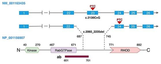 Fig. 1 Schematic representation of TBCK transcript and protein.1
Key structural properties of TBCK:
- Contains the conserved TBC domain
- Having multiple coiled-coil helical regions
- Specific phosphorylation recognition modules regulate mTOR signaling pathway activity
Functions of TBCK
The core function of the TBCK protein is to regulate the mTOR signaling pathway and neuronal development. However, this protein is also involved in a variety of cellular processes, including autophagy regulation, protein synthesis and cell proliferation.
| Function | Description |
| mTOR signal regulation | The activity of the mTORC1 pathway is negatively regulated through the TBC domain, influencing cell growth and metabolic processes. |
| Neural development | Maintaining the differentiation and migration of neurons, their functional deficiency leads to intellectual disability and delayed motor development. |
| Autophagy activation | After the inhibition of mTORC1 is lifted, it promotes the formation of autophagosomes and maintains the stability of the intracellular environment. |
| Regulation of protein synthesis | Regulate ribosome biosynthesis and translation initiation processes through the mTOR pathway. |
| Cell cycle regulation | Affect cell G1 / S phase transformation, its abnormal expression defects lead to proliferation. |
Unlike the complex network formed by other regulatory factors of the mTOR pathway, the TBCK protein directly acts on small G proteins through its unique TBC domain, demonstrating its specific localization and functional specificity in signal transduction.
Applications of TBCK and TBCK Antibody in Literature
1. Moreira, Danielle de Paula, et al. "Neuroprogenitor cells from patients with TBCK encephalopathy suggest deregulation of early secretory vesicle transport." Frontiers in cellular neuroscience 15 (2022): 803302. https://doi.org/10.3389/fncel.2021.803302
The article indicates that the biallelic variation of TBCK leads to IHPRF3. Research has found that the neural precursor cells derived from patients have impaired endoplasmic reticulum-Golgi apparatus transport and autophagy due to the absence of TBCK, resulting in a significant decline in cell migration ability. This may be the pathogenic mechanism of neural abnormalities.
2. Kim, Eun-Ae, et al. "MiR-1208 increases the sensitivity to cisplatin by targeting TBCK in renal cancer cells." International Journal of Molecular Sciences 20.14 (2019): 3540. https://doi.org/10.3390/ijms20143540
Studies have shown that microRNA-1208 plays a tumor suppressor role in renal cancer. It has therapeutic potential by directly targeting and inhibiting the expression of the TBCK gene, thereby enhancing the sensitivity of cancer cells to cisplatin and TRAIL and inducing apoptosis.
3. Liu, Yueli, Xiaoyi Yan, and Tianhua Zhou. "TBCK influences cell proliferation, cell size and mTOR signaling pathway." PloS one 8.8 (2013): e71349. https://doi.org/10.1371/journal.pone.0071349
Studies have shown that the TBCK protein affects cell proliferation and growth by regulating the mTOR pathway. Knocking down TBCK will reduce the protein levels of the components of the mTOR complex, inhibit their signaling activity, and lead to blocked cell proliferation, reduced volume and disordered actin structure.
4. Tan, Hao-Yi, Bin Wang, and Yuan-Zong Song. "Identification of a novel pathogenic TBCK variant in a Chinese patient with infantile hypotonia with psychomotor retardation and characteristic facies type 3 (IHPRF3): a case report." BMC pediatrics 22.1 (2022): 612. https://doi.org/10.1186/s12887-022-03672-w
This study reports for the first time the clinical and genetic characteristics of a Chinese patient with IHPRF3. The patient carries a novel homozygous TBCK gene variant c.247C>T, presenting with global developmental delay, hypotonia and distinctive facial features, providing a new basis for the diagnosis and genetic counseling of this disease.
5. Beck-Wödl, Stefanie, et al. "Homozygous TBC1 domain-containing kinase (TBCK) mutation causes a novel lysosomal storage disease–a new type of neuronal ceroid lipofuscinosis (CLN15)?" Acta neuropathologica communications 6.1 (2018): 145. https://doi.org/10.1186/s40478-018-0646-6
This study reveals for the first time that TBCK dysfunction is a novel lysosomal storage disorder. There is a large amount of lipofuscin deposition in the patient's neurons. The clinical and pathological features support the classification of it as a new subtype of neuronal wax lipofuscin deposition syndrome (CLN15), and the mechanism may be related to MTORC1-mediated autophagic flow disorder.
Creative Biolabs: TBCK Antibodies for Research
Creative Biolabs specializes in the production of high-quality TBCK antibodies for research and industrial applications. Our portfolio includes monoclonal antibodies tailored for ELISA, Flow Cytometry, Western blot, immunohistochemistry, and other diagnostic methodologies.
- Custom TBCK Antibody Development: Tailor-made solutions to meet specific research requirements.
- Bulk Production: Large-scale antibody manufacturing for industry partners.
- Technical Support: Expert consultation for protocol optimization and troubleshooting.
- Aliquoting Services: Conveniently sized aliquots for long-term storage and consistent experimental outcomes.
For more details on our TBCK antibodies, custom preparations, or technical support, contact us at email.
Reference
- Moreira, Danielle de Paula, et al. "Neuroprogenitor cells from patients with TBCK encephalopathy suggest deregulation of early secretory vesicle transport." Frontiers in cellular neuroscience 15 (2022): 803302. https://doi.org/10.3389/fncel.2021.803302
Anti-TBCK antibodies
 Loading...


Hot products 
-
Mouse Anti-ADIPOR2 Recombinant Antibody (V2-179983) (CBMAB-A1369-YC)

-
Mouse Anti-ALB Recombinant Antibody (V2-180650) (CBMAB-A2186-YC)

-
Human Anti-SARS-CoV-2 S1 Monoclonal Antibody (CBFYR-0120) (CBMAB-R0120-FY)

-
Mouse Anti-AK4 Recombinant Antibody (V2-180419) (CBMAB-A1891-YC)

-
Mouse Anti-BRD3 Recombinant Antibody (CBYY-0801) (CBMAB-0804-YY)

-
Mouse Anti-ESR1 Recombinant Antibody (Y31) (CBMAB-1208-YC)

-
Mouse Anti-FYN Recombinant Antibody (10) (CBMAB-S6332-CQ)

-
Rabbit Anti-CBL Recombinant Antibody (D4E10) (CBMAB-CP0149-LY)

-
Mouse Anti-CD63 Recombinant Antibody (CBXC-1200) (CBMAB-C1467-CQ)

-
Mouse Anti-CCDC25 Recombinant Antibody (CBLC132-LY) (CBMAB-C9786-LY)

-
Human Anti-SARS-CoV-2 Spike Recombinant Antibody (CR3022) (CBMAB-CR014LY)

-
Rabbit Anti-B2M Recombinant Antibody (CBYY-0059) (CBMAB-0059-YY)

-
Mouse Anti-ARSA Recombinant Antibody (CBYC-A799) (CBMAB-A3679-YC)

-
Mouse Anti-CDKL5 Recombinant Antibody (CBFYC-1629) (CBMAB-C1689-FY)

-
Rat Anti-C5AR1 Recombinant Antibody (8D6) (CBMAB-C9139-LY)

-
Mouse Anti-ADAM29 Recombinant Antibody (V2-179787) (CBMAB-A1149-YC)

-
Rat Anti-CCR2 Recombinant Antibody (475301) (CBMAB-C1338-LY)

-
Rabbit Anti-ATF4 Recombinant Antibody (D4B8) (CBMAB-A3872-YC)

-
Mouse Anti-ATG5 Recombinant Antibody (9H197) (CBMAB-A3945-YC)

-
Mouse Anti-DLG1 Monolconal Antibody (4F3) (CBMAB-0225-CN)

- AActivation
- AGAgonist
- APApoptosis
- BBlocking
- BABioassay
- BIBioimaging
- CImmunohistochemistry-Frozen Sections
- CIChromatin Immunoprecipitation
- CTCytotoxicity
- CSCostimulation
- DDepletion
- DBDot Blot
- EELISA
- ECELISA(Cap)
- EDELISA(Det)
- ESELISpot
- EMElectron Microscopy
- FFlow Cytometry
- FNFunction Assay
- GSGel Supershift
- IInhibition
- IAEnzyme Immunoassay
- ICImmunocytochemistry
- IDImmunodiffusion
- IEImmunoelectrophoresis
- IFImmunofluorescence
- IGImmunochromatography
- IHImmunohistochemistry
- IMImmunomicroscopy
- IOImmunoassay
- IPImmunoprecipitation
- ISIntracellular Staining for Flow Cytometry
- LALuminex Assay
- LFLateral Flow Immunoassay
- MMicroarray
- MCMass Cytometry/CyTOF
- MDMeDIP
- MSElectrophoretic Mobility Shift Assay
- NNeutralization
- PImmunohistologyp-Paraffin Sections
- PAPeptide Array
- PEPeptide ELISA
- PLProximity Ligation Assay
- RRadioimmunoassay
- SStimulation
- SESandwich ELISA
- SHIn situ hybridization
- TCTissue Culture
- WBWestern Blot
