ZAP70 Antibodies
Background
ZAP70 is a tyrosine kinase mainly expressed in mammalian lymphocytes, serving as a key mediator for T-cell receptor signal transduction. This protein regulates the development, activation and immune response functions of T cells by binding to the activated T-cell receptor complex and undergoing phosphorylation, thereby initiating a downstream signaling cascade reaction. The deficiency of ZAP70 is associated with severe combined immunodeficiency in humans, as its mutations can lead to the loss of T cell function. This gene was first identified in 1992. Its three-dimensional structure was analyzed by X-ray crystallography, revealing the specific interaction mechanism between the kinase domain and phosphorylated tyrosine. ZAP70 has become an important model in immunology and cancer research. The clarification of its mechanism of action has significantly promoted the development of drugs targeting immune signaling pathways and the advancement of treatment strategies for autoimmune diseases.
Structure of ZAP70
ZAP70 is a tyrosine kinase with a molecular weight of approximately 70 kDa. There are slight differences in molecular weight among different species, mainly due to subtle changes in amino acid sequences.
| Species | Human | Mouse | Rat |
| Molecular Weight (kDa) | Approximately 70 | Approximately 69 | Approximately 70 |
| Primary Structural Differences | Contains two SH2 domains and a kinase domain | Kinase domain active high structure, sequence is highly conserved | High homology with human ZAP70 in key functional domains |
The ZAP70 protein contains 619 amino acids, and its three-dimensional structure exhibits a typical kinase folding morphology. The core function of a protein depends on its two tandem SH2 domains and one kinase domain at the C-terminal. The SH2 domain is responsible for recognizing and binding to the phosphorylated ITAM motif on the T-cell receptor complex. This binding induces a conformational change in ZAP70 and its own phosphorylation, thereby activating its kinase function. The activated kinase domain catalyzes the tyrosine phosphorylation of downstream adaptor proteins (such as LAT and SLP-76), thereby initiating multiple signaling pathways such as calcium ion flow and MAPK, and ultimately driving the activation, proliferation and differentiation of T cells. The localization and activation of this protein at the immune synapse is a key switch for adaptive immune response.
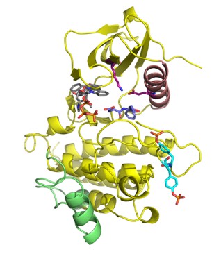 Fig. 1 Overview of ZAP–70 kinase domain.1
Key structural properties of ZAP70:
- Two tandem SH2 domains are used to identify phosphorylated ITAM motifs
- A C-terminal kinase domain is used for phosphorylating downstream signaling proteins
- The flexible hinge region between the SH2 domain and the kinase domain regulates conformational changes
- located in the hinge region (such as Tyr319) and the kinase domain activation loop (such as Tyr493), the degree of phosphorylation precisely regulates kinase activity and signal strength.
Functions of ZAP70
The main function of ZAP70 is to serve as a key switch and amplifier for T-cell receptor (TCR) signal transduction. However, it is also involved in regulating various physiological and pathological processes such as the development, differentiation and immune tolerance of T cells.
| Function | Description |
| Signal activation | Through its SH2 domain, it specifically binds to phosphorylated ITAM motifs on the TCR/CD3 complex, initiating downstream signaling cascades. |
| Signal Transduction and Amplification | After activation, downstream adaptor proteins (e.g., LAT, SLP-76) are phosphorylated, amplifying signals from membrane receptors and transmitting to multiple intracellular pathways. |
| T Cell Development | In thymus, its signal intensity is involved in the regulation of positive and negative selection, affecting the formation of functional T cell repertoire. |
| Immune Tolerance maintenance | By regulating the T-cell activation threshold, it participates in peripheral immune tolerance and prevents autoimmune reactions. |
| Disease Association | Its loss of function leads to severe combined immunodeficiency.However, it is abnormally high in some B-cell chronic lymphocytic leukemia, which is associated with poor prognosis. |
The activation kinetics of ZAP70 exhibits a strict phosphorylation cascade feature. Unlike the simple oxygen-binding curve of myoglobin, its activity is precisely regulated by the phosphorylation and dephosphorylation of multiple tyrosine sites (such as Tyr319 and Tyr493), forming a "molecular switch". Make sure that T cells will only be fully activated when they receive a strong enough antigenic stimulus.
Applications of ZAP70 and ZAP70 Antibody in Literature
1. Huber, Roland G., Hao Fan, and Peter J. Bond. "The structural basis for activation and inhibition of ZAP-70 kinase domain." PLoS computational biology 11.10 (2015): e1004560.https://doi.org/10.1371/journal.pcbi.1004560
This study revealed the molecular mechanism of phosphorylation activation and inhibitor regulation of the ZAP-70 kinase domain through molecular dynamics simulation, and discovered the existence of novel potential drug binding pockets near its activation loop, providing a new target for the treatment of immune deficiency.
2. Hu, Jing, Mengyue Wang, and Ruiyao Xiang. "ZAP70: A Key Gene Identified by Differential Expression Analysis for Early Diagnosis of Fetuses with Emanuel Syndrome." Biochemical Genetics 63.3 (2025): 2161-2171. https://doi.org/10.1007/s10528-024-10808-3
Based on transcriptome data analysis, this study, through multi-algorithm screening, found that the ZAP70 gene is a potential key biomarker for the early diagnosis of Emanuel syndrome caused by the translocation of fetal chromosomes 11 and 22.
3. Işıksaçan, Nilgün, et al. "Cytokine contents in chronic lymphocytic leukemia: association with ZAP70 expression." Turkish Journal of Hematology 33.3 (2016): 202. https://doi.org/10.4274/tjh.2014.0469
This study, through flow cytometry analysis, found that ZAP70 positivity was associated with disease progression in patients with chronic lymphocytic leukemia, and the secretion of IL-4 by T cells increased while IFN-γ decreased, suggesting that impaired cellular immunity and IL-4 may affect the disease process.
4. Schultz, Annika, et al. "A cysteine residue within the kinase domain of Zap70 regulates Lck activity and proximal TCR signaling." Cells 11.17 (2022): 2723. https://doi.org/10.3390/cells11172723
This study confirmed that the C564 site of the ZAP70 protein is not the key site for its palmitoylation. The C564A mutant can maintain T-cell receptor signal transduction, while the root cause of the functional loss of the C564R mutant lies in its abnormal enhancement of the activity of the upstream kinase Lck, which in turn interferes with the regulation of proximal signals.
5. Ren, Li, et al. "AQP9 and ZAP70 as immune-related prognostic biomarkers suppress proliferation, migration and invasion of laryngeal cancer cells." BMC cancer 22.1 (2022): 465. https://doi.org/10.1186/s12885-022-09458-8
This study, based on TCGA data analysis, found that ZAP70 and AQP9 are immune genes related to the prognosis of laryngeal cancer. Experiments have confirmed that low expression of both promotes the proliferation and migration of tumor cells, while upregulation inhibits malignant progression, suggesting their potential as new targets for immunotherapy of laryngeal cancer.
Creative Biolabs: ZAP70 Antibodies for Research
Creative Biolabs specializes in the production of high-quality ZAP70 antibodies for research and industrial applications. Our portfolio includes monoclonal antibodies tailored for ELISA, Flow Cytometry, Western blot, immunohistochemistry, and other diagnostic methodologies.
- Custom ZAP70 Antibody Development: Tailor-made solutions to meet specific research requirements.
- Bulk Production: Large-scale antibody manufacturing for industry partners.
- Technical Support: Expert consultation for protocol optimization and troubleshooting.
- Aliquoting Services: Conveniently sized aliquots for long-term storage and consistent experimental outcomes.
For more details on our ZAP70 antibodies, custom preparations, or technical support, contact us at email.
Reference
- Huber, Roland G., Hao Fan, and Peter J. Bond. "The structural basis for activation and inhibition of ZAP-70 kinase domain." PLoS computational biology 11.10 (2015): e1004560. https://doi.org/10.1371/journal.pcbi.1004560
Anti-ZAP70 antibodies
 Loading...


Hot products 
-
Mouse Anti-CDKL5 Recombinant Antibody (CBFYC-1629) (CBMAB-C1689-FY)

-
Mouse Anti-DLL4 Recombinant Antibody (D1090) (CBMAB-D1090-YC)

-
Rabbit Anti-ALDOA Recombinant Antibody (D73H4) (CBMAB-A2314-YC)

-
Mouse Anti-ENO1 Recombinant Antibody (CBYC-A950) (CBMAB-A4388-YC)

-
Mouse Anti-AKT1 (Phosphorylated S473) Recombinant Antibody (V2-505430) (PTM-CBMAB-0067LY)

-
Mouse Anti-GFAP Recombinant Antibody (24) (CBMAB-G2927-LY)

-
Mouse Anti-CD247 Recombinant Antibody (6B10.2) (CBMAB-C1583-YY)

-
Mouse Anti-FOXA3 Recombinant Antibody (2A9) (CBMAB-0377-YC)

-
Mouse Anti-APC Recombinant Antibody (CBYC-A661) (CBMAB-A3036-YC)

-
Mouse Anti-CCN2 Recombinant Antibody (CBFYC-2383) (CBMAB-C2456-FY)

-
Rabbit Anti-DLK1 Recombinant Antibody (9D8) (CBMAB-D1061-YC)

-
Rabbit Anti-CCL5 Recombinant Antibody (R0437) (CBMAB-R0437-CN)

-
Mouse Anti-ARHGAP5 Recombinant Antibody (54/P190-B) (CBMAB-P0070-YC)

-
Mouse Anti-CFL1 (Phospho-Ser3) Recombinant Antibody (CBFYC-1770) (CBMAB-C1832-FY)

-
Mouse Anti-8-oxoguanine Recombinant Antibody (V2-7697) (CBMAB-1869CQ)

-
Mouse Anti-FOSB Recombinant Antibody (CBXF-3593) (CBMAB-F2522-CQ)

-
Mouse Anti-ADAM29 Recombinant Antibody (V2-179787) (CBMAB-A1149-YC)

-
Mouse Anti-COL12A1 Recombinant Antibody (CBYY-C3117) (CBMAB-C4560-YY)

-
Mouse Anti-BRD3 Recombinant Antibody (CBYY-0801) (CBMAB-0804-YY)

-
Mouse Anti-AAV9 Recombinant Antibody (V2-634029) (CBMAB-AP023LY)

- AActivation
- AGAgonist
- APApoptosis
- BBlocking
- BABioassay
- BIBioimaging
- CImmunohistochemistry-Frozen Sections
- CIChromatin Immunoprecipitation
- CTCytotoxicity
- CSCostimulation
- DDepletion
- DBDot Blot
- EELISA
- ECELISA(Cap)
- EDELISA(Det)
- ESELISpot
- EMElectron Microscopy
- FFlow Cytometry
- FNFunction Assay
- GSGel Supershift
- IInhibition
- IAEnzyme Immunoassay
- ICImmunocytochemistry
- IDImmunodiffusion
- IEImmunoelectrophoresis
- IFImmunofluorescence
- IGImmunochromatography
- IHImmunohistochemistry
- IMImmunomicroscopy
- IOImmunoassay
- IPImmunoprecipitation
- ISIntracellular Staining for Flow Cytometry
- LALuminex Assay
- LFLateral Flow Immunoassay
- MMicroarray
- MCMass Cytometry/CyTOF
- MDMeDIP
- MSElectrophoretic Mobility Shift Assay
- NNeutralization
- PImmunohistologyp-Paraffin Sections
- PAPeptide Array
- PEPeptide ELISA
- PLProximity Ligation Assay
- RRadioimmunoassay
- SStimulation
- SESandwich ELISA
- SHIn situ hybridization
- TCTissue Culture
- WBWestern Blot
