AKT1 Antibodies
Background
AKT1 is a serine/threonine protein kinase that is widely present in eukaryotes as a core regulatory factor of cell signal transduction. This enzyme participates in regulating key biological processes such as cell proliferation, metabolism, survival and apoptosis through the PI3K-AKT signaling pathway, and plays an important role in maintaining the homeostasis of the body. Since its discovery in the 1970s, AKT1 has become a research hotspot due to its crucial role in cancer, diabetes and neurological diseases. The analysis of its tertiary structural characteristics (including the PH domain and kinase domain) has revealed the allosteric activation mechanism, providing a key theoretical basis for the development of targeted drugs. Continuous research on AKT1 has greatly advanced people's understanding of cell signal transduction, disease mechanisms, and targeted therapeutic strategies.
Structure of AKT1
AKT1 is a serine/threonine protein kinase with a molecular weight of approximately 57 kDa, and its molecular weight varies slightly among different species due to sequence differences.
| Species | Human | Mouse | Rat | Bovine |
| Molecular Weight (kDa) | 57 | 56.8 | 56.9 | 57.2 |
| Primary Structural Differences | With PH domain structure and the structure of kinase domain | Highly homologous to human AKT1 | Highly conserved structure of catalytic domain | There are minor variations in the amino acid sequence |
This protein is composed of approximately 480 amino acids, and its three-dimensional structure contains a characteristic PH domain and a kinase domain. After the activation loop (T-loop) in the kinase domain is phosphorylated at the Thr308 and Ser473 sites, the enzyme is fully activated, thereby regulating the downstream signaling pathways. This structural mechanism plays a core regulatory role in the survival, proliferation and metabolic processes of cells.
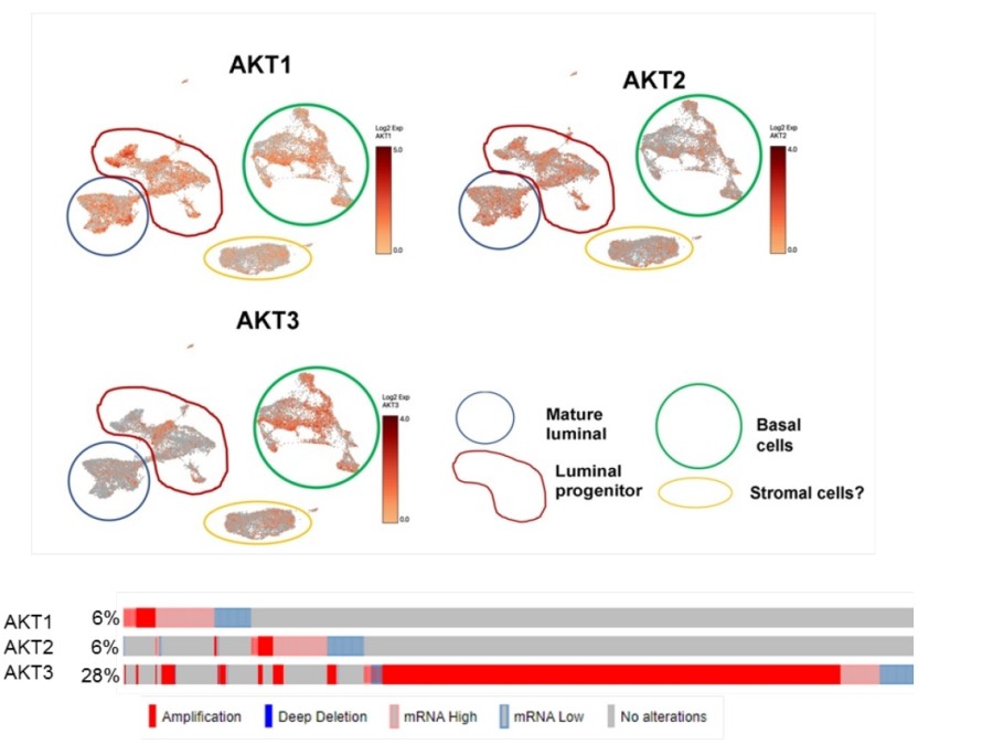 Fig. 1 Expression of AKT isoforms and experimental strategy.1
Key structural properties of AKT1:
- Conserved protein kinase structure consisting of a PH domain and a kinase domain
- Regulation of kinase activity dependent on phosphorylation (Thr308/Ser473)
- Substrate recognition and catalytic functions are achieved through ATP-binding pockets
Functions of AKT1
The core function of the AKT1 gene is to regulate cell survival, proliferation and metabolism. In addition, it is also widely involved in multiple physiological and pathological processes such as glycogen synthesis, apoptosis inhibition and angiogenesis.
| Function | Description |
| Regulation of cell survival | By phosphorylating and inhibiting pro-apoptotic proteins such as BAD and Caspase-9, the anti-apoptotic ability of cells is enhanced. |
| Regulation of glucose metabolism | Promote membrane translocation of the glucose transporter GLUT4 and glycogen synthesis, and maintain energy homeostasis. |
| Cell proliferation promotion | Activate downstream signals such as mTOR to promote the cell cycle process and protein synthesis. |
| Angiogenesis support | Up-regulate VEGF expression to promote endothelial cell proliferation and neovascularization. |
| Formation of chemotherapy resistance | In a wide variety of cancer high expression, mediated tumor of traditional chemotherapy drug resistance mechanisms. |
The activation of AKT1 depends on the phosphatidylinositol 3-kinase (PI3K) signaling pathway, and its activity is strictly regulated by phosphorylation at the Thr308 and Ser473 sites, demonstrating rapid and highly sensitive signal response characteristics and playing a core hub role in cell growth and metabolism.
Applications of AKT1 and AKT1 Antibody in Literature
1. Tian, Xueli, et al. "Costunolide is a dual inhibitor of MEK1 and AKT1/2 that overcomes osimertinib resistance in lung cancer." Molecular Cancer 21.1 (2022): 193. https://doi.org/10.1186/s12943-022-01662-1
This study discovered through phosphorylated proteomics that AKT1/2 was abnormally activated in osimertinib-resistant cells. Computational screening revealed that xylinolide can inhibit AKT1/2 kinase activity. Combined with osimertinib, it synergistically inhibits tumor growth in drug-resistant models, providing a potential therapeutic strategy for overcoming EGFR-TKI resistance.
2. George, Bijesh, et al. "AKT1 transcriptomic landscape in breast cancer cells." Cells 11.15 (2022): 2290. https://doi.org/10.3390/cells11152290
This study found through RNA sequencing that AKT1 not only positively regulates gene expression in breast cancer cells but also unexpectedly inhibits the transcription of some genes. After knockdown or inhibition of AKT1, these genes were significantly upregulated and consistent in clinical samples, expanding the understanding of the transcriptional regulatory function of AKT1.
3. Frederick, Mallory I., et al. "miRNA-dependent regulation of AKT1 phosphorylation." Cells 11.5 (2022): 821. https://doi.org/10.3390/cells11050821
This study found that in ovarian cancer, the let-7 miRNA family regulates molecules such as PI3KC2A, PDK1 and RICTOR, alters the phosphorylation patterns at the AKT1 T308 and S473 sites, affects their activity and downstream signals, and reveals a new mechanism by which miRNA regulates the AKT pathway.
4. Siddika, Tarana, et al. "Delivery of active AKT1 to human cells." Cells 11.23 (2022): 3834. https://doi.org/10.3390/cells11233834
This study developed a novel TAT-tag fused AKT1 protein delivery system, which can efficiently introduce active AKT1 with specific phosphorylation modifications into human cells without transfection reagents and continuously act for 24 hours, providing a new tool for studying AKT1-specific signal transduction.
5. Guirouilh-Barbat, Josée, Therese Wilhelm, and Bernard S. Lopez. "AKT1/BRCA1 in the control of homologous recombination and genetic stability: the missing link between hereditary and sporadic breast cancers." Oncotarget 1.8 (2010): 691. https://doi.org/10.18632/oncotarget.203
Research has found that AKT1 is closely related to DNA damage response (DDR) and homologous recombination (HR) repair in breast cancer. Excessive activation of AKT1 can inhibit the functions of BRCA1 and HR, but under physiological conditions, it may prevent excessive HR through moderate regulation to maintain genomic stability.
Creative Biolabs: AKT1 Antibodies for Research
Creative Biolabs specializes in the production of high-quality AKT1 antibodies for research and industrial applications. Our portfolio includes monoclonal antibodies tailored for ELISA, Flow Cytometry, Western blot, immunohistochemistry, and other diagnostic methodologies.
- Custom AKT1 Antibody Development: Tailor-made solutions to meet specific research requirements.
- Bulk Production: Large-scale antibody manufacturing for industry partners.
- Technical Support: Expert consultation for protocol optimization and troubleshooting.
- Aliquoting Services: Conveniently sized aliquots for long-term storage and consistent experimental outcomes.
For more details on our AKT1 antibodies, custom preparations, or technical support, contact us at email.
Reference
- George, Bijesh, et al. "AKT1 transcriptomic landscape in breast cancer cells." Cells 11.15 (2022): 2290. https://doi.org/10.3390/cells11152290
Anti-AKT1 antibodies
 Loading...


Hot products 
-
Mouse Anti-C5AR1 Recombinant Antibody (R63) (CBMAB-C9553-LY)

-
Mouse Anti-APOE Recombinant Antibody (A1) (CBMAB-0078CQ)

-
Mouse Anti-FOSB Recombinant Antibody (CBXF-3593) (CBMAB-F2522-CQ)

-
Mouse Anti-ALX1 Recombinant Antibody (96k) (CBMAB-C0616-FY)

-
Mouse Anti-DHFR Recombinant Antibody (D0821) (CBMAB-D0821-YC)

-
Mouse Anti-GLP1R Recombinant Antibody (4F3) (CBMAB-G0521-LY)

-
Mouse Anti-AMIGO2 Recombinant Antibody (CBYY-C0756) (CBMAB-C2192-YY)

-
Mouse Anti-ENO1 Recombinant Antibody (8G8) (CBMAB-E1329-FY)

-
Mouse Anti-CGAS Recombinant Antibody (CBFYM-0995) (CBMAB-M1146-FY)

-
Mouse Anti-CTCF Recombinant Antibody (CBFYC-2371) (CBMAB-C2443-FY)

-
Mouse Anti-CDKL5 Recombinant Antibody (CBFYC-1629) (CBMAB-C1689-FY)

-
Mouse Anti-GDF5 Recombinant Antibody (1F4) (CBMAB-G2740-LY)

-
Mouse Anti-CCDC6 Recombinant Antibody (CBXC-0106) (CBMAB-C5397-CQ)

-
Mouse Anti-BIRC5 Recombinant Antibody (6E4) (CBMAB-CP2646-LY)

-
Mouse Anti-FN1 Monoclonal Antibody (D6) (CBMAB-1240CQ)

-
Mouse Anti-CD33 Recombinant Antibody (6C5/2) (CBMAB-C8126-LY)

-
Mouse Anti-ENPP1 Recombinant Antibody (CBFYE-0159) (CBMAB-E0375-FY)

-
Mouse Anti-ARID3A Antibody (A4) (CBMAB-0128-YC)

-
Mouse Anti-ARID1B Recombinant Antibody (KMN1) (CBMAB-A3546-YC)

-
Rat Anti-EMCN Recombinant Antibody (28) (CBMAB-E0280-FY)

- AActivation
- AGAgonist
- APApoptosis
- BBlocking
- BABioassay
- BIBioimaging
- CImmunohistochemistry-Frozen Sections
- CIChromatin Immunoprecipitation
- CTCytotoxicity
- CSCostimulation
- DDepletion
- DBDot Blot
- EELISA
- ECELISA(Cap)
- EDELISA(Det)
- ESELISpot
- EMElectron Microscopy
- FFlow Cytometry
- FNFunction Assay
- GSGel Supershift
- IInhibition
- IAEnzyme Immunoassay
- ICImmunocytochemistry
- IDImmunodiffusion
- IEImmunoelectrophoresis
- IFImmunofluorescence
- IGImmunochromatography
- IHImmunohistochemistry
- IMImmunomicroscopy
- IOImmunoassay
- IPImmunoprecipitation
- ISIntracellular Staining for Flow Cytometry
- LALuminex Assay
- LFLateral Flow Immunoassay
- MMicroarray
- MCMass Cytometry/CyTOF
- MDMeDIP
- MSElectrophoretic Mobility Shift Assay
- NNeutralization
- PImmunohistologyp-Paraffin Sections
- PAPeptide Array
- PEPeptide ELISA
- PLProximity Ligation Assay
- RRadioimmunoassay
- SStimulation
- SESandwich ELISA
- SHIn situ hybridization
- TCTissue Culture
- WBWestern Blot
