ART3 Antibodies
Background
The ART3 gene encodes a glycosylation modification enzyme belonging to the ADP-ribosyltransferase protein family, which is mainly present in the testicular tissue of mammals. This gene participates in the regulation of spermatogenesis by catalyzing the ADP-ribose modification of target proteins and plays an important role in the normal development and functional maintenance of male germ cells. First identified through genomic screening technology in 1999, ART3 is one of the earliest members of the ART gene family to be confirmed to be involved in reproductive system functions. Its unique tissue-specific expression pattern and epigenetic regulatory mechanism make it an important model for studying the mechanisms of male reproductive disorders and immune immunity, providing a key molecular basis for exploring the functions of post-translational modifications of proteins in reproductive biology.
Structure of ART3
Adp-ribosyltransferase 3 encoded by the ART3 gene is a single-stranded transmembrane protein with a molecular weight of approximately 45 kDa. This value varies to some extent among different mammals, mainly due to the different degrees of glycosylation modification.
| Species | Human | Mouse | Rat | Rhesus monkey | Dog |
| Molecular Weight (kDa) | 45 | 44.5 | 44.8 | 45.2 | 44.7 |
| Primary Structural Differences | Contains conserved catalytic domains | The C-end sequence is slightly different | Retention of glycosylation sites | High homology with human | Highly conserved across the membrane area |
This protein is composed of 386 amino acid residues, and its N-terminal extracellular region contains a typical ADP-ribosyltransferase functional domain, which is responsible for recognizing substrates and catalyzing post-translational modification reactions. The core region of the protein is composed of six β -folds forming the catalytic active center, surrounded by five α -helices to maintain structural stability. The conserved "H-Y-E" catalytic triad (histidine - tyrosine - glutamic acid) is located within the active center, where histidine directly participates in NAD+ binding, tyrosine assists in substrate localization, and glutamic acid is responsible for initiating and completing the catalytic reaction. The hydrophobic region at the C-terminal forms a single transmembrane domain, anchoring the protein to the cell membrane.
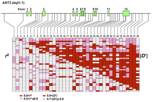 Fig. 1 Linkage Disequilibrium Pattern of ART3.1
Key structural properties of ART3:
- Typical ADP-ribosyl transferase catalytic domain
- Conservative NAD+ binding pocket and catalytic triad (H-Y-E)
- Single transmembrane anchoring of the domain
- The C-terminal intracellular segment contains multiple potential phosphorylation modification sites
Functions of ART3
The main function of the ART3 gene is to catalyze the ADP-ribonylation modification of the target protein. In addition, it is also involved in a variety of physiological and pathological processes, including germ cell development and immune regulation.
| Function | Description |
| Glycosylation catalysis | Using NAD+ as the substrate, the ADP-ribose group is transferred to a specific target protein to alter its activity. |
| Regulation of spermatogenesis | Specific expression in testicular germ cells, involved in sperm formation process of signal transduction and differentiated regulation. |
| Immune immunity is maintained | By modifying immune-related proteins in testicular tissue, it helps reproductive cells avoid attacks from the body's own immune system. |
| The influence of apoptosis | Mediate the modification of key proteins in the apoptotic pathway of germ cells and regulate the process of programmed cell death. |
| Association with cancer progression | Abnormal expression in prostate cancer and other tumors may be involved in disease development by affecting cell signaling pathways. |
The catalytic efficiency of this enzyme is strictly regulated by the concentration of NAD+ and the cellular microenvironment. Its activity in testicular tissue is significantly higher than that in other organs, demonstrating tissue-specific functional characteristics.
Applications of ART3 and ART3 Antibody in Literature
1. Tan, Ling, et al. "ART3 regulates triple-negative breast cancer cell function via activation of Akt and ERK pathways." Oncotarget 7.29 (2016): 46589. https://doi.org/10.18632/oncotarget.10306
Research has found that ART3 is highly expressed in refractory triple-negative breast cancer. Its overexpression can promote the proliferation, invasion and survival of cancer cells, and activate the AKT/ERK pathway, indicating that ART3 is a key marker of this cancer.
2. Kok, Dieuwertje E., et al. "Influence of maternal folate depletion on Art3 DNA methylation in the murine adult brain; potential consequences for brain and neurocognitive health." Mutagenesis 39.3 (2024): 196-204. https://doi.org/10.1093/mutage/geae007
Research has found that folic acid deficiency during pregnancy in female mice can lead to hypomethylation of the Art3 gene in the brains of their offspring in adulthood. Art3 is associated with neurocognition. This discovery may provide a key biomarker for the impact of early nutrition on lifelong brain health.
3. Okada, Hiroyuki, et al. "Genome-wide expression of azoospermia testes demonstrates a specific profile and implicates ART3 in genetic susceptibility." PLoS genetics 4.2 (2008): e26. https://doi.org/10.1371/journal.pgen.0040026
Research has found that the ADP-ribosyltransferase 3 (ART3) gene is a susceptibility gene for non-obstructive azoospermia (NOA). Its specific haplotype is significantly less frequent in patients and may play a protective role in the spermatogenic process by influencing testosterone levels.
4. Ma, Qinglan, et al. "Machine learning in identifying marker genes for congenital heart diseases of different cardiac cell types." Life 14.8 (2024): 1032. https://doi.org/10.3390/life14081032
Research has found that machine learning combined with single-cell analysis has identified genes such as ART3 as potential markers of congenital heart disease. Among them, ART3 is closely related to neonatal left heart dysplasia syndrome in cardiomyocytes.
5. Eckl, Miriam, et al. "Dosimetric benefits of daily treatment plan adaptation for prostate cancer stereotactic body radiotherapy." Radiation Oncology 16.1 (2021): 145. https://doi.org/10.1186/s13014-021-01872-9
Research shows that in hyperfractionated radiotherapy for prostate cancer, compared with conventional guided radiotherapy, all three adaptive radiotherapy strategies (ART1/2/3) can significantly reduce the radiation dose to the bladder and rectum. Among them, the completely re-optimized ART3 protocol has the best effect on protecting organs.
Creative Biolabs: ART3 Antibodies for Research
Creative Biolabs specializes in the production of high-quality ART3 antibodies for research and industrial applications. Our portfolio includes monoclonal antibodies tailored for ELISA, Flow Cytometry, Western blot, immunohistochemistry, and other diagnostic methodologies.
- Custom ART3 Antibody Development: Tailor-made solutions to meet specific research requirements.
- Bulk Production: Large-scale antibody manufacturing for industry partners.
- Technical Support: Expert consultation for protocol optimization and troubleshooting.
- Aliquoting Services: Conveniently sized aliquots for long-term storage and consistent experimental outcomes.
For more details on our ART3 antibodies, custom preparations, or technical support, contact us at email.
Reference
- Okada, Hiroyuki, et al. "Genome-wide expression of azoospermia testes demonstrates a specific profile and implicates ART3 in genetic susceptibility." PLoS genetics 4.2 (2008): e26. https://doi.org/10.1371/journal.pgen.0040026
Anti-ART3 antibodies
Products List
 Loading...


Hot products 
-
Rat Anti-EMCN Recombinant Antibody (28) (CBMAB-E0280-FY)

-
Mouse Anti-CORO1A Recombinant Antibody (4G10) (V2LY-1206-LY806)

-
Mouse Anti-FOXA3 Recombinant Antibody (2A9) (CBMAB-0377-YC)

-
Mouse Anti-EPO Recombinant Antibody (CBFYR0196) (CBMAB-R0196-FY)

-
Mouse Anti-ACTG1 Recombinant Antibody (V2-179597) (CBMAB-A0916-YC)

-
Mouse Anti-BIRC3 Recombinant Antibody (16E63) (CBMAB-C3367-LY)

-
Mouse Anti-AMOT Recombinant Antibody (CBYC-A564) (CBMAB-A2552-YC)

-
Mouse Anti-ATP1B3 Recombinant Antibody (1E9) (CBMAB-A4021-YC)

-
Mouse Anti-FN1 Monoclonal Antibody (71) (CBMAB-1241CQ)

-
Mouse Anti-CASQ1 Recombinant Antibody (CBFYC-0863) (CBMAB-C0918-FY)

-
Mouse Anti-BRD3 Recombinant Antibody (CBYY-0801) (CBMAB-0804-YY)

-
Mouse Anti-BLK Recombinant Antibody (CBYY-0618) (CBMAB-0621-YY)

-
Mouse Anti-BRCA2 Recombinant Antibody (CBYY-1728) (CBMAB-2077-YY)

-
Mouse Anti-APP Recombinant Antibody (5C2A1) (CBMAB-A3314-YC)

-
Mouse Anti-BCL2L1 Recombinant Antibody (H5) (CBMAB-1025CQ)

-
Mouse Anti-ALB Recombinant Antibody (V2-55272) (CBMAB-H0819-FY)

-
Mouse Anti-BACE1 Recombinant Antibody (61-3E7) (CBMAB-1183-CN)

-
Mouse Anti-CCDC25 Recombinant Antibody (CBLC132-LY) (CBMAB-C9786-LY)

-
Mouse Anti-CEMIP Recombinant Antibody (3C12) (CBMAB-K0296-LY)

-
Mouse Anti-AAV9 Recombinant Antibody (V2-634029) (CBMAB-AP023LY)

- AActivation
- AGAgonist
- APApoptosis
- BBlocking
- BABioassay
- BIBioimaging
- CImmunohistochemistry-Frozen Sections
- CIChromatin Immunoprecipitation
- CTCytotoxicity
- CSCostimulation
- DDepletion
- DBDot Blot
- EELISA
- ECELISA(Cap)
- EDELISA(Det)
- ESELISpot
- EMElectron Microscopy
- FFlow Cytometry
- FNFunction Assay
- GSGel Supershift
- IInhibition
- IAEnzyme Immunoassay
- ICImmunocytochemistry
- IDImmunodiffusion
- IEImmunoelectrophoresis
- IFImmunofluorescence
- IGImmunochromatography
- IHImmunohistochemistry
- IMImmunomicroscopy
- IOImmunoassay
- IPImmunoprecipitation
- ISIntracellular Staining for Flow Cytometry
- LALuminex Assay
- LFLateral Flow Immunoassay
- MMicroarray
- MCMass Cytometry/CyTOF
- MDMeDIP
- MSElectrophoretic Mobility Shift Assay
- NNeutralization
- PImmunohistologyp-Paraffin Sections
- PAPeptide Array
- PEPeptide ELISA
- PLProximity Ligation Assay
- RRadioimmunoassay
- SStimulation
- SESandwich ELISA
- SHIn situ hybridization
- TCTissue Culture
- WBWestern Blot
