CCM2 Antibodies
Background
The malcavernin protein encoded by the CCM2 gene is a key scaffold protein of the cytoplasmic signaling complex, and it is mainly expressed in vascular endothelial cells. This protein interacts with CCM1/KRIT1 and CCM3/PDCD10 through its phosphotyrosine binding domain to form an heterotrimeric complex, which maintains the integrity of the vascular barrier. Mutations in the CCM2 gene can lead to abnormal development of capillary walls, resulting in cavernous hemangiomas. This gene was first localized and cloned by Liquori et al. in 2003. The structural and functional studies of the encoded protein provide an important basis for revealing the molecular mechanism of cerebral vascular malformations.
Structure of CCM2
The malcavernin protein encoded by the CCM2 gene is a cytoplasmic scaffold protein with a molecular weight of approximately 45 kDa. This protein contains a characteristic phosphotyrosine-binding domain (PTB), which interacts with CCM1/KRIT1 and CCM3/PDCD10 through this domain to form an heterotrimeric complex. Malcavernin is highly expressed in vascular endothelial cells and is mainly located in the cytoplasm and cell junctions. Its C-terminal region mediates binding to downstream signaling molecules. The CCM2 protein sequence is highly conserved among different species, with a homology of over 95% between humans and mice, suggesting that it plays a crucial role in maintaining vascular integrity. The PTB domain of this protein consists of approximately 150 amino acid residues, forming a stable hydrophobic core that provides a structural basis for protein interactions.
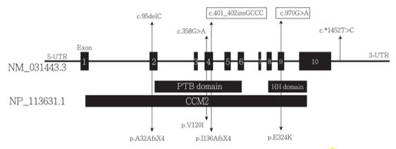 Fig. 1 Schematic representation of the CCM2 gene and protein structure.1
Key structural features of the protein encoded by the CCM2 gene:
- The N-terminal phosphotyrosine-binding domain mediates interaction with KRIT1
- The C-terminal region is involved in the binding with PDCD10 to form a trimeric complex
- The intermediate hinge region provides conformational flexibility
- Conserved hydrophobic residues maintain the stability of the scaffold protein
- Multiple phosphorylation sites regulate the signal transduction activity
Functions of CCM2
The malcavernin protein encoded by the CCM2 gene mainly participates in the regulation of vascular development signaling networks as a scaffold molecule. Its core function is to maintain the integrity of cerebral blood vessels. This protein exerts its effects through the following mechanisms:
| Function | Description |
| Protein complex assembly | By simultaneously binding CCM1 and CCM3 through the PTB domain, a ternary signaling complex is formed. |
| Endothelial junction stabilization | Regulates intercellular adhesion junctions, maintaining the integrity of the blood-brain barrier. |
| Vascular development regulation | Participates in the RhoA-ROCK signaling pathway, inhibiting abnormal vascular budding. |
| Oxidative stress response | Responds to changes in reactive oxygen levels, protecting endothelial cells from damage. |
| Inflammatory response regulation | Influences the expression of pro-inflammatory factors through the MEKK3 signaling pathway. |
The absence of malcavernin leads to loose intercellular connections in endothelial cells, resulting in dilated capillaries and the formation of cavernous hemangiomas. The differential expression of this protein in different tissues suggests that its function is cell type-specific.
Applications of CCM2 and CCM2 Antibody in Literature
1. Chang, Chun-Wei, et al. "CCM1 and CCM2 variants in patients with cerebral cavernous malformation in an ethnically Chinese population in Taiwan." Scientific Reports 9.1 (2019): 12387. https://doi.org/10.1038/s41598-019-48448-y
A study on cerebral cavernous vascular malformations in Taiwan region conducted genetic testing on 95 patients, and a pathogenic mutation was found in 6 individuals. Among them, a new insertion mutation in the CCM2 gene (c.401_402insGCCC) causes premature termination of protein translation.
2. Jiang, Xiaoting, et al. "Alternatively spliced isoforms reveal a novel type of PTB domain in CCM2 protein." Scientific Reports 9.1 (2019): 15808. https://doi.org/10.1038/s41598-019-52386-0
The research has revealed that the structure of the CCM2 gene is far more complex than previously known, containing 29 new exons and capable of generating up to 50 different splicing isoforms. These isoforms are distributed differently in various cells and contain a unique aPTB domain, which can simultaneously bind to CCM1 and CCM3 and is the core of the signaling complex.
3. Han, Guoqing, et al. "A novel CCM2 missense variant caused cerebral cavernous malformations in a Chinese family." Frontiers in Neuroscience 14 (2021): 604350. https://doi.org/10.3389/fnins.2020.604350
In a CCM family in China, a new mutation c.331G>C (p.A111P) of the CCM2 gene was discovered. This mutation would weaken the protein binding between KRIT1 and CCM2. Another patient in the family also carried a deletion of exon 13 of the KRIT1 gene. Both mutations coexisted.
4. Much, Christiane D., et al. "Novel pathogenic variants in a cassette exon of CCM2 in patients with cerebral cavernous malformations." Frontiers in neurology 10 (2019): 1219. https://doi.org/10.3389/fneur.2019.01219
The article indicates that the 5' end of the CCM2 gene has multiple alternative splicing events, which makes the interpretation of variations difficult. This study focused on the variable exon 3 region and discovered new pathogenic mutations. The amino acids encoded by this region are crucial for the function of CCM2. The CNV analysis combined with NGS technology can effectively shorten the diagnostic cycle.
5. Yang, Lipeng, Jian Wu, and Jing Zhang. "A novel CCM2 gene mutation associated with cerebral cavernous malformation." Frontiers in Neurology 11 (2020): 70. https://doi.org/10.3389/fneur.2020.00070
A novel deletion mutation c.755delC (p.S252fs*40X) of the CCM2 gene was identified in a Chinese family. This mutation results in a truncated protein, and the patient presents with multiple cerebral cavernous hemangiomas. This discovery has enriched the database of CCM2 gene mutations.
Creative Biolabs: CCM2 Antibodies for Research
Creative Biolabs specializes in the production of high-quality CCM2 antibodies for research and industrial applications. Our portfolio includes monoclonal and polyclonal antibodies tailored for ELISA, Flow Cytometry, Western blot, immunohistochemistry, and other diagnostic methodologies.
- Custom CCM2 Antibody Development: Tailor-made solutions to meet specific research requirements.
- Bulk Production: Large-scale antibody manufacturing for industry partners.
- Technical Support: Expert consultation for protocol optimization and troubleshooting.
- Aliquoting Services: Conveniently sized aliquots for long-term storage and consistent experimental outcomes.
For more details on our CCM2 antibodies, custom preparations, or technical support, contact us at email.
Reference
- Chang, Chun-Wei, et al. "CCM1 and CCM2 variants in patients with cerebral cavernous malformation in an ethnically Chinese population in Taiwan." Scientific Reports 9.1 (2019): 12387. Distributed under Open Access license CC BY 4.0, and cropped from the original figure. https://doi.org/10.1038/s41598-019-48448-y
Anti-CCM2 antibodies
 Loading...


Hot products 
-
Mouse Anti-ADAM12 Recombinant Antibody (V2-179752) (CBMAB-A1114-YC)

-
Mouse Anti-ENO2 Recombinant Antibody (H14) (CBMAB-E1341-FY)

-
Mouse Anti-ENPP1 Recombinant Antibody (CBFYE-0159) (CBMAB-E0375-FY)

-
Mouse Anti-CDK7 Recombinant Antibody (CBYY-C1783) (CBMAB-C3221-YY)

-
Rabbit Anti-ABL1 (Phosphorylated Y185) Recombinant Antibody (V2-443434) (PTM-CBMAB-0001YC)

-
Mouse Anti-ARID1B Recombinant Antibody (KMN1) (CBMAB-A3546-YC)

-
Mouse Anti-AFM Recombinant Antibody (V2-634159) (CBMAB-AP185LY)

-
Rabbit Anti-BAD (Phospho-Ser136) Recombinant Antibody (CAP219) (CBMAB-AP536LY)

-
Mouse Anti-APCS Recombinant Antibody (CBYC-A663) (CBMAB-A3054-YC)

-
Mouse Anti-CRYAB Recombinant Antibody (A4345) (CBMAB-A4345-YC)

-
Mouse Anti-AZGP1 Recombinant Antibody (CBWJZ-007) (CBMAB-Z0012-WJ)

-
Mouse Anti-CD2AP Recombinant Antibody (BR083) (CBMAB-BR083LY)

-
Rabbit Anti-ALOX5AP Recombinant Antibody (CBXF-1219) (CBMAB-F0750-CQ)

-
Rat Anti-AChR Recombinant Antibody (V2-12500) (CBMAB-0990-CN)

-
Rat Anti-4-1BB Recombinant Antibody (V2-1558) (CBMAB-0953-LY)

-
Mouse Anti-DDC Recombinant Antibody (8E8) (CBMAB-0992-YC)

-
Mouse Anti-Acetyl-α-Tubulin (Lys40) Recombinant Antibody (V2-623485) (CBMAB-CP2897-LY)

-
Mouse Anti-GFAP Recombinant Antibody (5) (CBMAB-G0346-LY)

-
Mouse Anti-AKT1/AKT2/AKT3 (Phosphorylated T308, T309, T305) Recombinant Antibody (V2-443454) (PTM-CBMAB-0030YC)

-
Mouse Anti-DMD Recombinant Antibody (D1190) (CBMAB-D1190-YC)

- AActivation
- AGAgonist
- APApoptosis
- BBlocking
- BABioassay
- BIBioimaging
- CImmunohistochemistry-Frozen Sections
- CIChromatin Immunoprecipitation
- CTCytotoxicity
- CSCostimulation
- DDepletion
- DBDot Blot
- EELISA
- ECELISA(Cap)
- EDELISA(Det)
- ESELISpot
- EMElectron Microscopy
- FFlow Cytometry
- FNFunction Assay
- GSGel Supershift
- IInhibition
- IAEnzyme Immunoassay
- ICImmunocytochemistry
- IDImmunodiffusion
- IEImmunoelectrophoresis
- IFImmunofluorescence
- IGImmunochromatography
- IHImmunohistochemistry
- IMImmunomicroscopy
- IOImmunoassay
- IPImmunoprecipitation
- ISIntracellular Staining for Flow Cytometry
- LALuminex Assay
- LFLateral Flow Immunoassay
- MMicroarray
- MCMass Cytometry/CyTOF
- MDMeDIP
- MSElectrophoretic Mobility Shift Assay
- NNeutralization
- PImmunohistologyp-Paraffin Sections
- PAPeptide Array
- PEPeptide ELISA
- PLProximity Ligation Assay
- RRadioimmunoassay
- SStimulation
- SESandwich ELISA
- SHIn situ hybridization
- TCTissue Culture
- WBWestern Blot
