CD103 Antibodies
Background
CD103, as an integrin protein, is mainly expressed on the surface of immune cells such as dendritic cells and T cells. This protein mediates the retention and interaction of immune cells in mucosal tissues by binding to E-cadherin on the surface of epithelial cells, thereby regulating the balance between local immune response and immune tolerance. In the tumor microenvironment, CD103+ T cells are regarded as an important subpopulation of tissue-resident memory T cells, and their existence is closely related to the improved prognosis of various cancers. Since its discovery in the 1990s, the functional research of CD103 has greatly promoted the understanding of the mechanisms of mucosal immunity, tumor immunity and autoimmune diseases, and its specific expression pattern has also provided new ideas for the development of targeted immunotherapy.
Structure of CD103
CD103 is a type I transmembrane glycoprotein with a molecular weight of approximately 150 kDa. There are slight differences in this molecular weight among different species, mainly due to the varying degrees of glycosylation modification.
| Species | Human | Mouse | Rat |
| Molecular Weight (kDa) | ~150 | ~145 | ~148 |
| Primary Structural Differences | Contains the αE domain | The αE domain is highly conserved | High homology with mouse |
The CD103 protein is composed of an α chain (encoded by ITGAE) and a β7 chain (encoded by ITGB7) linked by a disulfide bond to form a heterodimer. Its extracellular part contains an A domain that can specifically recognize E-cadherin. This domain adopts the Rossmann folding conformation and is mediated by metal ion-dependent adhesion sites (MIDAS) to bind to ligands. This specific interaction is the structural basis for CD103+ T cells to reside in epithelial tissues and participate in immune surveillance.
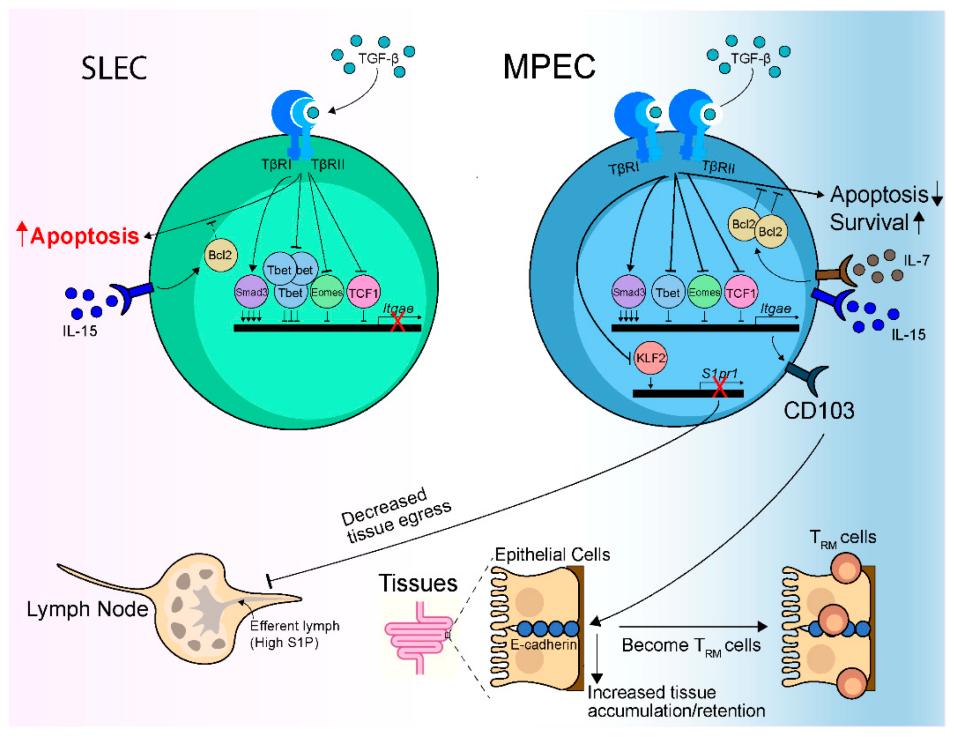 Fig. 1 TGF-β regulates the development and maintenance of CD103+ CD8 TRM cells through multiple mechanisms. 1
Key structural properties of CD103:
- Heterodimeric transmembrane structure consisting of αE and β7 subunits
- Extracellular section contains specific binding E - calcium mucins A domain structure
- Signal transduction is mediated by metal ion binding sites (MIDAS) dependent on divalent cations
Functions of CD103
The main function of CD103 (integrin αE) is to mediate the adhesion and retention of immune cells in epithelial tissues. However, it is also involved in a variety of immune regulatory processes, including immune surveillance, tolerance maintenance and inflammatory response regulation.
| Function | Description |
| Cell adhesion | CD103 binds to E-cadherin in epithelial cells, enabling immune cells to be localized to mucosal and barrier tissues. |
| Immune surveillance | Promote the recognition and clearance of infected or cancerous cells by intraepithelial T cells, and maintain tissue homeostasis. |
| Tolerance maintenance | Participate in regulatory T cell (Treg) function, inhibit excessive immune response, maintain autoimmune tolerance. |
| Inflammatory regulation | In the chronic inflammation environment affect lymphocyte migration and activation, participate in pathological process of inflammatory bowel disease. |
| Organizational resident memory formation | Support the formation of long-term resident memory by CD8+ T cells and provide rapid defense against local infections. |
The binding of CD103 to the ligand e-cadherin depends on divalent cations (such as Mg²⁺), and its affinity is precisely regulated by intracellular signals. This characteristic makes it a key regulatory molecule in mucosal immunity and immune responses in the epithelial barrier.
Applications of CD103 and CD103 Antibody in Literature
1. Qiu, Zhijuan, Timothy H. Chu, and Brian S. Sheridan. "TGF-β: many paths to CD103+ CD8 T cell residency." Cells 10.5 (2021): 989. https://doi.org/10.3390/cells10050989
The article indicates that CD103 is a key marker of CD8+ TRM cells, and its expression is regulated by TGF-β. TGF-β promotes the differentiation and residence of TRM cells by regulating the expression of Itgae and Klf2, and enhances local anti-infection and anti-tumor immune responses.
2. Kim, Younghoon, Yunjoo Shin, and Gyeong Hoon Kang. "Prognostic significance of CD103+ immune cells in solid tumor: a systemic review and meta-analysis." Scientific reports 9.1 (2019): 3808. https://doi.org/10.1038/s41598-019-40527-4
The article indicates that CD103+ immune cells are significantly associated with better overall survival and disease-free survival in patients with solid tumors, and they are important prognostic biomarkers. Its prognostic value is influenced by the assessment area (epithelium or stroma) and the detection method (whole section or tissue microarray).
3. Fukui, Takehito, et al. "Pivotal role of CD103 in the development of psoriasiform dermatitis." Scientific Reports 10.1 (2020): 8371. https://doi.org/10.1038/s41598-020-65355-9
The article indicates that CD103 inhibits skin inflammation by regulating the function of dendritic cells. Deficiency of CD103 can aggravate psoriasis-like dermatitis, promote the production of pro-inflammatory cytokines and IL-17, and lead to increased epidermal hyperplasia and infiltration of inflammatory cells.
4. Lai, Chester, et al. "CD8+ CD103+ tissue-resident memory T cells convey reduced protective immunity in cutaneous squamous cell carcinoma." Journal for Immunotherapy of Cancer 9.1 (2021): e001807. http://eprints.soton.ac.uk/id/eprint/445686
The article indicates that CD8+CD103+ TRM cells are enriched in cutaneous squamous cell carcinoma, highly express immune checkpoint molecules, and are significantly associated with tumor metastasis and poor prognosis of patients. This cell subpopulation may become an important prognostic biomarker.
5. Xu, Yu, et al. "CD103+ T Cells Eliminate Damaged Alveolar Epithelial Type II Cells Under Oxidative Stress to Prevent Lung Tumorigenesis." Advanced Science (2025): 2503557. https://doi.org/10.1002/advs.202503557
The article indicates that CD103+ T cells, especially the CD8+ subset, inhibit the occurrence of lung cancer by eliminating oxidized damaged alveolar epithelial cells. Their quantity decreases with age, leading to the accumulation of damaged cells and thereby driving the formation of KRAS-mutated tumors, revealing a new link between immunosenescence and the occurrence of lung cancer.
Creative Biolabs: CD103 Antibodies for Research
Creative Biolabs specializes in the production of high-quality CD103 antibodies for research and industrial applications. Our portfolio includes monoclonal antibodies tailored for ELISA, Flow Cytometry, Western blot, immunohistochemistry, and other diagnostic methodologies.
- Custom CD103 Antibody Development: Tailor-made solutions to meet specific research requirements.
- Bulk Production: Large-scale antibody manufacturing for industry partners.
- Technical Support: Expert consultation for protocol optimization and troubleshooting.
- Aliquoting Services: Conveniently sized aliquots for long-term storage and consistent experimental outcomes.
For more details on our CD103 antibodies, custom preparations, or technical support, contact us at email.
Reference
- Qiu, Zhijuan, Timothy H. Chu, and Brian S. Sheridan. "TGF-β: many paths to CD103+ CD8 T cell residency." Cells 10.5 (2021): 989. https://doi.org/10.3390/cells10050989
Anti-CD103 antibodies
 Loading...


Hot products 
-
Mouse Anti-APOA1 Monoclonal Antibody (CBFYR0637) (CBMAB-R0637-FY)

-
Mouse Anti-DISP2 Monoclonal Antibody (F66A4B1) (CBMAB-1112CQ)

-
Mouse Anti-DDC Recombinant Antibody (8E8) (CBMAB-0992-YC)

-
Mouse Anti-AQP2 Recombinant Antibody (G-3) (CBMAB-A3359-YC)

-
Mouse Anti-BACE1 Recombinant Antibody (CBLNB-121) (CBMAB-1180-CN)

-
Mouse Anti-CD33 Recombinant Antibody (P67.6) (CBMAB-C10189-LY)

-
Mouse Anti-BLNK Recombinant Antibody (CBYY-0623) (CBMAB-0626-YY)

-
Mouse Anti-14-3-3 Pan Recombinant Antibody (V2-9272) (CBMAB-1181-LY)

-
Mouse Anti-CD63 Recombinant Antibody (CBXC-1200) (CBMAB-C1467-CQ)

-
Mouse Anti-HTLV-1 gp46 Recombinant Antibody (CBMW-H1006) (CBMAB-V208-1154-FY)

-
Mouse Anti-AQP2 Recombinant Antibody (E-2) (CBMAB-A3358-YC)

-
Mouse Anti-ACTG1 Recombinant Antibody (V2-179597) (CBMAB-A0916-YC)

-
Mouse Anti-ADIPOR2 Recombinant Antibody (V2-179983) (CBMAB-A1369-YC)

-
Mouse Anti-ADIPOR1 Recombinant Antibody (V2-179982) (CBMAB-A1368-YC)

-
Mouse Anti-DLC1 Recombinant Antibody (D1009) (CBMAB-D1009-YC)

-
Mouse Anti-BACE1 Recombinant Antibody (61-3E7) (CBMAB-1183-CN)

-
Mouse Anti-CD1C Recombinant Antibody (L161) (CBMAB-C2173-CQ)

-
Mouse Anti-BAD (Phospho-Ser136) Recombinant Antibody (CBYY-0138) (CBMAB-0139-YY)

-
Human Anti-SARS-CoV-2 Spike Recombinant Antibody (CBC05) (CBMAB-CR005LY)

-
Mouse Anti-ACTN4 Recombinant Antibody (V2-6075) (CBMAB-0020CQ)

- AActivation
- AGAgonist
- APApoptosis
- BBlocking
- BABioassay
- BIBioimaging
- CImmunohistochemistry-Frozen Sections
- CIChromatin Immunoprecipitation
- CTCytotoxicity
- CSCostimulation
- DDepletion
- DBDot Blot
- EELISA
- ECELISA(Cap)
- EDELISA(Det)
- ESELISpot
- EMElectron Microscopy
- FFlow Cytometry
- FNFunction Assay
- GSGel Supershift
- IInhibition
- IAEnzyme Immunoassay
- ICImmunocytochemistry
- IDImmunodiffusion
- IEImmunoelectrophoresis
- IFImmunofluorescence
- IGImmunochromatography
- IHImmunohistochemistry
- IMImmunomicroscopy
- IOImmunoassay
- IPImmunoprecipitation
- ISIntracellular Staining for Flow Cytometry
- LALuminex Assay
- LFLateral Flow Immunoassay
- MMicroarray
- MCMass Cytometry/CyTOF
- MDMeDIP
- MSElectrophoretic Mobility Shift Assay
- NNeutralization
- PImmunohistologyp-Paraffin Sections
- PAPeptide Array
- PEPeptide ELISA
- PLProximity Ligation Assay
- RRadioimmunoassay
- SStimulation
- SESandwich ELISA
- SHIn situ hybridization
- TCTissue Culture
- WBWestern Blot
