CDK13 Antibodies
Background
The CDK13 gene encodes a cyclin-dependent kinase that mainly plays a role in cell cycle regulation and transcriptional extension. This protein participates in the processing and transcriptional regulation of precursor mRNA by phosphorylating the C-terminal domain of RNA polymerase II, which is of great significance for embryonic development and the maintenance of organ function. In 2016, multiple research teams, through whole exome sequencing technology, for the first time clearly identified that the loss-of-function mutation of this gene would lead to autosomal dominant neurodevelopmental disorder syndrome. Its highly conserved kinase domain and specific serine/threonine phosphorylation activity have become key models for studying transcriptional regulatory mechanisms and disease occurrence, providing a theoretical basis for the molecular diagnosis and therapeutic target development of related genetic diseases.
Structure of CDK13
CDK13 is a cyclin-dependent kinase with a molecular weight of approximately 160 kDa. There are slight differences in the molecular weight of this protein among different species, mainly due to the length variation of the C-terminal domain.
| Species | Human | Mouse | Zebrafish | Fruit fly |
| Molecular Weight (kDa) | ~160 | ~158 | ~155 | ~98 |
| Primary Structural Differences | Contains the long C-terminal regulatory domain | The C-terminal domain is short and functionally conserved | Core kinase domain structure highly homologous | Only the core kinase domain, the function is relatively basic |
The CDK13 protein is composed of a highly conserved kinase domain and a variable-length C-terminal extension region. In its three-dimensional structure, the kinase domain is composed of typical β -folding and α -helical ATP-substrate binding pockets. The conformation of the key activation loop (T-loop) is regulated by the phosphorylation state, and the "DFG motif" (aspartate - phenylalanine - glycine) located in the catalytic core is crucial for kinase activity. The C-terminal region is rich in proline and basic amino acids, mediating their specific interactions with transcriptional complexes and RNA processing factors.
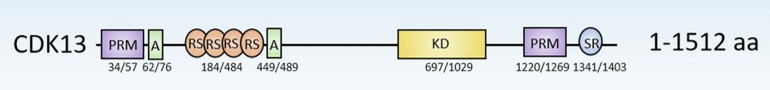 Fig. 1 Graphic representation of CDK13 domains.1
Key structural properties of CDK13:
- Core serine/threonine kinase domain
- The phosphorylation state of the activation loop (T-loop) regulates its kinase activity conformation
- "Conservative" DFG model body to maintain the catalytic center of metal ions and substrate recognition
Functions of CDK13
The main function of CDK13 is to regulate gene transcription and the progress of the cell cycle. However, it is also widely involved in a variety of key cell biological processes, including RNA processing and the regulation of embryonic development.
| Function | Description |
| Transcriptional regulation | By phosphorylating the C-terminal domain of RNA polymerase II, it regulates transcription initiation, extension and the processing of precursor mRNA. |
| Cell cycle regulation | As a cyclin-dependent kinase, it participates in the regulation of cell cycle checkpoints and affects cell proliferation and division. |
| Embryonic development | Its loss-of-function mutations can lead to severe neurodevelopmental disorders, indicating that it is crucial for the normal development of the nervous system and multiple organs. |
| RNA splicing regulation | By interacting with splicing factors, it participates in the selective splicing of specific pre-mrnas and affects proteome diversity. |
| Maintenance of genomic stability | Involved in DNA damage response pathway, and its dysfunction may be associated with genomic instability and disease. |
The phosphorylation of RNA polymerase II by CDK13 shows a high degree of substrate specificity and temporality, which contrasts with the mode of action of universal transcription factor kinases (such as CDK7/9), highlighting its unique role in fine coordinated transcriptional extension and co-transcriptional processing.
Applications of CDK13 and CDK13 Antibody in Literature
1. Tien, Jean Ching-Yi, et al. "CDK12 loss drives prostate cancer progression, transcription-replication conflicts, and synthetic lethality with paralog CDK13." Cell Reports Medicine 5.10 (2024). https://doi.org/10.1016/j.xcrm.2024.101758
The article indicates that the deletion of CDK12 can drive precancerous lesions of the prostate, cooperate with p53 to promote tumor development, and induce genomic instability. Research has found that the inhibition or degradation of CDK13 can specifically target CDK12-mutated tumors, providing a new therapeutic strategy for mCRPC.
2. Pitolli, Consuelo, et al. "Physiological and pathological roles of the transcriptional kinases CDK12 and CDK13 in the central nervous system." Cell Death & Differentiation 32.3 (2025): 371-381. https://doi.org/10.1038/s41418-024-01413-3
The article indicates that CDK12 and CDK13 affect gene expression by regulating the activity of RNA polymerase II, which is particularly crucial for the development and function of neurons. Its dysregulation of activity is involved in the occurrence of brain tumors, and related inhibitors show potential in tumor treatment.
3. Zhu, Tianyu, et al. "ARID1A loss promotes RNA editing of CDK13 in an ADAR1-dependent manner." BMC biology 22.1 (2024): 132. https://doi.org/10.1186/s12915-024-01927-9
The article indicates that the deletion of ARID1A affects the cell cycle by up-regulating ADAR1 and enhancing RNA editing of the CDK13 gene (at the Q113R and K117R loci). This mechanism makes ARID1A-deficient tumors sensitive to the CDK12/13 inhibitor SR-4835, suggesting the potential for targeted therapy.
4. Qi, Jin-Chun, et al. "CDK13 upregulation-induced formation of the positive feedback loop among circCDK13, miR-212-5p/miR-449a and E2F5 contributes to prostate carcinogenesis." Journal of Experimental & Clinical Cancer Research 40.1 (2021): 2. https://doi.org/10.1186/s13046-020-01814-5
Research has found that CDK13 is highly expressed in prostate cancer tissues and can interact with E2F5 to form a positive feedback loop. By adsorbing miRNA through circCDK13, it promotes the expression of E2F5 and drives the proliferation of cancer cells. This axis can provide new targets for the treatment of prostate cancer.
5. Wu, Zhouying, et al. "CDK13-mediated cell cycle disorder promotes tumorigenesis of high HMGA2 expression gastric cancer." Frontiers in Molecular Biosciences 8 (2021): 707295. https://doi.org/10.3389/fmolb.2021.707295
Studies have found that in gastric cancer, HMGA2 drives progression by promoting the S-G2/M phase transition, and CDK13 is its target gene. The co-high expression of both indicates a poor prognosis, and the combined inhibition of HMGA2 and CDK13 can become an effective treatment strategy.
Creative Biolabs: CDK13 Antibodies for Research
Creative Biolabs specializes in the production of high-quality CDK13 antibodies for research and industrial applications. Our portfolio includes monoclonal antibodies tailored for ELISA, Flow Cytometry, Western blot, immunohistochemistry, and other diagnostic methodologies.
- Custom CDK13 Antibody Development: Tailor-made solutions to meet specific research requirements.
- Bulk Production: Large-scale antibody manufacturing for industry partners.
- Technical Support: Expert consultation for protocol optimization and troubleshooting.
- Aliquoting Services: Conveniently sized aliquots for long-term storage and consistent experimental outcomes.
For more details on our CDK13 antibodies, custom preparations, or technical support, contact us at email.
Reference
- Pitolli, Consuelo, et al. "Physiological and pathological roles of the transcriptional kinases CDK12 and CDK13 in the central nervous system." Cell Death & Differentiation 32.3 (2025): 371-381. https://doi.org/10.1038/s41418-024-01413-3
Anti-CDK13 antibodies
 Loading...


Hot products 
-
Mouse Anti-BIRC3 Recombinant Antibody (315304) (CBMAB-1214-CN)

-
Mouse Anti-CD33 Recombinant Antibody (6C5/2) (CBMAB-C8126-LY)

-
Mouse Anti-ADGRE2 Recombinant Antibody (V2-261270) (CBMAB-C0813-LY)

-
Mouse Anti-ADAM29 Recombinant Antibody (V2-179787) (CBMAB-A1149-YC)

-
Mouse Anti-ACLY Recombinant Antibody (V2-179314) (CBMAB-A0610-YC)

-
Mouse Anti-CCND2 Recombinant Antibody (DCS-3) (CBMAB-G1318-LY)

-
Mouse Anti-ABL2 Recombinant Antibody (V2-179121) (CBMAB-A0364-YC)

-
Mouse Anti-ATP5F1A Recombinant Antibody (51) (CBMAB-A4043-YC)

-
Mouse Anti-CD33 Recombinant Antibody (P67.6) (CBMAB-C10189-LY)

-
Mouse Anti-AAV8 Recombinant Antibody (V2-634028) (CBMAB-AP022LY)

-
Mouse Anti-BHMT Recombinant Antibody (CBYY-0547) (CBMAB-0550-YY)

-
Mouse Anti-CD247 Recombinant Antibody (6B10.2) (CBMAB-C1583-YY)

-
Rat Anti-FABP3 Recombinant Antibody (CBXF-2299) (CBMAB-F1612-CQ)

-
Mouse Anti-BLK Recombinant Antibody (CBYY-0618) (CBMAB-0621-YY)

-
Mouse Anti-ALX1 Recombinant Antibody (96k) (CBMAB-C0616-FY)

-
Mouse Anti-FOSB Recombinant Antibody (CBXF-3593) (CBMAB-F2522-CQ)

-
Mouse Anti-EMP3 Recombinant Antibody (CBFYE-0100) (CBMAB-E0207-FY)

-
Mouse Anti-FOXL1 Recombinant Antibody (CBXF-0845) (CBMAB-F0462-CQ)

-
Mouse Anti-ACKR3 Recombinant Antibody (V2-261265) (CBMAB-C1023-LY)

-
Mouse Anti-AAV-5 Recombinant Antibody (V2-503417) (CBMAB-V208-1369-FY)

- AActivation
- AGAgonist
- APApoptosis
- BBlocking
- BABioassay
- BIBioimaging
- CImmunohistochemistry-Frozen Sections
- CIChromatin Immunoprecipitation
- CTCytotoxicity
- CSCostimulation
- DDepletion
- DBDot Blot
- EELISA
- ECELISA(Cap)
- EDELISA(Det)
- ESELISpot
- EMElectron Microscopy
- FFlow Cytometry
- FNFunction Assay
- GSGel Supershift
- IInhibition
- IAEnzyme Immunoassay
- ICImmunocytochemistry
- IDImmunodiffusion
- IEImmunoelectrophoresis
- IFImmunofluorescence
- IGImmunochromatography
- IHImmunohistochemistry
- IMImmunomicroscopy
- IOImmunoassay
- IPImmunoprecipitation
- ISIntracellular Staining for Flow Cytometry
- LALuminex Assay
- LFLateral Flow Immunoassay
- MMicroarray
- MCMass Cytometry/CyTOF
- MDMeDIP
- MSElectrophoretic Mobility Shift Assay
- NNeutralization
- PImmunohistologyp-Paraffin Sections
- PAPeptide Array
- PEPeptide ELISA
- PLProximity Ligation Assay
- RRadioimmunoassay
- SStimulation
- SESandwich ELISA
- SHIn situ hybridization
- TCTissue Culture
- WBWestern Blot
