DLK1 Antibodies
Background
DLK1, as a transmembrane glycoprotein, is mainly present in the embryonic development tissues, placenta, and some adult stem cells of mammals. This protein plays a crucial role in the formation of skeletal muscles, differentiation of adipocytes, and neural development by regulating cell proliferation and differentiation. The high expression of DLK1 during the fetal stage is essential for the normal construction of tissues and organs, while its expression level significantly decreases in the adult stage. This dynamic change is closely related to the metabolic requirements of the body during different developmental stages. It was first identified by Danish scientists in 1990 in the fat tissues of mammals. As a non-classical ligand of the Notch signaling pathway, DLK1, with its unique epidermal growth factor-like repeat domain, provides an important model for studying the mechanism of cell fate determination. The discovery of this protein and its role in stem cell regulation have laid a molecular foundation for understanding the basic laws of developmental biology and exploring intervention strategies in regenerative medicine.
Structure of DLK1
The molecular weight of the DLK1 protein is approximately 38 kDa. However, due to glycosylation modifications, specific bands are often observed at 50-60 kDa during actual detection. This protein exhibits high sequence conservation across different species.
| Species | Human | Mouse | Rat | Pig | Bovine |
| Number of amino acids | 383 | 385 | 383 | 384 | 383 |
| Primary Structural Differences | Classic EGF repeat | Conservation of the EGF domain | Extremely similar extracellular regions | There is a slight variation in the intracellular region | Conservation of signal peptide sequence |
DLK1 is a single transmembrane protein consisting of 383 amino acids. Its extracellular region contains 6 epidermal growth factor-like repeat domains, which mediate protein-protein interactions. This characteristic structure can be cleaved by proteases after synthesis to release a soluble form, which exerts paracrine regulatory effects. DLK1 belongs to the non-classical Notch ligand family. Its intracellular region lacks the classic signal transduction domains and mainly regulates cell differentiation direction by competing with Notch receptors. This protein is highly expressed in undifferentiated cells and its expression level gradually decreases as the cell differentiation process progresses. This expression pattern change is closely related to the temporal sequence of embryonic development.
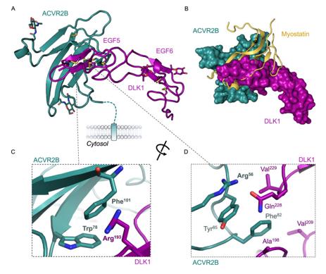 Fig. 1 Crystal structure of DLK1 EGF5-6 bound to ACVR2B.1
Key structural properties of DLK1:
- Extracellular domain containing six EGF-like repeating domains
- Protease cleavage site at juxtamembrane
- Single transmembrane domain
- Short intracellular tails lacking signal transduction activity
Functions of DLK1
The core function of DLK1 is to regulate cell differentiation and proliferation. Additionally, this gene is involved in various physiological processes, including fat formation, neural development, and skeletal muscle regeneration.
| Function | Description |
| Cell Differentiation Regulation | DLK1 inhibits the Notch signaling pathway, maintaining the undifferentiated state of precursor cells and regulating the processes of myogenic and adipogenic differentiation. |
| Embryonic Development Support | It is highly expressed during the embryonic development stage in placental and fetal tissues, and participates in organ formation and tissue construction. |
| Maintenance of adult stem cells | Expressed in adult stem cells such as skeletal muscle satellite cells, it regulates the balance between the quiescent and activated states of the stem cells. |
| Metabolic regulatory effect | It participates in energy metabolism regulation through its soluble form, influencing the distribution of adipose tissue and insulin sensitivity. |
| Paracrine signal transmission | The extracellular domains released by protease cleavage can act as ligands and influence the proliferation and differentiation fate of adjacent cells through paracrine means. |
The expression pattern of DLK1 shows stage-dependent downregulation during development, which is closely related to the process of cells transitioning from proliferation to differentiation. Its extracellular domain is structurally similar to that of the Notch receptor, but lacks the intracellular signaling domain. It exerts negative regulatory effects by competitively binding.
Applications of DLK1 and DLK1 Antibody in Literature
1. Antfolk, Daniel, et al. "Molecular mechanism of Activin receptor inhibition by DLK1." Nature Communications 16.1 (2025): 5976. https://doi.org/10.1038/s41467-025-60634-3
This study found that DLK1 does not bind to the Notch receptor, but instead mimics the TGF-β ligand to competitively bind to ACVR2B, antagonizing the myostatin signaling and thereby promoting myogenic differentiation. Its effect on Notch stems from interfering with the formation of the NICD-SMAD complex, revealing a new mechanism for muscle development.
2. Grassi, Elisa Stellaria, and Alexander Pietras. "Emerging roles of DLK1 in the stem cell niche and cancer stemness." Journal of Histochemistry & Cytochemistry 70.1 (2022): 17-28. https://doi.org/10.1369/00221554211048951
This study reveals that DLK1, as a maternal imprinted gene, regulates stem cell differentiation through spatiotemporal-specific expression and splicing, in a NOTCH-dependent or -independent manner. It also maintains the characteristics of cancer stem cells in various tumors and has potential as a therapeutic target.
3. Lu, Hsin-Pin, et al. "TRIM28 regulates Dlk1 expression in adipogenesis." International journal of molecular sciences 21.19 (2020): 7245. https://doi.org/10.3390/ijms21197245
The study found that TRIM28 regulates Dlk1 expression by recruiting E2f1. The absence of TRIM28 alters the histone methylation at the Dlk1 promoter (increased H3K4/H3K27 decreased), upregulates its expression, and thereby inhibits adipogenic differentiation. Knockdown of Dlk1 can rescue differentiation, demonstrating that the TRIM28-DLK1 axis is crucial for adipogenesis.
4. Huang, Deyu, et al. "Dlk1 maintains adult mice long-term HSCs by activating Notch signaling to restrict mitochondrial metabolism." Experimental Hematology & Oncology 12.1 (2023): 11. https://doi.org/10.1186/s40164-022-00369-9
The study found that adult mouse hematopoietic stem cells highly express Dlk1. Although knocking out Dlk1 increased the number of LT-HSCs, it caused these cells to enter the cell cycle and enhance metabolic activity (increasing ROS), thereby damaging the long-term reconstitution ability. Mechanistically, it is regulated by the Notch signaling pathway, revealing the crucial role of Dlk1 in maintaining the quiescent state of hematopoietic stem cells.
5. Dai, Rujuan, Zhuang Wang, and S. Ansar Ahmed. "Epigenetic contribution and genomic imprinting Dlk1-Dio3 miRNAs in systemic lupus erythematosus." Genes 12.5 (2021): 680. https://doi.org/10.3390/genes12050680
This review explores the mutual regulation between DNA methylation and miRNAs in SLE: Abnormal methylation affects miRNA expression, while miRNAs target methylation enzymes. The focus is on the dysregulation of the imprinted Dlk1-Dio3 miRNA cluster, which is regulated by methylation, in autoimmune diseases such as SLE.
Creative Biolabs: DLK1 Antibodies for Research
Creative Biolabs specializes in the production of high-quality DLK1 antibodies for research and industrial applications. Our portfolio includes monoclonal and polyclonal antibodies tailored for ELISA, Flow Cytometry, Western blot, immunohistochemistry, and other diagnostic methodologies.
- Custom DLK1 Antibody Development: Tailor-made solutions to meet specific research requirements.
- Bulk Production: Large-scale antibody manufacturing for industry partners.
- Technical Support: Expert consultation for protocol optimization and troubleshooting.
- Aliquoting Services: Conveniently sized aliquots for long-term storage and consistent experimental outcomes.
For more details on our DLK1 antibodies, custom preparations, or technical support, contact us at email.
Reference
- Antfolk, Daniel, et al. "Molecular mechanism of Activin receptor inhibition by DLK1." Nature Communications 16.1 (2025): 5976. Distributed under Open Access license CC BY 4.0. Cropped from the original figure. https://doi.org/10.1038/s41467-025-60634-3
Anti-DLK1 antibodies
 Loading...


Hot products 
-
Mouse Anti-NSUN6 Recombinant Antibody (D-5) (CBMAB-N3674-WJ)

-
Rat Anti-4-1BB Recombinant Antibody (V2-1558) (CBMAB-0953-LY)

-
Mouse Anti-BAX Recombinant Antibody (CBYY-0216) (CBMAB-0217-YY)

-
Mouse Anti-FLT1 Recombinant Antibody (11) (CBMAB-V0154-LY)

-
Mouse Anti-CHRNA9 Recombinant Antibody (8E4) (CBMAB-C9161-LY)

-
Mouse Anti-AMACR Recombinant Antibody (CB34A) (CBMAB-CA034LY)

-
Rabbit Anti-ADRA1A Recombinant Antibody (V2-12532) (CBMAB-1022-CN)

-
Mouse Anti-CTNND1 Recombinant Antibody (CBFYC-2414) (CBMAB-C2487-FY)

-
Mouse Anti-ARID3A Antibody (A4) (CBMAB-0128-YC)

-
Mouse Anti-ESR1 Recombinant Antibody (Y31) (CBMAB-1208-YC)

-
Mouse Anti-BHMT Recombinant Antibody (CBYY-0547) (CBMAB-0550-YY)

-
Rabbit Anti-AKT3 Recombinant Antibody (V2-12567) (CBMAB-1057-CN)

-
Rabbit Anti-ATF4 Recombinant Antibody (D4B8) (CBMAB-A3872-YC)

-
Mouse Anti-AOC3 Recombinant Antibody (CBYY-0014) (CBMAB-0014-YY)

-
Mouse Anti-CCND2 Recombinant Antibody (DCS-3) (CBMAB-G1318-LY)

-
Mouse Anti-G6PD Recombinant Antibody (13B331) (CBMAB-G1553-LY)

-
Mouse Anti-DHFR Recombinant Antibody (D0821) (CBMAB-D0821-YC)

-
Mouse Anti-ENO2 Recombinant Antibody (85F11) (CBMAB-0276CQ)

-
Rat Anti-ADAM10 Recombinant Antibody (V2-179741) (CBMAB-A1103-YC)

-
Mouse Anti-FLI1 Recombinant Antibody (CBXF-0733) (CBMAB-F0435-CQ)

- AActivation
- AGAgonist
- APApoptosis
- BBlocking
- BABioassay
- BIBioimaging
- CImmunohistochemistry-Frozen Sections
- CIChromatin Immunoprecipitation
- CTCytotoxicity
- CSCostimulation
- DDepletion
- DBDot Blot
- EELISA
- ECELISA(Cap)
- EDELISA(Det)
- ESELISpot
- EMElectron Microscopy
- FFlow Cytometry
- FNFunction Assay
- GSGel Supershift
- IInhibition
- IAEnzyme Immunoassay
- ICImmunocytochemistry
- IDImmunodiffusion
- IEImmunoelectrophoresis
- IFImmunofluorescence
- IGImmunochromatography
- IHImmunohistochemistry
- IMImmunomicroscopy
- IOImmunoassay
- IPImmunoprecipitation
- ISIntracellular Staining for Flow Cytometry
- LALuminex Assay
- LFLateral Flow Immunoassay
- MMicroarray
- MCMass Cytometry/CyTOF
- MDMeDIP
- MSElectrophoretic Mobility Shift Assay
- NNeutralization
- PImmunohistologyp-Paraffin Sections
- PAPeptide Array
- PEPeptide ELISA
- PLProximity Ligation Assay
- RRadioimmunoassay
- SStimulation
- SESandwich ELISA
- SHIn situ hybridization
- TCTissue Culture
- WBWestern Blot
