H2AX Antibodies
Background
The H2AX gene is responsible for encoding an important variant of histone H2A, which plays a core role in the chromatin structure of eukaryotic cells. When double-strand breaks occur in cellular DNA, H2AX is rapidly phosphorylated to form γH2AX markers. This modification can serve as a molecular beacon for DNA damage responses, initiating the recruitment of repair proteins and signal transduction pathways. This gene was first identified in 1998, and its phosphorylation phenomenon has become the gold standard biomarker for detecting DNA damage. Through continuous research on the formation of γH2AX foci, scientists have deepened their understanding of genomic stability regulation, the mechanism of cancer occurrence, and the response to radiotherapy, providing an important theoretical basis for the diagnosis and treatment of related diseases.
Structure of H2AX
H2AX is a core histone variant with a molecular weight of approximately 15.4 kDa. Its molecular weight varies slightly among different species, mainly depending on the sequence length and modification status of the H2AX-specific C-terminal tail.
| Species | Human | Mouse | Rat | African clawed toad | Fruit fly |
| Molecular Weight (kDa) | 15.4 | 15.4 | 15.4 | 15.2 | 15.8 |
| Primary Structural Differences | The C-terminal contains the SQEY motif | The C-terminal is highly homologous to humans | Highly conserved sequence | Contains similar phosphorylation sites | Have C-end tails with similar functions |
This protein is composed of approximately 142 amino acids and is assembled into a nucleosome together with other core histones through its spherical core domain. The most distinctive feature of H2AX is its unique C-terminal tail, which contains a conserved serine (Ser139 in humans). When cells encounter a double-strand break in DNA, this site is rapidly phosphorylated to form a landmark modification known as γH2AX, thereby initiating the recruitment of repair proteins and signal amplification at the damage site.
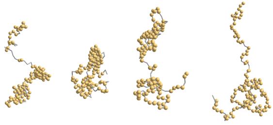 Fig. 1 Snapshots of the histone H2AX.1
Key structural properties of H2AX:
- Conserved histone folding domains
- Unique C-terminal tail sequence (including SQEY motif)
- The phosphorylable Ser139 site
Functions of H2AX
The core function of H2AX is to serve as a molecular beacon for DNA damage responses. However, it is also involved in various cellular processes such as the maintenance of chromatin structure and the determination of cell fate.
| Function | Description |
| DNA damage markers | After a double-strand break in DNA, serine at the C-terminal of H2AX is rapidly phosphorylated to form γH2AX, which serves as an early marker of the damage site. |
| Repair platform assembly | γH2AX directly recruits repair proteins such as MDC1 and BRCA1 to the damage site, forming protein aggregation foci visible under a microscope. |
| Signal amplification and transmission | By establishing a broad phosphorylation modification network, the initial damage signal is amplified and downstream cell cycle checkpoints are activated. |
| Cell fate determination | Continuous γH2AX signaling may guide cells towards apoptosis or senescence, preventing the further transmission of damaged DNA. |
| Chromatin structure regulation | Phosphorylation locally alters chromatin conformation, making tightly wound DNA more accessible to repair machinery. |
Unlike the common histone H2A which plays a structural role in the nucleosome, H2AX, through its reversible post-translational modification characteristics, acts as a dynamic regulator in maintaining genomic stability.
Applications of H2AX and H2AX Antibody in Literature
1. Turinetto, Valentina, and Claudia Giachino. "Multiple facets of histone variant H2AX: a DNA double-strand-break marker with several biological functions." Nucleic acids research 43.5 (2015): 2489-2498. https://doi.org/10.1093/nar/gkv061
The article indicates that the histone variant H2AX and its phosphorylation (γH2AX) are not only markers of DNA double-strand breaks but also play a key role in "non-classical" functions such as stem cell development, cellular senescence, and chromosomal inactivation.
2. Prabhu, Kirti S., et al. "H2AX: A key player in DNA damage response and a promising target for cancer therapy." Biomedicine & Pharmacotherapy 175 (2024): 116663. https://doi.org/10.1016/j.biopha.2024.116663
The article indicates that H2AX phosphorylation (γH2AX) is a core signal of DNA damage, capable of convening repair factors and regulating cell fate (repair or apoptosis), which makes it a highly promising biomarker and therapeutic target in cancer diagnosis and treatment.
3. Noubissi, Felicite K., et al. "Detection and quantification of γ-H2AX using a dissociation enhanced lanthanide fluorescence immunoassay." Scientific reports 11.1 (2021): 8945. https://doi.org/10.1038/s41598-021-88296-3
This study developed a novel method based on time-resolved fluorescence for the quantitative detection of the DNA double-strand break marker γ-H2AX. The new method features nanomolar-level ultra-high sensitivity and a convenient process, significantly outperforming traditional techniques.
4. Du, Changzheng, et al. "A PRMT5-RNF168-SMURF2 axis controls h2ax proteostasis." Cell reports 28.12 (2019): 3199-3211. https://doi.org/10.1016/j.celrep.2019.08.031
This study reveals that the homeostasis of H2AX protein is regulated by the PRMT5-RNF168-SMURF2 signaling axis: RNF168 plays a stabilizing role, while SMURF2 promotes its degradation. This pathway is dysregulated in MTAP-deficient glioblastoma, resulting in decreased H2AX levels and defective DNA damage responses.
5. Zhao, Baobing, et al. "H2AX deficiency is associated with erythroid dysplasia and compromised haematopoietic stem cell function." Scientific reports 6.1 (2016): 19589. https://doi.org/10.1038/srep19589
Research has found that the absence of H2AX can impair the function of hematopoietic stem cells and lead to dissonation of red blood cells, which triggers pathological hematopoiesis in both animal models and human MDS patients, revealing the crucial role of H2AX in maintaining normal hematopoiesis.
Creative Biolabs: H2AX Antibodies for Research
Creative Biolabs specializes in the production of high-quality H2AX antibodies for research and industrial applications. Our portfolio includes monoclonal antibodies tailored for ELISA, Flow Cytometry, Western blot, immunohistochemistry, and other diagnostic methodologies.
- Custom H2AX Antibody Development: Tailor-made solutions to meet specific research requirements.
- Bulk Production: Large-scale antibody manufacturing for industry partners.
- Technical Support: Expert consultation for protocol optimization and troubleshooting.
- Aliquoting Services: Conveniently sized aliquots for long-term storage and consistent experimental outcomes.
For more details on our H2AX antibodies, custom preparations, or technical support, contact us at email.
Reference
- Fritsche, Miriam, et al. "Variation in structure of a protein (H2AX) with knowledge-based interactions." PLoS One 8.5 (2013): e64507. https://doi.org/10.1371/journal.pone.0064507
Anti-H2AX antibodies
Products List
 Loading...


Hot products 
-
Mouse Anti-AQP2 Recombinant Antibody (G-3) (CBMAB-A3359-YC)

-
Mouse Anti-ADRB2 Recombinant Antibody (V2-180026) (CBMAB-A1420-YC)

-
Rat Anti-4-1BB Recombinant Antibody (V2-1558) (CBMAB-0953-LY)

-
Mouse Anti-BCL6 Recombinant Antibody (CBYY-0435) (CBMAB-0437-YY)

-
Rabbit Anti-AP2M1 (Phosphorylated T156) Recombinant Antibody (D4F3) (PTM-CBMAB-0610LY)

-
Mouse Anti-AMH Recombinant Antibody (5/6) (CBMAB-A2527-YC)

-
Mouse Anti-ADV Recombinant Antibody (V2-503423) (CBMAB-V208-1364-FY)

-
Mouse Anti-APOA1 Monoclonal Antibody (CBFYR0637) (CBMAB-R0637-FY)

-
Rabbit Anti-DLK1 Recombinant Antibody (9D8) (CBMAB-D1061-YC)

-
Mouse Anti-AGO2 Recombinant Antibody (V2-634169) (CBMAB-AP203LY)

-
Mouse Anti-ACE2 Recombinant Antibody (V2-179293) (CBMAB-A0566-YC)

-
Mouse Anti-CCT6A/B Recombinant Antibody (CBXC-0168) (CBMAB-C5570-CQ)

-
Mouse Anti-C5B-9 Recombinant Antibody (CBFYA-0216) (CBMAB-X0304-FY)

-
Mouse Anti-AKT1/AKT2/AKT3 (Phosphorylated T308, T309, T305) Recombinant Antibody (V2-443454) (PTM-CBMAB-0030YC)

-
Mouse Anti-BANF1 Recombinant Antibody (3F10-4G12) (CBMAB-A0707-LY)

-
Mouse Anti-AGK Recombinant Antibody (V2-258056) (CBMAB-M0989-FY)

-
Mouse Anti-ADGRL2 Recombinant Antibody (V2-58519) (CBMAB-L0166-YJ)

-
Mouse Anti-FOXL1 Recombinant Antibody (CBXF-0845) (CBMAB-F0462-CQ)

-
Mouse Anti-14-3-3 Pan Recombinant Antibody (V2-9272) (CBMAB-1181-LY)

-
Rat Anti-ADAM10 Recombinant Antibody (V2-179741) (CBMAB-A1103-YC)

- AActivation
- AGAgonist
- APApoptosis
- BBlocking
- BABioassay
- BIBioimaging
- CImmunohistochemistry-Frozen Sections
- CIChromatin Immunoprecipitation
- CTCytotoxicity
- CSCostimulation
- DDepletion
- DBDot Blot
- EELISA
- ECELISA(Cap)
- EDELISA(Det)
- ESELISpot
- EMElectron Microscopy
- FFlow Cytometry
- FNFunction Assay
- GSGel Supershift
- IInhibition
- IAEnzyme Immunoassay
- ICImmunocytochemistry
- IDImmunodiffusion
- IEImmunoelectrophoresis
- IFImmunofluorescence
- IGImmunochromatography
- IHImmunohistochemistry
- IMImmunomicroscopy
- IOImmunoassay
- IPImmunoprecipitation
- ISIntracellular Staining for Flow Cytometry
- LALuminex Assay
- LFLateral Flow Immunoassay
- MMicroarray
- MCMass Cytometry/CyTOF
- MDMeDIP
- MSElectrophoretic Mobility Shift Assay
- NNeutralization
- PImmunohistologyp-Paraffin Sections
- PAPeptide Array
- PEPeptide ELISA
- PLProximity Ligation Assay
- RRadioimmunoassay
- SStimulation
- SESandwich ELISA
- SHIn situ hybridization
- TCTissue Culture
- WBWestern Blot
