
Histone H3 Antibodies
Background
Histone H3 is a type of core histone. As the basic structural unit of the nucleosome, it, together with DNA, constitutes the basic building blocks of eukaryotic chromatin. This protein wraps around DNA by forming an octamer structure, not only maintaining the higher structure of chromatin but also directly regulating the transcriptional activity of genes through epigenetic modifications such as methylation and acetylation. As one of the earliest discovered histones, the high conservation of histone H3 in evolution reflects its core position in cell division, DNA repair and gene expression regulation. The systematic study of its modification patterns has greatly promoted the development of the epigenetics field and provided a key molecular basis for understanding the gene expression regulation mechanisms during cell differentiation, tumorigenesis and development.
Structure of Histone H3
Histone H3 is a core histone with a molecular weight of approximately 15.4 kDa. Its molecular weight varies slightly among different species, mainly due to variations in the N-terminal tail of the amino acid sequence. The molecular weight and characteristics of histone H3 in humans and common model organisms are as follows:
| Species | Human | Mouse | Fruit fly | Yeast |
| Molecular Weight (kDa) | 15.4 | 15.3 | 15.5 | 15.2 |
| Primary Structural Differences | Highly conserved globular core domain with variable N-terminal tail | High homology with humans | The core area is conserved, while the tail sequence is different | The sequence variation is significant and the function is conserved |
This protein is composed of 135 amino acids, forming a typical long N-terminal tail-spherical core structure. Its spherical domain forms histone folding through a helical-ring-helical motif, mediating the assembly of H3-H4 tetramers. The extended N-terminal tail segment presents a disordered conformation, providing a molecular basis for histone modification. In the spatial configuration, this protein forms a triple helix bundle through a helical arrangement of α 1-ring -α2-α3. Among them, the L1 ring and the α2 helix directly participate in the interaction of the DNA phosphate skeleton, while the 116th cysteine reversibly oxidizes and modifies to regulate the higher structure of chromatin. This structural characteristic makes histone H3 a key carrier for epigenetic regulation.
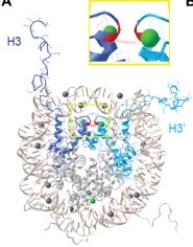 Fig. 1 Structure of histone H3 in the histone octamer.1
Key structural properties of Histone H3:
- Conserved histone folding domains, linked by three sets of α-helices via short loops
- Distribution of positively charged amino acids
- Flexible N-terminal tail structure, providing binding sites for epigenetic modification
Functions of Histone H3
The core function of histone H3 is to participate in nucleosome assembly and regulate gene expression. Its specific functions include:
| Function | Description |
| Nucleosome assembly | As a core histone, it forms an octamer together with H2A, H2B and H4, providing a structural scaffold for DNA entangling and maintaining genomic stability. |
| Epigenetic regulation | The chemical modifications at its N-terminal tail (such as methylation and acetylation) form a "histone code", directly controlling the open state of chromatin and the binding of transcription factors. |
| Transcriptional activation and silencing | Modifications such as H3K4me3 mark active genes, while modifications such as H3K9me3 promote heterochromatin formation, achieving heritable gene silencing. |
| Cell fate determination | By establishing specific modification patterns, it guides the establishment and maintenance of cell identity during processes such as stem cell differentiation and embryonic development. |
| DNA damage repair | Specific H3 modifications (such as H3K79me) recruit repair proteins to the damage site, participating in the repair process of DNA double-strand breaks. |
Unlike the histone variant H3.3 which replaces the classical H3.1 in the transcriptional active region, the classical subtype of H3 mainly incorporates into chromatin during the DNA replication period. This difference reflects its unique role in maintaining epigenetic memory.
Applications of Histone H3 and Histone H3 Antibody in Literature
1. Lai, Po Man, Xiaoxiang Gong, and Kui Ming Chan. "Roles of Histone H2B, H3 and H4 Variants in cancer development and prognosis." International Journal of Molecular Sciences 25.17 (2024): 9699. https://doi.org/10.3390/ijms25179699
The article indicates that histone H3 variants play a significant role in key biological processes such as DNA damage repair and transcriptional regulation. Recent studies have revealed novel variants of them in various cancers, and their expression levels have significant prognostic assessment value.
2. Collins, Bridget E., et al. "Histone H3 lysine K4 methylation and its role in learning and memory." Epigenetics & chromatin 12.1 (2019): 7. https://doi.org/10.1186/s13072-018-0251-8
The article indicates that the methylation of histone H3 lysine 4 (H3K4) is a key epigenetic mechanism in the formation of learning and memory. The abnormal reading, writing and erasing of this modification are directly related to cognitive disorders such as hippocampus memory function deficiency and human intellectual disability. This review will explore its regulatory mechanism and its association with neurocognitive impairment.
3. Klein, Rachel H., and Paul S. Knoepfler. "Knockout tales: the versatile roles of histone H3. 3 in development and disease." Epigenetics & Chromatin 16.1 (2023): 38. https://doi.org/10.1186/s13072-023-00512-8
The article indicates that the histone variant H3.3 plays a key role in gamete formation, brain-lung development and other processes, and is crucial for maintaining genomic stability. The deletion or mutation of the H3.3 encoding genes H3f3a and H3f3b can damage chromatin organization, leading to developmental defects and being closely related to human diseases such as pediatric tumors and neurodevelopmental syndromes.
4. Hillje, Roman, et al. "Time makes histone H3 modifications drift in mouse liver." Aging (Albany NY) 14.12 (2022): 4959. https://doi.org/10.18632/aging.204107
The article indicates that to detect the drift of epigenetics over time, the study found that the modifications of multiple histone H3 sites in the liver of mice would be redistributed across the entire genome with age, especially in the intergenic regions and near the transcription start sites. Calorie-restricted diets can effectively slow down such H3 modification changes related to aging.
5. Gleason, Ryan J., et al. "Developmentally programmed histone H3 expression regulates cellular plasticity at the parental-to-early embryo transition." Science advances 9.14 (2023): eadh0411. https://doi.org/10.1126/sciadv.adh0411
This study reveals that during embryonic development, the genomic landscape of histone H3 expression undergoes significant changes, that is, it shifts from a state rich in H3.3 to one rich in conventional H3. This transformation is regulated by the differential expression of different histone gene clusters, thereby limiting developmental plasticity and regulating the function of germline chromatin.
Creative Biolabs: Histone H3 Antibodies for Research
Creative Biolabs specializes in the production of high-quality Histone H3 antibodies for research and industrial applications. Our portfolio includes monoclonal antibodies tailored for ELISA, Flow Cytometry, Western blot, immunohistochemistry, and other diagnostic methodologies.
- Custom Histone H3 Antibody Development: Tailor-made solutions to meet specific research requirements.
- Bulk Production: Large-scale antibody manufacturing for industry partners.
- Technical Support: Expert consultation for protocol optimization and troubleshooting.
- Aliquoting Services: Conveniently sized aliquots for long-term storage and consistent experimental outcomes.
For more details on our Histone H3 antibodies, custom preparations, or technical support, contact us at email.
Reference
- Gleason, Ryan J., et al. "Developmentally programmed histone H3 expression regulates cellular plasticity at the parental-to-early embryo transition." Science advances 9.14 (2023): eadh0411. https://doi.org/10.1126/sciadv.adh0411
Anti-Histone H3 antibodies
Products List
 Loading...
 Compare




Hot products 
-
Mouse Anti-CD24 Recombinant Antibody (ALB9) (CBMAB-0176CQ)

-
Mouse Anti-AGK Recombinant Antibody (V2-258056) (CBMAB-M0989-FY)

-
Mouse Anti-CASP8 Recombinant Antibody (CBYY-C0987) (CBMAB-C2424-YY)

-
Mouse Anti-GLP1R Recombinant Antibody (4F3) (CBMAB-G0521-LY)

-
Mouse Anti-ARIH1 Recombinant Antibody (C-7) (CBMAB-A3563-YC)

-
Mouse Anti-FN1 Monoclonal Antibody (D6) (CBMAB-1240CQ)

-
Mouse Anti-C1QC Recombinant Antibody (CBFYC-0600) (CBMAB-C0654-FY)

-
Rabbit Anti-ABL1 (Phosphorylated Y185) Recombinant Antibody (V2-443434) (PTM-CBMAB-0001YC)

-
Mouse Anti-CD33 Recombinant Antibody (6C5/2) (CBMAB-C8126-LY)

-
Mouse Anti-FeLV g27 Recombinant Antibody (1) (CBMAB-V208-1714-FY)

-
Mouse Anti-CCNH Recombinant Antibody (CBFYC-1054) (CBMAB-C1111-FY)

-
Mouse Anti-ACTG1 Recombinant Antibody (V2-179597) (CBMAB-A0916-YC)

-
Mouse Anti-ENO2 Recombinant Antibody (85F11) (CBMAB-0276CQ)

-
Mouse Anti-AFM Recombinant Antibody (V2-634159) (CBMAB-AP185LY)

-
Mouse Anti-CTNND1 Recombinant Antibody (CBFYC-2414) (CBMAB-C2487-FY)

-
Mouse Anti-AHCYL1 Recombinant Antibody (V2-180270) (CBMAB-A1703-YC)

-
Mouse Anti-AP4E1 Recombinant Antibody (32) (CBMAB-A2996-YC)

-
Mouse Anti-CD24 Recombinant Antibody (SN3) (CBMAB-C1037-CQ)

-
Mouse Anti-BLK Recombinant Antibody (CBYY-0618) (CBMAB-0621-YY)

-
Mouse Anti-CRYAB Recombinant Antibody (A4345) (CBMAB-A4345-YC)

- AActivation
- AGAgonist
- APApoptosis
- BBlocking
- BABioassay
- BIBioimaging
- CImmunohistochemistry-Frozen Sections
- CIChromatin Immunoprecipitation
- CTCytotoxicity
- CSCostimulation
- DDepletion
- DBDot Blot
- EELISA
- ECELISA(Cap)
- EDELISA(Det)
- ESELISpot
- EMElectron Microscopy
- FFlow Cytometry
- FNFunction Assay
- GSGel Supershift
- IInhibition
- IAEnzyme Immunoassay
- ICImmunocytochemistry
- IDImmunodiffusion
- IEImmunoelectrophoresis
- IFImmunofluorescence
- IGImmunochromatography
- IHImmunohistochemistry
- IMImmunomicroscopy
- IOImmunoassay
- IPImmunoprecipitation
- ISIntracellular Staining for Flow Cytometry
- LALuminex Assay
- LFLateral Flow Immunoassay
- MMicroarray
- MCMass Cytometry/CyTOF
- MDMeDIP
- MSElectrophoretic Mobility Shift Assay
- NNeutralization
- PImmunohistologyp-Paraffin Sections
- PAPeptide Array
- PEPeptide ELISA
- PLProximity Ligation Assay
- RRadioimmunoassay
- SStimulation
- SESandwich ELISA
- SHIn situ hybridization
- TCTissue Culture
- WBWestern Blot
