HIVEP2 Antibodies
Background
HIVEP2 is a multifunctional transcription factor that is expressed in multiple organs of the human body. This protein recognizes and binds to specific DNA sequences through its zinc finger domain, regulating the expression of downstream genes. In the nervous system, HIVEP2 participates in neuronal development and the regulation of synaptic plasticity; in immune cells, it affects the expression of inflammatory factors. Studies have shown that mutations in the HIVEP2 gene can lead to autosomal dominant intellectual disability type 43, characterized by overall developmental delay. Moreover, abnormal expression of this gene is also associated with schizophrenia, substance use disorders, and other diseases, suggesting that it plays an important role in the pathogenesis of neuro-psychiatric diseases.
Structure of HIVEP2
HIVEP2 is a large-molecular-weight transcription factor, approximately 270 kDa in size. This protein contains multiple zinc finger domains and an acidic activation region, which regulate gene expression by recognizing specific DNA sequences. The molecular weight and structural characteristics of HIVEP2 in different species are as follows:
| Species | Humans | Mice | Rats | Simian |
| Molecular Weight (kDa) | 270 | 268 | 269 | 271 |
| Primary Structural Differences | Zinc finger domains are highly conserved | Acidic activation region has slight differences | DNA binding domain is similar | Has the highest homology with humans |
The HIVEP2 protein contains approximately 2,400 amino acids, and its structure includes multiple zinc finger motifs that form the DNA-binding domain and the acidic region responsible for transcriptional activation. These zinc finger structures can specifically recognize the κB sites in the promoter regions of target genes, thereby regulating the expression of immune-related genes and genes related to neural development. This protein functions in the cell nucleus and participates in important biological processes such as inflammatory responses and neuronal differentiation through interactions with other transcription factors.
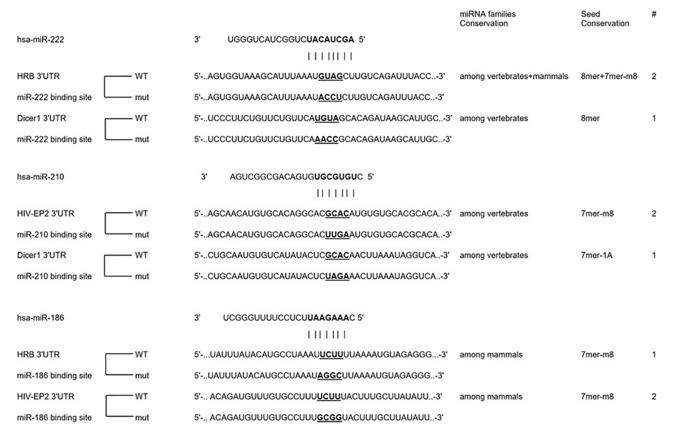 Fig. 1 miRNA-186, 210 and 222 directly regulate Dicer1, HRB and HIV-EP21
The key structural characteristics of HIVEP2:
- Multiple zinc finger domains mediate specific recognition of DNA sequences
- Acidic transcriptional activation region regulates downstream gene expression
- Serine-rich region participates in protein interactions
- Nuclear localization signal ensures its localization within the cell nucleus and functional expression
Functions of HIVEP2
The main function of HIVEP2 is to regulate gene expression as a transcription factor, playing a crucial role in immunity and the nervous system.
| Function | Description |
| Transcriptional regulation | By binding to specific DNA sequences through the zinc finger domain, it activates or inhibits the transcription of downstream target genes. |
| Immune regulation | Regulates the expression of genes related to inflammation, participating in T-cell activation and the immune response process. |
| Neurodevelopment | Influences neuronal differentiation and synaptic plasticity, playing an important role in nervous system development. |
| Stress response | Regulates the cell's adaptability to oxidative stress and hypoxic environments. |
| Disease association | Gene mutations can lead to intellectual disability, and abnormal expression is related to schizophrenia, substance use disorders, etc. |
The DNA binding kinetics of HIVEP2 show that it has high specificity for the κB site, achieving precise regulation of gene expression by recruiting co-activators or inhibitors.
Applications of HIVEP2 and HIVEP2 Antibody in Literature
1. Modai, Shira, et al. "HIV-1 infection increases microRNAs that inhibit Dicer1, HRB and HIV-EP2, thereby reducing viral replication." PloS one 14.1 (2019): e0211111. https://doi.org/10.1371/journal.pone.0211111
The article indicates that after HIV-1 infects Sup-T1 cells, the expressions of miR-186, 210 and 222 increase, thereby inhibiting the host target genes Dicer1, HRB and HIVEP2. These miRNAs can down-regulate viral expression and may play a key role in HIV-1 replication and miRNA generation, providing new ideas for treatment.
2. Zhao, Juan, et al. "Identification of HIVEP2 as a dopaminergic transcription factor related to substance use disorders in rats and humans." Translational Psychiatry 9.1 (2019): 247. https://doi.org/10.1038/s41398-019-0573-8
The research has found that HIVEP2 is a key factor regulating dopamine neurons and can activate the SLC6A3 gene. This mechanism plays a role in substance use disorders and shows gender differences, providing a new perspective for understanding addiction.
3. Abreu, Maria, et al. "Parental gonadossomatic mosaicism in HIVEP2-related intellectual disability and impact on genetic counseling–case report." Frontiers in Genetics 14 (2023): 1156847. https://doi.org/10.3389/fgene.2023.1156847
The study identified a case of autosomal dominant intellectual disability type 43 (MRD43) caused by a HIVEP2 gene mutation. The patient's mother was a healthy mosaic individual, raising the recurrence risk to 50%, highlighting the importance of parental testing in genetic counseling.
4. Murphy, Caitlin E., et al. "Nuclear factor kappa B activation appears weaker in schizophrenia patients with high brain cytokines than in non-schizophrenic controls with high brain cytokines." Journal of Neuroinflammation 17.1 (2020): 215. https://doi.org/10.1186/s12974-020-01890-6
The study found that in patients with schizophrenia accompanied by high neuroinflammation, the expression of HIVEP2 in the prefrontal cortex was decreased, and the transcription of its upstream activation factor NF-κB was weaker than that of the high inflammation control group. This suggests that the absence of HIVEP2 may be related to the weakened regulation of inflammation.
5. Karaoglu, Ismail Can, et al. "Single–gene knockout of RNLS or HIVEP2 are insufficient to protect β–cell spheroids from allo–and xeno–rejection." Frontiers in Immunology 17 (2026): 1759835. https://doi.org/10.3389/fimmu.2026.1759835
The study found that knocking out the RNLS or HIVEP2 genes failed to protect pancreatic β-cell grafts from immune rejection. This indicates that the effect of a single gene editing is limited and that other strategies need to be combined to achieve the goal of transplant therapy for type 1 diabetes.
Creative Biolabs: HIVEP2 Antibodies for Research
Creative Biolabs specializes in the production of high-quality HIVEP2 antibodies for research and industrial applications. Our portfolio includes monoclonal and polyclonal antibodies tailored for ELISA, Flow Cytometry, Western blot, immunohistochemistry, and other diagnostic methodologies.
- Custom HIVEP2 Antibody Development: Tailor-made solutions to meet specific research requirements.
- Bulk Production: Large-scale antibody manufacturing for industry partners.
- Technical Support: Expert consultation for protocol optimization and troubleshooting.
- Aliquoting Services: Conveniently sized aliquots for long-term storage and consistent experimental outcomes.
For more details on our HIVEP2 antibodies, custom preparations, or technical support, contact us at email.
Reference
- Modai, Shira, et al. "HIV-1 infection increases microRNAs that inhibit Dicer1, HRB and HIV-EP2, thereby reducing viral replication." PloS one 14.1 (2019): e0211111. Distributed under Open Access license CC BY 4.0, and cropped from the original figure. https://doi.org/10.1371/journal.pone.0211111
Anti-HIVEP2 antibodies
 Loading...


Hot products 
-
Rat Anti-EMCN Recombinant Antibody (28) (CBMAB-E0280-FY)

-
Mouse Anti-CD19 Recombinant Antibody (CBXC-1224) (CBMAB-C1491-CQ)

-
Rat Anti-CD63 Recombinant Antibody (7G4.2E8) (CBMAB-C8725-LY)

-
Mouse Anti-ARID1B Recombinant Antibody (KMN1) (CBMAB-A3546-YC)

-
Rabbit Anti-B2M Recombinant Antibody (CBYY-0059) (CBMAB-0059-YY)

-
Mouse Anti-Acetyl SMC3 (K105/K106) Recombinant Antibody (V2-634053) (CBMAB-AP052LY)

-
Mouse Anti-ARHGDIA Recombinant Antibody (CBCNA-009) (CBMAB-R0415-CN)

-
Rabbit Anti-ADRA1A Recombinant Antibody (V2-12532) (CBMAB-1022-CN)

-
Mouse Anti-BMI1 Recombinant Antibody (CBYC-P026) (CBMAB-P0108-YC)

-
Mouse Anti-ADRB2 Recombinant Antibody (V2-180026) (CBMAB-A1420-YC)

-
Mouse Anti-CRTAM Recombinant Antibody (CBFYC-2235) (CBMAB-C2305-FY)

-
Mouse Anti-dsRNA Recombinant Antibody (2) (CBMAB-D1807-YC)

-
Mouse Anti-GFP Recombinant Antibody (28) (CBMAB-G3038-LY)

-
Mouse Anti-FPR2 Recombinant Antibody (1D6) (CBMAB-F2628-CQ)

-
Mouse Anti-FTH1 Recombinant Antibody (CBXF-1896) (CBMAB-F3426-CQ)

-
Mouse Anti-AAV-5 Recombinant Antibody (V2-503417) (CBMAB-V208-1369-FY)

-
Mouse Anti-FeLV g27 Recombinant Antibody (1) (CBMAB-V208-1714-FY)

-
Mouse Anti-BIRC5 Recombinant Antibody (6E4) (CBMAB-CP2646-LY)

-
Mouse Anti-CAT Recombinant Antibody (724810) (CBMAB-C8431-LY)

-
Mouse Anti-CD33 Recombinant Antibody (P67.6) (CBMAB-C10189-LY)

- AActivation
- AGAgonist
- APApoptosis
- BBlocking
- BABioassay
- BIBioimaging
- CImmunohistochemistry-Frozen Sections
- CIChromatin Immunoprecipitation
- CTCytotoxicity
- CSCostimulation
- DDepletion
- DBDot Blot
- EELISA
- ECELISA(Cap)
- EDELISA(Det)
- ESELISpot
- EMElectron Microscopy
- FFlow Cytometry
- FNFunction Assay
- GSGel Supershift
- IInhibition
- IAEnzyme Immunoassay
- ICImmunocytochemistry
- IDImmunodiffusion
- IEImmunoelectrophoresis
- IFImmunofluorescence
- IGImmunochromatography
- IHImmunohistochemistry
- IMImmunomicroscopy
- IOImmunoassay
- IPImmunoprecipitation
- ISIntracellular Staining for Flow Cytometry
- LALuminex Assay
- LFLateral Flow Immunoassay
- MMicroarray
- MCMass Cytometry/CyTOF
- MDMeDIP
- MSElectrophoretic Mobility Shift Assay
- NNeutralization
- PImmunohistologyp-Paraffin Sections
- PAPeptide Array
- PEPeptide ELISA
- PLProximity Ligation Assay
- RRadioimmunoassay
- SStimulation
- SESandwich ELISA
- SHIn situ hybridization
- TCTissue Culture
- WBWestern Blot
