IL2RA Antibodies
Background
The IL2RA gene encodes the alpha chain of the interleukin-2 receptor (also known as CD25), which is a transmembrane protein mainly expressed on the surface of immune cells (such as regulatory T cells and activated T cells). This protein participates in regulating the proliferation, differentiation, and immune tolerance of T cells, playing a crucial role in maintaining immune homeostasis. Abnormalities in its function are closely related to various autoimmune diseases (such as type 1 diabetes, multiple sclerosis) and organ transplant rejection reactions. This gene was first identified in 1984, and subsequent studies gradually revealed its core position in the immune signaling pathway, providing a molecular basis for targeted therapy (such as using anti-CD25 monoclonal antibodies), which has greatly promoted the development of immune regulation mechanisms and treatment strategies for autoimmune diseases.
Structure of IL2RA
The protein encoded by the IL2RA gene (interleukin-2 receptor alpha chain, CD25) has a molecular weight of approximately 55-60 kDa. The exact size may vary slightly due to post-translational modifications and species differences.
| Species | Human | Mouse | Rat |
| Molecular Weight (kDa) | ~55 | ~58 | ~57 |
| Primary Structural Differences | Contains the signal peptide, transmembrane region and extracellular domain structure, is the key of IL - 2 high affinity receptor complexes | Highly homologous to human IL2RA structure, it is an important model for autoimmunity research | Often used in experimental immunology research, with functional conservation |
This protein is composed of approximately 272 amino acids (in its precursor form), and its extracellular part contains a cysteine-rich domain and a complement control protein (CCP)-like domain, which are crucial for ligand binding. Its secondary structure is mainly composed of β-sheets, forming a rigid extracellular ligand-binding interface. The transmembrane region anchors the protein to the cell membrane, while the shorter intracellular tail, although without catalytic activity, plays an important role in the internalization of the receptor complex and the regulation of signal transduction.
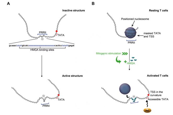 Fig. 1 Tentative model for the activation of IL2RA gene expression.1
Key structural properties of IL2RA:
- The extracellular region is dominated by β-folded and rigid domains, which form the ligand-binding interface
- Cysteine-rich domains stabilize extracellular conformations
- Intracellular structure domain extremely short, mainly responsible for signal transduction and receptor memory
Functions of IL2RA
The CD25 protein encoded by the IL2RA gene, whose main function is to serve as a key subunit of the interleukin-2 receptor, participates in the precise regulation of the immune system. The specific physiological processes involving this protein are as follows:
| Function | Description |
| Forming high-affinity receptors | CD25, together with the IL-2Rβ chain and the γc chain, assemble to form a complete IL-2 high-affinity receptor, enabling effective reception of the IL-2 signal. |
| T Cell Activation and Proliferation | By transmitting the IL-2 signal, it drives the clonal expansion of antigen-activated T cells (especially effector T cells). |
| Maintenance of immune tolerance | It is constitutively highly expressed on regulatory T cells and is a key molecule that maintains the immunosuppressive function and prevents autoimmune reactions. |
| Immune Homeostasis Regulation | By competitively consuming IL-2, it regulates the balance among different T cell subsets, influencing the intensity and duration of the immune response. |
| Disease Association | Its expression and functional abnormalities are directly related to autoimmune diseases, transplant rejection, and certain lymphoproliferative disorders. |
Unlike myoglobin which has a single function, CD25 does not directly bind to signaling molecules. Its function is entirely dependent on the complex formed with IL-2 and other receptor chains. The differential expression and function of CD25 on regulatory T cells and effector T cells reflect its dual precise regulatory role in the immune system, which both promotes responses and maintains tolerance.
Applications of IL2RA and IL2RA Antibody in Literature
1. Milani, Pascale, et al. "Mechanics of the IL2RA gene activation revealed by modeling and atomic force microscopy." Plos one 6.4 (2011): e18811. https://doi.org/10.1371/journal.pone.0018811
Through microscopic techniques and model simulations, this study discovered that the PRRII region of the IL2RA promoter contains a natural negative superhelix. Its dynamic curvature causes the vertex to gradually approach the transcription initiation site, promoting local DNA unwinding and mediating the specific binding of transcription factors, thereby revealing the crucial role of the dynamic changes in the promoter conformation at the transcription initiation stage.
2. Andrabi, Syed Bilal Ahmad, et al. "Long noncoding RNA LIRIL2R modulates FOXP3 levels and suppressive function of human CD4+ regulatory T cells by regulating IL2RA." Proceedings of the National Academy of Sciences 121.23 (2024): e2315363121. https://doi.org/10.1073/pnas.2315363121
The research has found that the long non-coding RNA LIRIL2R, which is highly expressed during the differentiation process of regulatory T cells, can bind to the upstream region of the IL2RA gene. By reshaping the epigenetic state, it positively regulates the expression of this gene, which is crucial for maintaining the inhibitory function of T cells.
3. Buhelt, Sophie, et al. "IL2RA methylation and gene expression in relation to the multiple sclerosis-associated gene variant rs2104286 and soluble IL-2Rα in CD8+ T cells." Frontiers in Immunology 12 (2021): 676141. https://doi.org/10.3389/fimmu.2021.676141
The research found that in the study of multiple sclerosis, a specific CpG site within the IL2RA gene, related to the risk SNP rs2104286, exhibited an allelic-specific methylation pattern. However, this site did not directly affect the expression of IL2RA. The level of soluble IL-2Rα was negatively correlated with the expression of various immune genes, suggesting that it may have an inhibitory effect on the CD8+ T cell response.
4. Gil-Quiñones, S. R., et al. "Effect of PTPN22, FAS/FASL, IL2RA and CTLA4 genetic polymorphisms on the risk of developing alopecia areata: A systematic review of the literature and meta-analysis." PLoS One 16.11 (2021): e0258499. https://doi.org/10.1371/journal.pone.0258499
This systematic review evaluated the association between multiple genes and the risk of alopecia areata. The analysis indicated that the rs2476601 polymorphism (T allele) of the PTPN22 gene was a definite risk factor for alopecia areata, while variations in the FAS, FASL, CTLA4, and IL2RA genes did not show significant correlations. Further research is needed to validate these findings.
5. Du, Wen, et al. "High IL2RA mRNA expression is an independent adverse prognostic biomarker in core binding factor and intermediate-risk acute myeloid leukemia." Journal of translational medicine 17.1 (2019): 191. https://doi.org/10.1186/s12967-019-1926-z
This study has confirmed that in acute myeloid leukemia, the highly expressed IL2RA mRNA is an independent predictor of poor prognosis. It is significantly correlated with shorter relapse-free survival and overall survival periods of patients, and its prognostic value is particularly prominent in CBF-AML and intermediate-risk AML patients.
Creative Biolabs: IL2RA Antibodies for Research
Creative Biolabs specializes in the production of high-quality IL2RA antibodies for research and industrial applications. Our portfolio includes monoclonal antibodies tailored for ELISA, Flow Cytometry, Western blot, immunohistochemistry, and other diagnostic methodologies.
- Custom IL2RA Antibody Development: Tailor-made solutions to meet specific research requirements.
- Bulk Production: Large-scale antibody manufacturing for industry partners.
- Technical Support: Expert consultation for protocol optimization and troubleshooting.
- Aliquoting Services: Conveniently sized aliquots for long-term storage and consistent experimental outcomes.
For more details on our IL2RA antibodies, custom preparations, or technical support, contact us at email.
Reference
- Milani, Pascale, et al. "Mechanics of the IL2RA gene activation revealed by modeling and atomic force microscopy." Plos one 6.4 (2011): e18811. https://doi.org/10.1371/journal.pone.0018811
Anti-IL2RA antibodies
 Loading...


Hot products 
-
Mouse Anti-ALB Recombinant Antibody (V2-180650) (CBMAB-A2186-YC)

-
Mouse Anti-ACTN4 Recombinant Antibody (V2-6075) (CBMAB-0020CQ)

-
Mouse Anti-4-Hydroxynonenal Recombinant Antibody (V2-502280) (CBMAB-C1055-CN)

-
Mouse Anti-ADV Recombinant Antibody (V2-503423) (CBMAB-V208-1364-FY)

-
Mouse Anti-AHCYL1 Recombinant Antibody (V2-180270) (CBMAB-A1703-YC)

-
Mouse Anti-ALB Recombinant Antibody (V2-55272) (CBMAB-H0819-FY)

-
Mouse Anti-DDC Recombinant Antibody (8E8) (CBMAB-0992-YC)

-
Rabbit Anti-AKT3 Recombinant Antibody (V2-12567) (CBMAB-1057-CN)

-
Mouse Anti-CFL1 (Phospho-Ser3) Recombinant Antibody (CBFYC-1770) (CBMAB-C1832-FY)

-
Mouse Anti-AGO2 Recombinant Antibody (V2-634169) (CBMAB-AP203LY)

-
Mouse Anti-ALPL Antibody (B4-78) (CBMAB-1009CQ)

-
Mouse Anti-ARHGDIA Recombinant Antibody (CBCNA-009) (CBMAB-R0415-CN)

-
Mouse Anti-CD24 Recombinant Antibody (HIS50) (CBMAB-C10123-LY)

-
Rat Anti-ABCC11 Recombinant Antibody (V2-179001) (CBMAB-A0236-YC)

-
Mouse Anti-AAV9 Recombinant Antibody (V2-634029) (CBMAB-AP023LY)

-
Mouse Anti-DLC1 Recombinant Antibody (D1009) (CBMAB-D1009-YC)

-
Mouse Anti-CD24 Recombinant Antibody (SN3) (CBMAB-C1037-CQ)

-
Mouse Anti-ALB Recombinant Antibody (V2-363290) (CBMAB-S0173-CQ)

-
Mouse Anti-AKT1/AKT2/AKT3 (Phosphorylated T308, T309, T305) Recombinant Antibody (V2-443454) (PTM-CBMAB-0030YC)

-
Mouse Anti-ATM Recombinant Antibody (2C1) (CBMAB-A3970-YC)

- AActivation
- AGAgonist
- APApoptosis
- BBlocking
- BABioassay
- BIBioimaging
- CImmunohistochemistry-Frozen Sections
- CIChromatin Immunoprecipitation
- CTCytotoxicity
- CSCostimulation
- DDepletion
- DBDot Blot
- EELISA
- ECELISA(Cap)
- EDELISA(Det)
- ESELISpot
- EMElectron Microscopy
- FFlow Cytometry
- FNFunction Assay
- GSGel Supershift
- IInhibition
- IAEnzyme Immunoassay
- ICImmunocytochemistry
- IDImmunodiffusion
- IEImmunoelectrophoresis
- IFImmunofluorescence
- IGImmunochromatography
- IHImmunohistochemistry
- IMImmunomicroscopy
- IOImmunoassay
- IPImmunoprecipitation
- ISIntracellular Staining for Flow Cytometry
- LALuminex Assay
- LFLateral Flow Immunoassay
- MMicroarray
- MCMass Cytometry/CyTOF
- MDMeDIP
- MSElectrophoretic Mobility Shift Assay
- NNeutralization
- PImmunohistologyp-Paraffin Sections
- PAPeptide Array
- PEPeptide ELISA
- PLProximity Ligation Assay
- RRadioimmunoassay
- SStimulation
- SESandwich ELISA
- SHIn situ hybridization
- TCTissue Culture
- WBWestern Blot
