IRS1 Antibodies
Background
IRS1 is a crucial adaptor protein that is mainly present in insulin-sensitive tissues and is highly expressed in the liver, skeletal muscle, and adipocytes. This protein binds to the insulin receptor through multiple tyrosine phosphorylation sites, activating downstream signaling pathways such as PI3K-AKT, thereby regulating glucose uptake, lipid synthesis, and protein metabolism in cells. As early as 1993, scientists discovered that the deficiency of the IRS1 gene could lead to insulin resistance. This groundbreaking discovery laid the foundation for subsequent research on the mechanisms of type 2 diabetes. Due to its central position in insulin signal transduction, IRS1 has become a research hotspot for exploring the mechanisms of metabolic diseases and developing new therapeutic targets.
Structure of IRS1
IRS1 is a signaling adaptor protein with a molecular weight of approximately 131 kDa. This value may vary depending on different subtypes and post-translational modification states.
| Species | Human | Mouse | Rat |
| Molecular Weight (kDa) | ~131 | ~132 | ~131 |
| Primary Structural Differences | Contains over 1200 amino acids, multiple tyrosine/serine phosphorylation sites | Highly homologous to humans, with slightly different phosphorylation regulatory patterns | Similar to mice, often used in metabolic disease models |
The IRS1 protein contains a pleckstrin homology domain and a phosphotyrosine binding domain, both of which are responsible for recognizing and binding to the activated insulin receptor. Its core structure contains multiple conserved tyrosine phosphorylation sites, which can recruit downstream effector molecules with SH2 domains after being phosphorylated. The serine/threonine phosphorylation sites of IRS1 act as a negative feedback regulatory hub. When these sites are over-phosphorylated, it will prevent tyrosine phosphorylation and thereby weaken insulin signal transduction.
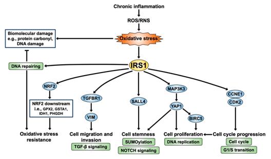 Fig. 1 IRS1 signaling pathway: a perspective on CCA progression.1
Structural characteristics of IRS1:
- PH domain: Anchors the cell membrane and recognizes the insulin receptor
- PTB domain: Bonds to the receptor and initiates signal transduction
- Tyrosine site: Recruits molecules such as PI3K and activates the pathway
- Serine site: Feedback regulation to control the intensity of the signal
Functions of IRS1
The core function of IRS1 is to conduct insulin signals and regulate cellular metabolism. However, it is also involved in various physiological processes, including cell growth, differentiation, and stress responses.
| Function | Description |
| Signal Transduction | As a linker protein, it connects the insulin receptor with downstream pathways such as PI3K-AKT and MAPK |
| Glucose Metabolism | Mediates insulin-stimulated glucose uptake, promotes glycogen synthesis and glycolysis |
| Lipid Regulation | Participates in the balance regulation of fat synthesis and decomposition, affecting lipid storage |
| Cell Growth | Activates proliferation-promoting signals, regulates cell cycle and protein synthesis |
| Stress Protection | Regulates the balance of cell survival and apoptosis under conditions such as oxidative stress |
The activation of IRS1 follows a dose-dependent pattern: physiological levels of insulin induce moderate phosphorylation to maintain metabolic homeostasis; while long-term hyperinsulinemia triggers negative feedback regulation, resulting in excessive phosphorylation of serine and ultimately inducing insulin resistance.
Applications of IRS1 and IRS1 Antibody in Literature
1. Kaewlert, Waleeporn, et al. "Overexpression of Insulin Receptor Substrate 1 (IRS1) relates to poor prognosis and promotes proliferation, stemness, migration, and oxidative stress resistance in cholangiocarcinoma." International Journal of Molecular Sciences 24.3 (2023): 2428. https://doi.org/10.3390/ijms24032428
The study found that oxidative stress promotes the progression of cholangiocarcinoma by upregulating the expression of IRS1. High expression of IRS1 is associated with a shorter survival period of patients, and it enhances the proliferation, migration, invasion and stress-resistance ability of cancer cells. IRS1 and its related genes can be used as prognostic markers and therapeutic targets for cholangiocarcinoma.
2. Rohde, Kerstin, et al. "IRS1 DNA promoter methylation and expression in human adipose tissue are related to fat distribution and metabolic traits." Scientific reports 7.1 (2017): 12369. https://doi.org/10.1038/s41598-017-12393-5
The study found that the rs2943650 variation near the IRS1 gene is associated with increased methylation of the IRS1 promoter and decreased expression in the visceral fat of obese individuals, which in turn affects waist circumference and glucose metabolism indicators. This suggests that DNA methylation may mediate the effect of this variation on metabolic phenotypes.
3. Keshavarzi, Fatemeh, and Shadi Golsheh. "IRS 1‐rs10498210 G/A and CCR 5‐59029 A/G polymorphisms in patients with type 2 diabetes in Kurdistan." Molecular Genetics & Genomic Medicine 7.5 (2019): e631. https://doi.org/10.1002/mgg3.631
The study found that the AA genotype of the IRS1 and CCR5 genes was significantly associated with type 2 diabetes in the Kurdish population, and carriers had an increased risk of the disease (IRS1: OR = 3.3; CCR5: OR = 2.9). Other genotypes did not show this association.
4. Gómez-Vilarrubla, Ariadna, et al. "Longitudinal Analysis of Placental IRS1 DNA Methylation and Childhood Obesity." International Journal of Molecular Sciences 26.7 (2025): 3141. https://doi.org/10.3390/ijms26073141
The study found that the methylation level of the placental IRS1 gene was significantly correlated with obesity and metabolic risks in children at the age of 6, and could effectively predict obesity in future generations. This suggests that IRS1 can serve as an early biomarker for predicting childhood obesity.
5. Martínez Báez, Anabel, et al. "Phosphorylation codes in IRS-1 and IRS-2 are associated with the activation/inhibition of insulin canonical signaling pathways." Current issues in molecular biology 46.1 (2024): 634-649. https://doi.org/10.3390/cimb46010041
The review states that IRS1/2, as a signaling adaptor protein, is precisely regulated by phosphorylation: tyrosine phosphorylation activates PI3K to transmit insulin signals, while serine phosphorylation has an inhibitory effect. More than 50 key phosphorylation sites have been identified.
Creative Biolabs: IRS1 Antibodies for Research
Creative Biolabs specializes in the production of high-quality IRS1 antibodies for research and industrial applications. Our portfolio includes monoclonal and polyclonal antibodies tailored for ELISA, Flow Cytometry, Western blot, immunohistochemistry, and other diagnostic methodologies.
- Custom IRS1 Antibody Development: Tailor-made solutions to meet specific research requirements.
- Bulk Production: Large-scale antibody manufacturing for industry partners.
- Technical Support: Expert consultation for protocol optimization and troubleshooting.
- Aliquoting Services: Conveniently sized aliquots for long-term storage and consistent experimental outcomes.
For more details on our IRS1 antibodies, custom preparations, or technical support, contact us at email.
Reference
- Kaewlert, Waleeporn, et al. "Overexpression of Insulin Receptor Substrate 1 (IRS1) relates to poor prognosis and promotes proliferation, stemness, migration, and oxidative stress resistance in cholangiocarcinoma." International Journal of Molecular Sciences 24.3 (2023): 2428. Distributed under Open Access license CC BY 4.0, without modification. https://doi.org/10.3390/ijms24032428
Anti-IRS1 antibodies
 Loading...


Hot products 
-
Rat Anti-CD63 Recombinant Antibody (7G4.2E8) (CBMAB-C8725-LY)

-
Mouse Anti-BRCA2 Recombinant Antibody (CBYY-0790) (CBMAB-0793-YY)

-
Mouse Anti-ABL2 Recombinant Antibody (V2-179121) (CBMAB-A0364-YC)

-
Mouse Anti-CFL1 Recombinant Antibody (CBFYC-1771) (CBMAB-C1833-FY)

-
Rat Anti-(1-5)-α-L-Arabinan Recombinant Antibody (V2-501861) (CBMAB-XB0003-YC)

-
Mouse Anti-COL1A2 Recombinant Antibody (CF108) (V2LY-1206-LY626)

-
Rat Anti-CD34 Recombinant Antibody (MEC 14.7) (CBMAB-C10196-LY)

-
Mouse Anti-AK4 Recombinant Antibody (V2-180419) (CBMAB-A1891-YC)

-
Mouse Anti-CSPG4 Recombinant Antibody (CBFYM-1050) (CBMAB-M1203-FY)

-
Mouse Anti-B2M Recombinant Antibody (CBYY-0050) (CBMAB-0050-YY)

-
Rat Anti-ABCC11 Recombinant Antibody (V2-179001) (CBMAB-A0236-YC)

-
Mouse Anti-ALPL Antibody (B4-78) (CBMAB-1009CQ)

-
Mouse Anti-ABIN2 Recombinant Antibody (V2-179106) (CBMAB-A0349-YC)

-
Mouse Anti-AGO2 Recombinant Antibody (V2-634169) (CBMAB-AP203LY)

-
Mouse Anti-ADGRE2 Recombinant Antibody (V2-261270) (CBMAB-C0813-LY)

-
Mouse Anti-C5B-9 Recombinant Antibody (CBFYA-0216) (CBMAB-X0304-FY)

-
Mouse Anti-8-oxoguanine Recombinant Antibody (V2-7719) (CBMAB-1898CQ)

-
Mouse Anti-GDF5 Recombinant Antibody (1F4) (CBMAB-G2740-LY)

-
Mouse Anti-FOXL1 Recombinant Antibody (CBXF-0845) (CBMAB-F0462-CQ)

-
Mouse Anti-CD24 Recombinant Antibody (ALB9) (CBMAB-0176CQ)

- AActivation
- AGAgonist
- APApoptosis
- BBlocking
- BABioassay
- BIBioimaging
- CImmunohistochemistry-Frozen Sections
- CIChromatin Immunoprecipitation
- CTCytotoxicity
- CSCostimulation
- DDepletion
- DBDot Blot
- EELISA
- ECELISA(Cap)
- EDELISA(Det)
- ESELISpot
- EMElectron Microscopy
- FFlow Cytometry
- FNFunction Assay
- GSGel Supershift
- IInhibition
- IAEnzyme Immunoassay
- ICImmunocytochemistry
- IDImmunodiffusion
- IEImmunoelectrophoresis
- IFImmunofluorescence
- IGImmunochromatography
- IHImmunohistochemistry
- IMImmunomicroscopy
- IOImmunoassay
- IPImmunoprecipitation
- ISIntracellular Staining for Flow Cytometry
- LALuminex Assay
- LFLateral Flow Immunoassay
- MMicroarray
- MCMass Cytometry/CyTOF
- MDMeDIP
- MSElectrophoretic Mobility Shift Assay
- NNeutralization
- PImmunohistologyp-Paraffin Sections
- PAPeptide Array
- PEPeptide ELISA
- PLProximity Ligation Assay
- RRadioimmunoassay
- SStimulation
- SESandwich ELISA
- SHIn situ hybridization
- TCTissue Culture
- WBWestern Blot
