MARK3 Antibodies
Background
MARK3 gene is a serine/threonine protein kinase, which is mainly involved in the regulation of microtubule dynamics and cytoskeletal reorganization. The protein encoded by this gene affects key biological processes such as neuronal morphogenesis, synaptic plasticity and cell migration by binding to microtubules and phosphorylating microtubule-associated proteins. Research has found that MARK3 may play a significant role in neurodegenerative diseases such as Alzheimer's disease, as it can regulate the phosphorylation state of tau protein and thereby affect the formation of neurofibrillary tangles. This gene was first identified by the Drewes team in 1996. Its three-dimensional structure analysis revealed unique kinase domain characteristics, providing an important theoretical basis for the development of targeted drugs for neurodegenerative diseases. Unlike myoglobin, MARK3 participates in the regulation of multiple cellular functions through a complex signaling network, demonstrating the functional diversity of multi-domain proteins in higher organisms.
Structure of MARK3
MARK3 is a serine/threonine protein kinase with a molecular weight of approximately 90 kDa, and its precise molecular weight varies slightly among different species.
| Species | Human | Mouse | Rat | Fruit fly |
| Molecular Weight (kDa) | 89.8 | 90.2 | 89.6 | 88.3 |
| Primary Structural Differences | N-terminal kinase domain structure and the C regulation area | Highly conserved kinase domain | Species-specific variation existed in the regulatory region | Structure more concise, function is simple |
MARK3 is composed of multiple domains, including the N-terminal kinase domain and the C-terminal microtubule binding region, forming a functional spatial conformation through α -helix and β -folding. Its catalytic core contains a conserved ATP-binding site (Gly-X-Gly-X-X-Gly motif) and an activation loop (T-loop), while the C-terminal domain regulates kinase activity through phosphorylation modification. The unique KA1 (kinase-associated 1) domain of this protein is involved in membrane localization. This modular design enables it to coordinate cytoskeletal recombination and polarity establishment.
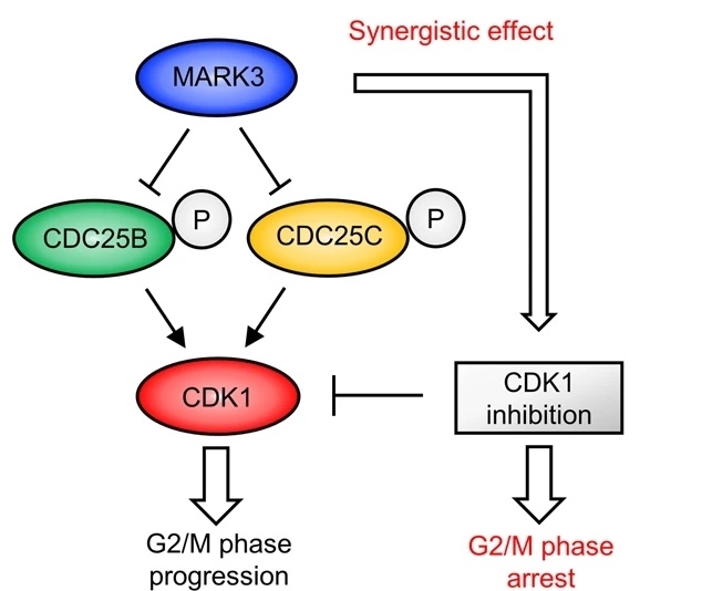 Fig. 1 Schematic of the effect of MARK3 inducing G2/M phase arrest with CDK1 inhibition.1
Key structural properties of MARK3:
- Modular kinase domain (N-terminal)
- Unique KA1 domain (C-end)
- Regulating element
- Key functional residues
Functions of MARK3
The core function of the MARK3 gene is to regulate microtubule stability and cell polarity, and it is also involved in a variety of important cellular processes.
| Function | Description |
| Microtubule dynamic regulation | Regulating microtubule assembly/depolymerization by phosphorylating microtubule-associated proteins (such as Tau and MAP2/4) affects neuronal morphogenesis. |
| Establishment of cell polarity | Asymmetric cell division is regulated in epithelial cells through the Par3/Par6/aPKC complex. |
| Regulation of synaptic plasticity | In the regulation of dendritic spines in the hippocampal neurons form, affect learning and memory function. |
| Regulation of cell migration | Rho GTPases signaling pathway affects the invasion and metastasis of tumor cells. |
| Energy metabolism sensing | As an AMPK-related kinase, it responds to changes in the energy state of cells. |
The enzyme activity curve of MARK3 exhibits typical biphasic characteristics: it shows Michaelis-Menten kinetics at low substrate concentrations, while it shows allostrucic regulatory properties at high substrate concentrations. This unique dynamic feature enables it to precisely respond to a variety of cellular signals (including phosphorylation modifications and protein interactions), thereby playing the role of a "molecular switch" in processes such as neurodevelopment and tumor metastasis.
Applications of MARK3 and MARK3 Antibody in Literature
1. Wang, Yudan, and Liyuan Guo. "Mark3 a Prognostic Marker for the Endometrial Cancer." Current Oncology 32.3 (2025): 157.https://doi.org/10.3390/curroncol32030157
The article indicates that MARK3 inhibits cell proliferation and migration in endometrial cancer, and its overexpression reduces colony formation and viability of Ishikawa and HEC-1B cells, possibly exerting its effect by regulating the pAKT pathway. Bioinformatics analysis suggests its potential as a prognostic marker and therapeutic target.
2. Machino, Hidenori, et al. "The metabolic stress-activated checkpoint LKB1-MARK3 axis acts as a tumor suppressor in high-grade serous ovarian carcinoma." Communications Biology 5.1 (2022): 39.https://doi.org/10.1038/s42003-021-02992-4
The article indicates that MARK3 is down-regulated in high-grade serous ovarian cancer (HGSOC) and is associated with a poor prognosis. Overexpression of MARK3 can inhibit cancer cell proliferation and angiogenesis, induce G2/M phase arrest through the LKB1-MARK3-CDC25 pathway, and down-regulate AP-1 and Hippo signaling target genes. Metabolic stress inducers targeting this pathway may become potential therapies.
3. Aparicio-Bautista, Diana I., et al. "Interaction between MARK3 (rs11623869), PLCB4 (rs6086746) and GEMIN2 (rs2277458) variants with bone mineral density and serum 25-hidroxivitamin D levels in Mexican Mestizo women." Frontiers in Endocrinology 15 (2024): 1392063.https://doi.org/10.3389/fendo.2024.1392063
Research has found that in Mexican women, the MARK3 gene variation (rs11623869) interacts with PLCB4 and GEMIN2, affecting bone mineral density (BMD) in the hip and femoral neck as well as vitamin D levels. The polygenic risk score (GRS) indicates that the combined effect of the three is significant, which may provide a new target for the early intervention of osteoporosis.
4. Lund, Harald, et al. "MARK4 and MARK3 associate with early tau phosphorylation in Alzheimer's disease granulovacuolar degeneration bodies." Acta neuropathologica communications 2.1 (2014): 22.https://doi.org/10.1186/2051-5960-2-22
Studies have found that in the hippocampus of patients with Alzheimer's disease (AD), the phosphorylated forms of MARK3 and MARK4 are co-localized with p-tau Ser262 in granular vacuolar denaturates (GVDs), suggesting that neurons may clear overactivated MARK kinases through GVDs, thereby reducing abnormal phosphorylation of tau protein. Slow down the progression of neurodegenerative diseases.
5. Calabrese, Gina M., et al. "Integrating GWAS and co-expression network data identifies bone mineral density genes SPTBN1 and MARK3 and an osteoblast functional module." Cell systems 4.1 (2017): 46-59.https://doi.org/10.1016/j.cels.2016.10.014
Research has found that the MARK3 gene is associated with the pathogenesis of osteoporosis, and its single nucleotide variation (rs11623869) affects bone mineral density (BMD) and vitamin D levels in Mexican women through intergene interactions. This gene may become a new target for the early intervention of osteoporosis.
Creative Biolabs: MARK3 Antibodies for Research
Creative Biolabs specializes in the production of high-quality MARK3 antibodies for research and industrial applications. Our portfolio includes monoclonal antibodies tailored for ELISA, Flow Cytometry, Western blot, immunohistochemistry, and other diagnostic methodologies.
- Custom MARK3 Antibody Development: Tailor-made solutions to meet specific research requirements.
- Bulk Production: Large-scale antibody manufacturing for industry partners.
- Technical Support: Expert consultation for protocol optimization and troubleshooting.
- Aliquoting Services: Conveniently sized aliquots for long-term storage and consistent experimental outcomes.
For more details on our MARK3 antibodies, custom preparations, or technical support, contact us at email.
Reference
- Machino, Hidenori, et al. "The metabolic stress-activated checkpoint LKB1-MARK3 axis acts as a tumor suppressor in high-grade serous ovarian carcinoma." Communications Biology 5.1 (2022): 39.https://doi.org/10.1038/s42003-021-02992-4
Anti-MARK3 antibodies
 Loading...


Hot products 
-
Mouse Anti-BRCA2 Recombinant Antibody (CBYY-0790) (CBMAB-0793-YY)

-
Mouse Anti-ACLY Recombinant Antibody (V2-179314) (CBMAB-A0610-YC)

-
Mouse Anti-APOH Recombinant Antibody (4D9A4) (CBMAB-A3249-YC)

-
Rat Anti-CD63 Recombinant Antibody (7G4.2E8) (CBMAB-C8725-LY)

-
Rat Anti-4-1BB Recombinant Antibody (V2-1558) (CBMAB-0953-LY)

-
Rabbit Anti-ABL1 (Phosphorylated Y185) Recombinant Antibody (V2-443434) (PTM-CBMAB-0001YC)

-
Mouse Anti-ALDOA Recombinant Antibody (A2) (CBMAB-A2316-YC)

-
Mouse Anti-ENO1 Recombinant Antibody (8G8) (CBMAB-E1329-FY)

-
Mouse Anti-DMD Recombinant Antibody (D1190) (CBMAB-D1190-YC)

-
Mouse Anti-GIPC2 Recombinant Antibody (10) (CBMAB-G0476-LY)

-
Rat Anti-EMCN Recombinant Antibody (28) (CBMAB-E0280-FY)

-
Mouse Anti-ENO2 Recombinant Antibody (85F11) (CBMAB-0276CQ)

-
Mouse Anti-ADGRE2 Recombinant Antibody (V2-261270) (CBMAB-C0813-LY)

-
Mouse Anti-BAX Recombinant Antibody (CBYY-0216) (CBMAB-0217-YY)

-
Mouse Anti-AMOT Recombinant Antibody (CBYC-A564) (CBMAB-A2552-YC)

-
Mouse Anti-DLG1 Monolconal Antibody (4F3) (CBMAB-0225-CN)

-
Human Anti-SARS-CoV-2 Spike Recombinant Antibody (CBC05) (CBMAB-CR005LY)

-
Rat Anti-EPO Recombinant Antibody (16) (CBMAB-E1578-FY)

-
Mouse Anti-ADAM29 Recombinant Antibody (V2-179787) (CBMAB-A1149-YC)

-
Mouse Anti-BIRC3 Recombinant Antibody (315304) (CBMAB-1214-CN)

- AActivation
- AGAgonist
- APApoptosis
- BBlocking
- BABioassay
- BIBioimaging
- CImmunohistochemistry-Frozen Sections
- CIChromatin Immunoprecipitation
- CTCytotoxicity
- CSCostimulation
- DDepletion
- DBDot Blot
- EELISA
- ECELISA(Cap)
- EDELISA(Det)
- ESELISpot
- EMElectron Microscopy
- FFlow Cytometry
- FNFunction Assay
- GSGel Supershift
- IInhibition
- IAEnzyme Immunoassay
- ICImmunocytochemistry
- IDImmunodiffusion
- IEImmunoelectrophoresis
- IFImmunofluorescence
- IGImmunochromatography
- IHImmunohistochemistry
- IMImmunomicroscopy
- IOImmunoassay
- IPImmunoprecipitation
- ISIntracellular Staining for Flow Cytometry
- LALuminex Assay
- LFLateral Flow Immunoassay
- MMicroarray
- MCMass Cytometry/CyTOF
- MDMeDIP
- MSElectrophoretic Mobility Shift Assay
- NNeutralization
- PImmunohistologyp-Paraffin Sections
- PAPeptide Array
- PEPeptide ELISA
- PLProximity Ligation Assay
- RRadioimmunoassay
- SStimulation
- SESandwich ELISA
- SHIn situ hybridization
- TCTissue Culture
- WBWestern Blot
