MATK Antibodies
Background
MATK could identify genetic encoding of a protein known as macrophages activated kinase, mainly distributed in immune cells and the nervous system. This protein plays a key role in maintaining the immune balance of the body by regulating immune responses and inflammatory reactions through participating in cellular signaling pathways. Research has found that the MATK gene is associated with certain autoimmune diseases and neurodegenerative disorders, and its mutations may affect T-cell receptor signal transduction. As an important regulatory factor of Src family kinases, the molecular structure characteristics of MATK have been resolved, which includes typical SH3 and SH2 protein interaction domains. In-depth research on this gene provides an important theoretical basis for understanding the immune regulatory mechanism and developing targeted treatments for related diseases. Currently, it remains one of the hot research objects in the field of biomedicine.
Structure of MATK
MATK is a tyrosine kinase with a molecular weight of approximately 56 kDa. Its molecular weight remains relatively stable across different species, with the main differences coming from minor variations in the regulatory domain.
| Species | Human | Mouse | Rat |
| Molecular Weight (kDa) | 56 | 55.8 | 55.9 |
| Primary Structural Differences | Containing SH3 and SH2 domains, it regulates immune signals | Highly conserved, with over 90% homology to human MATK | Kinase domain structure similar to that of C terminal is slightly different |
MATK is composed of approximately 500 amino acids and has a typical kinase folding structure, including the N-terminal SH3 domain (mediating protein interactions), the central SH2 domain (binding phosphorylated tyrosine), and the C-terminal kinase domain (catalytic phosphorylation). Its activity is regulated by its own phosphorylation and plays a key role in the T-cell receptor signaling pathway. The crystal structure analysis of MATK reveals its self-inhibition mechanism, providing a structural basis for the design of targeted drugs.
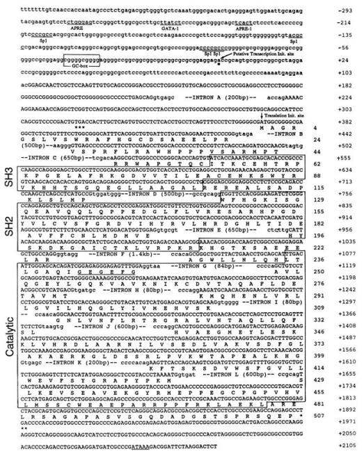 Fig. 1 Nucleotide and deduced amino acid sequence of the human MATK gene.1
Key structural properties of MATK:
- SH3-SH2- kinase domain modular architecture
- Self-inhibitory conformation
- Key phosphorylation sites (Tyr394/Tyr504)
Functions of MATK
The core function of the MATK gene is to regulate immune cell signal transduction and neural development processes, and it is also involved in multiple pathophysiological mechanisms.
| Function | Description |
| Immune regulation | By inhibiting the activity of Src family kinases and negatively regulating the T-cell receptor (TCR) signaling pathway, immune homeostasis is maintained. |
| Neural development | In the process of neuronal differentiation, regulating the growth cone guide effect axons extends and synaptic plasticity. |
| Tumor suppression | Expressed in various types of cancer, its absence leads to abnormal cell proliferation and metastasis potential increase. |
| Inflammatory regulation | Inhibiting excessive inflammatory response through NF-κB signaling pathway plays a protective role in autoimmune diseases. |
| Hematopoietic regulation | Regulating the differentiation of balance in the bone marrow hematopoietic stem cells, affect the myeloid and lymphoid cell development. |
The kinase activity of MATK exhibits typical substrate-specific phosphorylation characteristics. Compared with other tyrosine kinases (such as Lck and Fyn), its signal kinetics shows more precise threshold regulation characteristics, which makes it a precise regulator of immune synaptic signal transduction. The latest research has found that MATK forms signal micro-regions through a phase separation mechanism, further enhancing the specificity of its signal transduction.
Applications of MATK and MATK Antibody in Literature
1. Maybee, Deanna V., et al. "MMP-2 regulates Src activation via repression of the CHK/MATK tumor suppressor in osteosarcoma." Cancer Reports 7.2 (2024): e1946. https://doi.org/10.1002/cnr2.1946
This study reveals that MMP-2 regulates Src kinase activity and promotes osteosarcoma cell migration by inhibiting its endogenous inhibitor CHK/MATK. Experiments have shown that inhibiting MMP-2 or overexpressing CHK/MATK can reverse doxorubicin-induced migration, suggesting that targeting the MMP-2-CHK/MATK pathway can enhance the efficacy of doxorubicin and inhibit osteosarcoma metastasis.
2. Zhong, Cheng, et al. "Prognostic Function and Immunologic Landscape of a Predictive Model Based on Five Senescence-Related Genes in IPF Bronchoalveolar Lavage Fluid." Biomedicines 12.6 (2024): 1246. https://doi.org/10.3390/biomedicines12061246
This study constructed a prognostic model of idiopathic pulmonary fibrosis based on five aging-related genes (including megakaryocyte-associated tyrosine kinase MATK), verifying that the model can effectively predict the risk stratification of patients and revealing that the increased infiltration of neutrophils and other immune cells in the alveolar lavage fluid of high-risk group patients is associated with a poor prognosis.
3. Deng, Bijia, et al. "Structural and functional studies of the intracellular tyrosine kinase MATK gene and its translated product." Journal of Biological Chemistry 270.4 (1995): 1833-1842. https://doi.org/10.1074/jbc.270.4.1833
In this study, human and murine MATK genes (megakaryocyte-associated tyrosine kinases) were successfully cloned and identified, revealing that they regulate the activity of Src family kinases through the SH3/SH2 domain and ATP binding sites. Experiments have confirmed that MATK can phosphorylate the C-terminal tyrosine of Src protein, and antisense oligonucleotide inhibition significantly reduces the proliferation of megakaryocyte progenitor cells, indicating that it plays a key regulatory role in the megakaryocyte growth and differentiation signaling pathway.
4. Advani, Gahana, et al. "Csk-homologous kinase (Chk) is an efficient inhibitor of Src-family kinases but a poor catalyst of phosphorylation of their C-terminal regulatory tyrosine." Cell Communication and Signaling 15.1 (2017): 29. https://doi.org/10.1186/s12964-017-0186-x
This study reveals the dual mechanism differences in the inhibition of Src family kinases (SFKs) by Csk and Chk (MATK) : Csk mainly inhibits SFKs by catalyzing C-terminal tyrosine phosphorylation, while Chk (MATK) relies on high-affinity binding in the kinase domain to achieve non-catalytic inhibition. In colorectal cancer cells, the absence of Chk leads to the excessive activation of SFKs, confirming the key role of its non-catalytic inhibitory mechanism in regulating tumor growth.
5. Zrihan-Licht, Sheila, et al. "Association of csk-homologous kinase (CHK)(formerly MATK) with HER-2/ErbB-2 in breast cancer cells." Journal of Biological Chemistry 272.3 (1997): 1856-1863. https://doi.org/10.1074/jbc.272.3.1856
This study reveals that CHK (MATK) is a tyrosine kinase that is highly expressed in breast cancer tissues but not detected in normal tissues. Research has found that CHK specifically binds to Heregulin-activated ErbB-2 through its SH2 domain and may be involved in breast cancer signaling.
Creative Biolabs: MATK Antibodies for Research
Creative Biolabs specializes in the production of high-quality MATK antibodies for research and industrial applications. Our portfolio includes monoclonal antibodies tailored for ELISA, Flow Cytometry, Western blot, immunohistochemistry, and other diagnostic methodologies.
- Custom MATK Antibody Development: Tailor-made solutions to meet specific research requirements.
- Bulk Production: Large-scale antibody manufacturing for industry partners.
- Technical Support: Expert consultation for protocol optimization and troubleshooting.
- Aliquoting Services: Conveniently sized aliquots for long-term storage and consistent experimental outcomes.
For more details on our MATK antibodies, custom preparations, or technical support, contact us at email.
Reference
- Deng, Bijia, et al. "Structural and functional studies of the intracellular tyrosine kinase MATK gene and its translated product." Journal of Biological Chemistry 270.4 (1995): 1833-1842. https://doi.org/10.1074/jbc.270.4.1833
Anti-MATK antibodies
 Loading...


Hot products 
-
Mouse Anti-APOA1 Monoclonal Antibody (CBFYR0637) (CBMAB-R0637-FY)

-
Mouse Anti-ATP5F1A Recombinant Antibody (51) (CBMAB-A4043-YC)

-
Mouse Anti-FYN Recombinant Antibody (10) (CBMAB-S6332-CQ)

-
Mouse Anti-ACKR3 Recombinant Antibody (V2-261265) (CBMAB-C1023-LY)

-
Mouse Anti-BBS2 Recombinant Antibody (CBYY-0253) (CBMAB-0254-YY)

-
Mouse Anti-ACTG1 Recombinant Antibody (V2-179597) (CBMAB-A0916-YC)

-
Mouse Anti-DES Monoclonal Antibody (440) (CBMAB-AP1857LY)

-
Mouse Anti-NSUN6 Recombinant Antibody (D-5) (CBMAB-N3674-WJ)

-
Mouse Anti-AOC3 Recombinant Antibody (CBYY-0014) (CBMAB-0014-YY)

-
Mouse Anti-DLC1 Recombinant Antibody (D1009) (CBMAB-D1009-YC)

-
Mouse Anti-ADRB2 Recombinant Antibody (V2-180026) (CBMAB-A1420-YC)

-
Mouse Anti-ATM Recombinant Antibody (2C1) (CBMAB-A3970-YC)

-
Mouse Anti-CD59 Recombinant Antibody (CBXC-2097) (CBMAB-C4421-CQ)

-
Mouse Anti-CORO1A Recombinant Antibody (4G10) (V2LY-1206-LY806)

-
Mouse Anti-AKR1C3 Recombinant Antibody (V2-12560) (CBMAB-1050-CN)

-
Mouse Anti-ACE2 Recombinant Antibody (V2-179293) (CBMAB-A0566-YC)

-
Mouse Anti-ADIPOR1 Recombinant Antibody (V2-179982) (CBMAB-A1368-YC)

-
Mouse Anti-CFL1 (Phospho-Ser3) Recombinant Antibody (CBFYC-1770) (CBMAB-C1832-FY)

-
Mouse Anti-ACO2 Recombinant Antibody (V2-179329) (CBMAB-A0627-YC)

-
Mouse Anti-AFM Recombinant Antibody (V2-634159) (CBMAB-AP185LY)

- AActivation
- AGAgonist
- APApoptosis
- BBlocking
- BABioassay
- BIBioimaging
- CImmunohistochemistry-Frozen Sections
- CIChromatin Immunoprecipitation
- CTCytotoxicity
- CSCostimulation
- DDepletion
- DBDot Blot
- EELISA
- ECELISA(Cap)
- EDELISA(Det)
- ESELISpot
- EMElectron Microscopy
- FFlow Cytometry
- FNFunction Assay
- GSGel Supershift
- IInhibition
- IAEnzyme Immunoassay
- ICImmunocytochemistry
- IDImmunodiffusion
- IEImmunoelectrophoresis
- IFImmunofluorescence
- IGImmunochromatography
- IHImmunohistochemistry
- IMImmunomicroscopy
- IOImmunoassay
- IPImmunoprecipitation
- ISIntracellular Staining for Flow Cytometry
- LALuminex Assay
- LFLateral Flow Immunoassay
- MMicroarray
- MCMass Cytometry/CyTOF
- MDMeDIP
- MSElectrophoretic Mobility Shift Assay
- NNeutralization
- PImmunohistologyp-Paraffin Sections
- PAPeptide Array
- PEPeptide ELISA
- PLProximity Ligation Assay
- RRadioimmunoassay
- SStimulation
- SESandwich ELISA
- SHIn situ hybridization
- TCTissue Culture
- WBWestern Blot
