MLKL Antibodies
Background
MLKL is a crucial effector protein that is mainly present in various cell types of vertebrates. This protein plays a central role in the cell programmed necrosis (necroptosis) signaling pathway. When activated by upstream kinases through phosphorylation, MLKL undergoes oligomerization and translocates to the cell membrane, resulting in increased membrane permeability and cell lysis. This process is crucial for the body's resistance to pathogen infections, inflammation regulation, and certain pathological conditions. As the terminal effector molecule of programmed necrosis, MLKL was simultaneously identified by multiple research teams in 2012. The elucidation of its mechanism has revolutionized the traditional understanding of cell death mechanisms. In-depth studies on the structure of this protein and its regulatory network have greatly advanced the development of cell death biology, infection immunity, and related disease treatment strategies.
Structure of MLKL
MLKL is a protein kinase-like domain protein with crucial functions. Its molecular weight varies depending on the subtype and post-translational modification status, and is approximately 54-55 kDa in general. This molecular weight shows some differences among different species, as shown in the following table:
| Species | Human | Mouse | Rat |
| Molecular Weight (kDa) | ~54 | ~54 | ~54.5 |
| Primary Structural Differences | Containing the N-terminal four-helix bundle and the C-terminal pseudokinase domain | False kinase domain highly conserved structure, similar regulatory mechanism | Overall domain composition with functional regions homologous to human |
The full-length of the MLKL protein typically consists of approximately 470 amino acids, and its three-dimensional structure presents a unique dimeric configuration. The core of this structure lies in its C-terminal pseudokinase domain, which does not have catalytic activity but is responsible for receiving upstream signals; while the N-terminal forms a four-helix bundle execution domain. After the activation of the cell programmed necrosis signaling pathway, specific phosphorylation events will induce conformational changes in MLKL, exposing its N-terminal domain. Subsequently, MLKL undergoes oligomerization and is transported to the cell membrane, where it executes the final cell lysis function by directly disrupting membrane integrity or forming channels. This process is a crucial irreversible step in programmed necrosis.
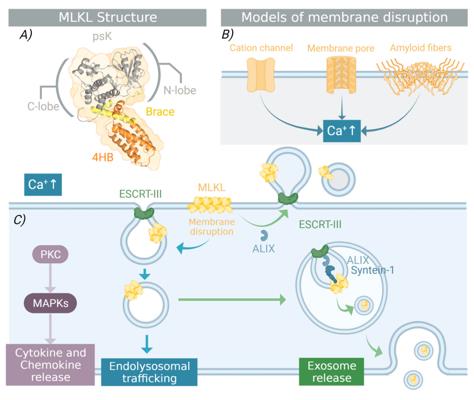 Fig. 1 Structure and function of MLKL.1
Key structural properties of MLKL:
- Junctional kinase domain
- Four-helix beam execution domain
- Hinge region connection domain
Functions of MLKL
The main function of the MLKL gene is to mediate programmed cell necrosis. However, it is also involved in the regulation of various other pathological and physiological processes in the body.
| Function | Description |
| Execution of Programmed Necrosis | After being phosphorylated by the upstream RIPK3 and oligomerized, MLKL translocates to the cell membrane, leading to increased membrane permeability and cell lysis, which is the final step of programmed necrosis. |
| Inflammation Regulation | By mediating controlled cell necrosis and releasing intracellular signaling molecules (such as DAMPs), it initiates and regulates the inflammatory immune response. |
| Anti-infection Defense | In certain scenarios of viral infection, cells activate MLKL-dependent programmed necrosis to limit viral replication and eliminate infected cells. |
| Disease Association | Its abnormal regulation is associated with a variety of diseases, including ischemia-reperfusion injury, neurodegenerative diseases, inflammatory diseases, and the development of certain tumors. |
| Cell fate determination | It acts in balance with other forms of cell death such as apoptosis and pyroptosis, jointly determining the fate of cells under stress conditions. |
Unlike the "silent" clearance of apoptosis, the programmed necrosis mediated by MLKL has obvious pro-inflammatory properties. Its activation process involves a strict multi-level regulation from self-inhibition to oligomerization, which determines its dual roles in physiological and pathological conditions.
Applications of MLKL and MLKL Antibody in Literature
1. Martinez-Osorio, Veronica, Yasmin Abdelwahab, and Uris Ros. "The many faces of MLKL, the executor of necroptosis." International Journal of Molecular Sciences 24.12 (2023): 10108. https://doi.org/10.3390/ijms241210108
The article indicates that the key mechanism by which MLKL mediates membrane rupture during necroptosis remains unclear. This paper reviews its activation pathways, structural functions and disease effects, and summarizes the research progress of related inhibitors.
2. Liu, Shuzhen, et al. "MLKL polymerization-induced lysosomal membrane permeabilization promotes necroptosis." Cell Death & Differentiation 31.1 (2024): 40-52. https://doi.org/10.1038/s41418-023-01237-7
The research found that MLKL forms polymers during necroptosis and transfers to the lysosomal membrane, causing the lysosomal membrane to become permeable, and promoting the release of cathepsin B into the cytoplasm, thereby executing cell death. This reveals a new mechanism by which MLKL polymerization induces lysosomal damage.
3. Geng, Lu, et al. "MLKL deficiency alleviates neuroinflammation and motor deficits in the α-synuclein transgenic mouse model of Parkinson's disease." Molecular Neurodegeneration 18.1 (2023): 94. https://doi.org/10.1186/s13024-023-00686-5
This study reveals that inhibiting or knocking out the key protein MLKL involved in necroptosis can significantly improve the motor symptoms of the Parkinson's disease model, reduce the deposition of α-synuclein and the loss of dopaminergic neurons, and inhibit neuroinflammation, indicating that MLKL is a potential therapeutic target for Parkinson's disease.
4. Zhan, Chaoning, et al. "MLKL: Functions beyond serving as the Executioner of Necroptosis." Theranostics 11.10 (2021): 4759. https://doi.org/10.7150/thno.54072
In addition to mediating necroptosis, the non-apoptotic functions of MLKL have gained increasing attention and are involved in the regulation of various diseases. This article reviews its structure, signaling pathways, and associations with other forms of cell death, with a focus on its new functions and inhibitor progress, providing a reference for clinical targeted therapy.
5. Liccardi, Gianmaria, and Alessandro Annibaldi. "MLKL post-translational modifications: road signs to infection, inflammation and unknown destinations." Cell Death & Differentiation 30.2 (2023): 269-278. https://doi.org/10.1038/s41418-022-01061-5
The article indicates that MLKL is the execution protein for necroptosis, and the activation mechanism of it remains unclear. Besides the known phosphorylation pathway of RIPK3, new post-translational modifications (such as ubiquitination) and the differences between human and mouse MLKL suggest that there are more complex regulatory mechanisms and potential non-apoptotic inflammatory functions of it.
Creative Biolabs: MLKL Antibodies for Research
Creative Biolabs specializes in the production of high-quality MLKL antibodies for research and industrial applications. Our portfolio includes monoclonal antibodies tailored for ELISA, Flow Cytometry, Western blot, immunohistochemistry, and other diagnostic methodologies.
- Custom MLKL Antibody Development: Tailor-made solutions to meet specific research requirements.
- Bulk Production: Large-scale antibody manufacturing for industry partners.
- Technical Support: Expert consultation for protocol optimization and troubleshooting.
- Aliquoting Services: Conveniently sized aliquots for long-term storage and consistent experimental outcomes.
For more details on our MLKL antibodies, custom preparations, or technical support, contact us at email.
Reference
- Martinez-Osorio, Veronica, Yasmin Abdelwahab, and Uris Ros. "The many faces of MLKL, the executor of necroptosis." International Journal of Molecular Sciences 24.12 (2023): 10108. Distributed under Open Access license CC BY 4.0, without modification. https://doi.org/10.3390/ijms241210108
Anti-MLKL antibodies
 Loading...


Hot products 
-
Mouse Anti-DHFR Recombinant Antibody (D0821) (CBMAB-D0821-YC)

-
Rabbit Anti-DLK1 Recombinant Antibody (9D8) (CBMAB-D1061-YC)

-
Rabbit Anti-B2M Recombinant Antibody (CBYY-0059) (CBMAB-0059-YY)

-
Rabbit Anti-BAD (Phospho-Ser136) Recombinant Antibody (CAP219) (CBMAB-AP536LY)

-
Mouse Anti-CCL18 Recombinant Antibody (64507) (CBMAB-C7910-LY)

-
Mouse Anti-ABCA3 Recombinant Antibody (V2-178911) (CBMAB-A0145-YC)

-
Mouse Anti-CD19 Recombinant Antibody (CBXC-1224) (CBMAB-C1491-CQ)

-
Mouse Anti-CD24 Recombinant Antibody (SN3) (CBMAB-C1037-CQ)

-
Mouse Anti-CD83 Recombinant Antibody (HB15) (CBMAB-C1765-CQ)

-
Mouse Anti-CRTAM Recombinant Antibody (CBFYC-2235) (CBMAB-C2305-FY)

-
Mouse Anti-CALR Recombinant Antibody (CBFYC-0763) (CBMAB-C0818-FY)

-
Mouse Anti-ADGRL2 Recombinant Antibody (V2-58519) (CBMAB-L0166-YJ)

-
Mouse Anti-ATG5 Recombinant Antibody (9H197) (CBMAB-A3945-YC)

-
Mouse Anti-CDK7 Recombinant Antibody (CBYY-C1783) (CBMAB-C3221-YY)

-
Mouse Anti-ATP1B3 Recombinant Antibody (1E9) (CBMAB-A4021-YC)

-
Mouse Anti-CD63 Recombinant Antibody (CBXC-1200) (CBMAB-C1467-CQ)

-
Mouse Anti-AFDN Recombinant Antibody (V2-58751) (CBMAB-L0408-YJ)

-
Human Anti-SARS-CoV-2 S1 Monoclonal Antibody (CBFYR-0120) (CBMAB-R0120-FY)

-
Rabbit Anti-Acetyl-Histone H4 (Lys16) Recombinant Antibody (V2-623415) (CBMAB-CP1021-LY)

-
Mouse Anti-FPR2 Recombinant Antibody (1D6) (CBMAB-F2628-CQ)

- AActivation
- AGAgonist
- APApoptosis
- BBlocking
- BABioassay
- BIBioimaging
- CImmunohistochemistry-Frozen Sections
- CIChromatin Immunoprecipitation
- CTCytotoxicity
- CSCostimulation
- DDepletion
- DBDot Blot
- EELISA
- ECELISA(Cap)
- EDELISA(Det)
- ESELISpot
- EMElectron Microscopy
- FFlow Cytometry
- FNFunction Assay
- GSGel Supershift
- IInhibition
- IAEnzyme Immunoassay
- ICImmunocytochemistry
- IDImmunodiffusion
- IEImmunoelectrophoresis
- IFImmunofluorescence
- IGImmunochromatography
- IHImmunohistochemistry
- IMImmunomicroscopy
- IOImmunoassay
- IPImmunoprecipitation
- ISIntracellular Staining for Flow Cytometry
- LALuminex Assay
- LFLateral Flow Immunoassay
- MMicroarray
- MCMass Cytometry/CyTOF
- MDMeDIP
- MSElectrophoretic Mobility Shift Assay
- NNeutralization
- PImmunohistologyp-Paraffin Sections
- PAPeptide Array
- PEPeptide ELISA
- PLProximity Ligation Assay
- RRadioimmunoassay
- SStimulation
- SESandwich ELISA
- SHIn situ hybridization
- TCTissue Culture
- WBWestern Blot
