MYF5 Antibodies
Background
The MYF5 gene encodes a basic helical-ring-helix (bHLH) transcription factor belonging to the myogenic regulatory factor family, which is mainly present in the skeletal muscle precursor cells of vertebrates. This gene dominates the formation and differentiation of skeletal muscle during embryonic development by regulating the expression of downstream target genes, and is crucial for the determination of muscle cell lineages and the early development of muscle tissue. Since its identification in the early 1990s, MYF5 has been widely studied due to its core role in muscle formation and has become one of the key models for researching the molecular mechanisms of muscle development. The research on the function and regulatory network of this gene has deepened our understanding of cell fate determination, tissue-specific differentiation, and the pathogenesis of related muscle diseases.
Structure of MYF5
The protein encoded by the MYF5 gene is a relatively small transcription factor with a molecular weight of approximately 25 kDa. Its molecular weight may fluctuate slightly among different species due to differences in specific amino acid sequences, but overall it belongs to a conserved bHLH domain protein.
| Species | Human | Mouse | Rat | Chicken | Zebrafish |
| Molecular Weight (kDa) | Approximately 25.0 | Approximately 24.8 | Approximately 25.1 | Approximately 24.6 | Approximately 24.2 |
| Primary Structural Differences | Contains the bHLH domain, which is a key regulator of myoblast differentiation | Highly conserved in function, it is often used in models of muscle development | The structure and function are highly similar to those of mice | Plays an early determining role in skeletal muscle pattern formation | Regulating the specialization of myoblasts in body segments shows evolutionary conservation |
This protein is composed of approximately 255 amino acids, and its functional core lies in a basic helical-ring-helix (bHLH) domain made up of 60 to 70 amino acids. This domain interacts with a linker ring region through two α -helices (one rich in basic amino acids for DNA recognition binding and the other involved in dimerization). This bHLH domain can form a heterodimer with E proteins (such as E12/E47), thereby specifically recognizing and binding to the E-box sequence (CANNTG) in the promoter region of downstream target genes, activating or inhibiting the expression of genes related to muscle differentiation. Therefore, although the MYF5 protein itself is small, its highly specialized structure makes it a key molecular switch that initiates the entire skeletal muscle generation cascade reaction.
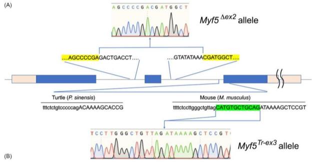 Fig. 1 Schematic representation of the Myf5 gene.1
Key structural properties of MYF5:
- Contains the conserved basic helix-loop-helix (bHLH) domain
- The bHLH domain, which consists of two α-helices and a connecting ring, is responsible for DNA recognition and dimerization
- Protein N-terminal region containing alkaline amino acid, specific binding target genes of DNA sequences
- The C-terminal region contains the transcriptional activation domain
Functions of MYF5
The main function of the MYF5 gene is to regulate the development and differentiation of skeletal muscle. In addition, it is also involved in a variety of cell biological processes, including the fate determination of myoblasts, the maintenance of muscle stem cells (satellite cells), and its significant role in muscle regeneration.
| Function | Description |
| Myogenic determination | In the early stage of embryonic development, the expression of MYF5 determines the differentiation of mesodermal cells into myoblast lineages and serves as a key switch to initiate the muscle-generating cascade reaction. |
| Regulation of muscle differentiation | As a transcription factor, MYF5 forms a heterodimer with the E protein and binds to the E-box sequence of downstream target genes (such as myoietin MyoD), activating the muscle-specific gene expression network. |
| Muscle pattern formation | During the development of body segments, the expression of MYF5 has precise spatiotemporal specificity and is responsible for regulating the localization and formation of different muscle groups (such as paraxial muscles and limb muscles). |
| Satellite cell function | In adult muscles, MYF5 is continuously expressed in both resting and activated satellite cells and plays a regulatory role in maintaining the proliferation and differentiation potential of muscle stem cells. |
| Muscle regeneration participation | After muscle injury, MYF5 is reactivated to promote the differentiation of satellite cells into myoblasts, thereby driving the repair and regeneration of damaged muscle fibers. |
The expression of MYF5 usually occurs earlier than that of other myogenic regulatory factors (such as MyoD), and its kinetics shows a pulse-like activation pattern, which is consistent with its "pioneer" function as an early determinant and setting the cellular state for subsequent differentiation programs. The functional deficiency of MYF5 can lead to severe developmental defects in specific muscle groups, especially the intercostal muscles and some axial muscles.
Applications of MYF5 and MYF5 Antibody in Literature
1. Dong, Liying, et al. "miR-9-5p promotes myogenic differentiation via the Dlx3/Myf5 axis." PeerJ 10 (2022): e13360. https://doi.org/10.7717/peerj.13360
Studies have found that miR-9-5p promotes myogenic differentiation by targeting and inhibiting the expression of Dlx3, thereby relieving its transcriptional inhibition of Myf5. This axis can serve as a potential target for treating muscle dysfunction.
2. Cai, Shufang, et al. "MLL1 promotes myogenesis by epigenetically regulating Myf5." Cell Proliferation 53.2 (2020): e12744. https://doi.org/10.1111/cpr.12744
Research has found that MLL1 epigenetically regulates Myf5 expression by mediating H3K4me3 modification in the Myf5 promoter region, thereby promoting the proliferation of myoblasts and PAx7-positive satellite cells and regulating the muscle regeneration process.
3. Tingler, Melanie, et al. "dmrt2 and myf5 link early somitogenesis to left-right Axis determination in Xenopus laevis." Frontiers in Cell and Developmental Biology 10 (2022): 858272.https://doi.org/10.3389/fcell.2022.858272
Research has found that in African clawing frogs, the transcription factor Dmrt2 regulates the expression of the myogenic factor Myf5, influencing somatogenesis and the fate of sensing cells in the LR tissue organ, thereby linking the establishment of left-right asymmetry of the body axis to the process of somatogenesis.
4. Wang, Shirong, et al. "Dnmt3b deficiency in Myf5+-brown fat precursor cells promotes obesity in female mice." Biomolecules 11.8 (2021): 1087. https://doi.org/10.3390/biom11081087
Research has found that conditional knockout of Dnmt3b using Myf5-Cre leads to myogenic transformation of brown fat in female mice, impaired thermogenesis and susceptibility to obesity, revealing the key role of DNA methylation in regulating the fate and energy metabolism of brown fat.
5. Li, Feng, et al. "Maternal nutrition altered embryonic MYOD1, MYF5, and MYF6 gene expression in genetically fat and lean lines of chickens." Animal bioscience 35.8 (2022): 1223. https://doi.org/10.5713/ab.21.0521
Research has found that the feed intake of hens during the laying period significantly affects the expression patterns of MyoD1, Myf5 and Myf6 genes in their offspring embryos, indicating that maternal nutritional restriction and the selection of genetic strains in chickens jointly regulate the process of embryonic muscle formation.
Creative Biolabs: MYF5 Antibodies for Research
Creative Biolabs specializes in the production of high-quality MYF5 antibodies for research and industrial applications. Our portfolio includes monoclonal antibodies tailored for ELISA, Flow Cytometry, Western blot, immunohistochemistry, and other diagnostic methodologies.
- Custom MYF5 Antibody Development: Tailor-made solutions to meet specific research requirements.
- Bulk Production: Large-scale antibody manufacturing for industry partners.
- Technical Support: Expert consultation for protocol optimization and troubleshooting.
- Aliquoting Services: Conveniently sized aliquots for long-term storage and consistent experimental outcomes.
For more details on our MYF5 antibodies, custom preparations, or technical support, contact us at email.
Reference
- Tekko, Triin, et al. "Assessing Myf5 and Lbx1 contribution to carapace development by reproducing their turtle‐specific signatures in mouse embryos." Developmental Dynamics 251.10 (2022): 1698-1710. https://doi.org/10.1002/dvdy.502
Anti-MYF5 antibodies
 Loading...


Hot products 
-
Mouse Anti-ACE2 Recombinant Antibody (V2-179293) (CBMAB-A0566-YC)

-
Mouse Anti-BLK Recombinant Antibody (CBYY-0618) (CBMAB-0621-YY)

-
Mouse Anti-FLT1 Recombinant Antibody (11) (CBMAB-V0154-LY)

-
Mouse Anti-ASB9 Recombinant Antibody (1D8) (CBMAB-A0529-LY)

-
Mouse Anti-G6PD Recombinant Antibody (13B331) (CBMAB-G1553-LY)

-
Mouse Anti-ALB Recombinant Antibody (V2-180650) (CBMAB-A2186-YC)

-
Human Anti-SARS-CoV-2 Spike Recombinant Antibody (CBC05) (CBMAB-CR005LY)

-
Mouse Anti-APP Recombinant Antibody (DE2B4) (CBMAB-1122-CN)

-
Mouse Anti-BIRC3 Recombinant Antibody (16E63) (CBMAB-C3367-LY)

-
Mouse Anti-FeLV g27 Recombinant Antibody (1) (CBMAB-V208-1714-FY)

-
Mouse Anti-FOXL1 Recombinant Antibody (CBXF-0845) (CBMAB-F0462-CQ)

-
Mouse Anti-ENPP1 Recombinant Antibody (CBFYE-0159) (CBMAB-E0375-FY)

-
Mouse Anti-ARID1B Recombinant Antibody (KMN1) (CBMAB-A3546-YC)

-
Mouse Anti-ATM Recombinant Antibody (2C1) (CBMAB-A3970-YC)

-
Mouse Anti-ACLY Recombinant Antibody (V2-179314) (CBMAB-A0610-YC)

-
Mouse Anti-AKT1 (Phosphorylated S473) Recombinant Antibody (V2-505430) (PTM-CBMAB-0067LY)

-
Mouse Anti-EMP3 Recombinant Antibody (CBFYE-0100) (CBMAB-E0207-FY)

-
Mouse Anti-AMIGO2 Recombinant Antibody (CBYY-C0756) (CBMAB-C2192-YY)

-
Mouse Anti-CIITA Recombinant Antibody (CBLC160-LY) (CBMAB-C10987-LY)

-
Mouse Anti-CECR2 Recombinant Antibody (CBWJC-2465) (CBMAB-C3533WJ)

- AActivation
- AGAgonist
- APApoptosis
- BBlocking
- BABioassay
- BIBioimaging
- CImmunohistochemistry-Frozen Sections
- CIChromatin Immunoprecipitation
- CTCytotoxicity
- CSCostimulation
- DDepletion
- DBDot Blot
- EELISA
- ECELISA(Cap)
- EDELISA(Det)
- ESELISpot
- EMElectron Microscopy
- FFlow Cytometry
- FNFunction Assay
- GSGel Supershift
- IInhibition
- IAEnzyme Immunoassay
- ICImmunocytochemistry
- IDImmunodiffusion
- IEImmunoelectrophoresis
- IFImmunofluorescence
- IGImmunochromatography
- IHImmunohistochemistry
- IMImmunomicroscopy
- IOImmunoassay
- IPImmunoprecipitation
- ISIntracellular Staining for Flow Cytometry
- LALuminex Assay
- LFLateral Flow Immunoassay
- MMicroarray
- MCMass Cytometry/CyTOF
- MDMeDIP
- MSElectrophoretic Mobility Shift Assay
- NNeutralization
- PImmunohistologyp-Paraffin Sections
- PAPeptide Array
- PEPeptide ELISA
- PLProximity Ligation Assay
- RRadioimmunoassay
- SStimulation
- SESandwich ELISA
- SHIn situ hybridization
- TCTissue Culture
- WBWestern Blot
