PBX1 Antibodies
Background
The PBX1 gene belongs to the TALE homeobox gene family and encodes a transcription factor that is crucial for embryonic development and organ formation. This protein recognizes specific DNA sequences through its highly conserved homology domain and mainly regulates cell proliferation, differentiation, and precursor cell fate determination. It plays a central role in the morphogenesis of the pancreas, kidneys, and central nervous system. Initially, this gene was discovered in pre-B cell leukemia and had a chromosomal translocation with the E2A gene, which gave it its name. Subsequent studies further revealed its pathogenic mutations in congenital malformation syndromes such as renal-ocular syndrome. As a key component of the homeobox protein complex, PBX1 can collaborate with HOX and other auxiliary factors. The structural-function analysis provides a classic model for understanding the spatiotemporal specificity regulation of the multi-protein-DNA interaction network and expands our understanding of developmental biology and disease molecular mechanisms.
Structure of PBX1
The PBX1 encoded protein has a molecular weight of approximately 46.7 kDa. The homologous domains among different species are highly conserved, but the full-length sequences vary.
| Species | Human | Mouse | Zebrafish | Fruit fly | African clawed frog |
| Molecular Weight (kDa) | 46.7 | 46.5 | 47.2 | 53.1 | 46.8 |
| Primary Structural Differences | Containing a typical tri-amino acid ring | Extremely similar to humans in origin | Two splicing variants | Containing a longer C-terminal region | High expression during the oocyte stage |
PBX1 contains over 430 amino acids and has a core TALE homologous domain. Its characteristic is that a tri-amino acid ring is inserted between the DNA binding helices. The upstream of this domain contains the PBC domain, which mediates heterodimerization with HOX proteins and members of the Prep/Meis family. PBX1 regulates downstream gene expression by binding to specific DNA sequences. The N-terminal self-inhibitory region of PBX1 changes conformation after binding to a cofactor, thereby activating transcription.
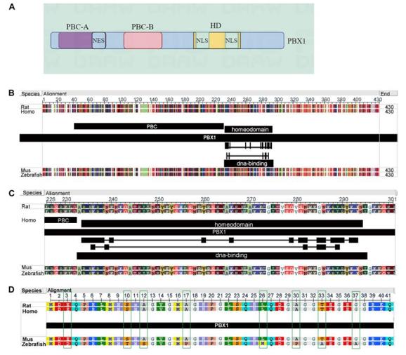 Fig. 1 Structure of PBX1and amino acid sequences in different species.1
Key structural properties of PBX1:
- TALE homologous domain, containing a three-amino-acid loop insertion
- PBC domain mediates heterodimerization with HOX proteins
- N-terminal autonomous inhibitory region, released upon binding of cofactors
- DNA binding region recognizes the core motif of 5’-TGAT-3’
Functions of PBX1
The core function of PBX1 is to act as a transcription factor, regulating embryonic development and organ formation. Additionally, it is involved in various pathological and physiological processes, including tumor occurrence and hematopoietic regulation.
| Function | Description |
| Development patterning | PBX1 works in synergy with HOX proteins to regulate the formation of the anterior-posterior body axis and the establishment of organ boundaries. |
| Transcriptional regulation | It binds to DNA and recruits co-activators or co-inhibitors to regulate the expression of downstream target genes. |
| Cell fate determination | It maintains the undifferentiated state of precursor cells or promotes their differentiation into specific lineages. |
| Tumor-related | In leukemia, an E2A-PBX1 fusion occurs, which abnormally activates proliferation-promoting genes. |
| Congenital malformation pathogenesis | Gene mutations or insufficient haploid dosage lead to multi-organ developmental defects such as renal-ocular syndrome. |
The DNA binding of PBX1 is sequence-specific and occurs in an heterodimeric form, recognizing the TGAT motif. The binding affinity with HOX proteins is significantly enhanced when they are co-bound.
Applications of PBX1 and PBX1 Antibody in Literature
1. Liu, Mingsheng, et al. "Comprehensive summary: the role of PBX1 in development and cancers." Frontiers in cell and developmental biology 12 (2024): 1442052. https://doi.org/10.3389/fcell.2024.1442052
The article indicates that PBX1 is a carcinogenic transcription factor, and its fusion protein E2A-PBX1 is associated with childhood leukemia. They drive the malignant progression by regulating tumor growth, metastasis and drug resistance, making them important potential therapeutic targets. They can also serve as prenatal screening markers to reduce the risk of childhood hematological malignancies.
2. Wang, Xinyue, et al. "Rescue RM/CS-AKI by blocking strategy with one-dose anti-myoglobin RabMAb." Nature Communications 16.1 (2025): 1044. https://doi.org/10.1016/j.bbcan.2024.189085
The article indicates that PBX1 is a key transcription factor that regulates cell fate, and it mediates multiple malignant processes such as proliferation, metastasis, and immune evasion in cancer. Its expression is influenced by a multi-level regulatory network and has the potential to serve as a prognostic marker and therapeutic target, especially providing new intervention strategies for high-risk and drug-resistant tumors.
3. Crisafulli, Laura, et al. "PBX1: a TALE of two seasons—key roles during development and in cancer." Frontiers in Cell and Developmental Biology 12 (2024): 1372873. https://doi.org/10.3389/fcell.2024.1372873
The article indicates that PBX1 is a transcription factor that plays a crucial role in embryonic development, and its abnormal expression is associated with various cancers. It can act as a cancer gene to promote the progression of leukemia, breast cancer, etc., but in colorectal cancer, it exhibits a tumor suppressor function, demonstrating the complexity of its role.
4. Grebbin, Britta M., and Dorothea Schulte. "PBX1 as pioneer factor: a case still open." Frontiers in cell and developmental biology 5 (2017): 9. https://doi.org/10.3389/fcell.2017.00009
The article indicates that PBX1, as a pioneer transcription factor, can recognize and open the closed chromatin structure, thereby initiating cell fate transformation and gene expression. The mechanism of its action is currently an important frontier and hotspot in research.
5. Chen, Hao, et al. "Research progress of PBX1 in developmental and regenerative medicine." International Journal of Medical Sciences 20.2 (2023): 225. https://doi.org/10.7150/ijms.80262
The article indicates that PBX1 is a structurally conserved transcription factor that regulates embryonic development and various pathological and physiological processes. Studying its mechanism of action in regenerative medicine will provide new targets for maintaining cellular homeostasis and disease treatment.
Creative Biolabs: PBX1 Antibodies for Research
Creative Biolabs specializes in the production of high-quality PBX1 antibodies for research and industrial applications. Our portfolio includes monoclonal and polyclonal antibodies tailored for ELISA, Flow Cytometry, Western blot, immunohistochemistry, and other diagnostic methodologies.
- Custom PBX1 Antibody Development: Tailor-made solutions to meet specific research requirements.
- Bulk Production: Large-scale antibody manufacturing for industry partners.
- Technical Support: Expert consultation for protocol optimization and troubleshooting.
- Aliquoting Services: Conveniently sized aliquots for long-term storage and consistent experimental outcomes.
For more details on our PBX1 antibodies, custom preparations, or technical support, contact us at info@creative-biolabs.com.
Reference
- Liu, Mingsheng, et al. "Comprehensive summary: the role of PBX1 in development and cancers." Frontiers in cell and developmental biology 12 (2024): 1442052. Distributed under Open Access license CC BY 4.0, without modification. https://doi.org/10.3389/fcell.2024.1442052
Anti-PBX1 antibodies
 Loading...


Hot products 
-
Mouse Anti-AQP2 Recombinant Antibody (E-2) (CBMAB-A3358-YC)

-
Mouse Anti-ASB9 Recombinant Antibody (1D8) (CBMAB-A0529-LY)

-
Mouse Anti-CDK7 Recombinant Antibody (CBYY-C1783) (CBMAB-C3221-YY)

-
Mouse Anti-HTLV-1 gp46 Recombinant Antibody (CBMW-H1006) (CBMAB-V208-1154-FY)

-
Mouse Anti-CA9 Recombinant Antibody (CBXC-2079) (CBMAB-C0131-CQ)

-
Mouse Anti-ESR1 Recombinant Antibody (Y31) (CBMAB-1208-YC)

-
Rat Anti-4-1BB Recombinant Antibody (V2-1558) (CBMAB-0953-LY)

-
Mouse Anti-BBS2 Recombinant Antibody (CBYY-0253) (CBMAB-0254-YY)

-
Rabbit Anti-ABL1 (Phosphorylated Y185) Recombinant Antibody (V2-443434) (PTM-CBMAB-0001YC)

-
Mouse Anti-EPO Recombinant Antibody (CBFYR0196) (CBMAB-R0196-FY)

-
Mouse Anti-ACVR1C Recombinant Antibody (V2-179685) (CBMAB-A1041-YC)

-
Rabbit Anti-ATF4 Recombinant Antibody (D4B8) (CBMAB-A3872-YC)

-
Rat Anti-CD63 Recombinant Antibody (7G4.2E8) (CBMAB-C8725-LY)

-
Mouse Anti-AFDN Recombinant Antibody (V2-58751) (CBMAB-L0408-YJ)

-
Mouse Anti-AAV-5 Recombinant Antibody (V2-503416) (CBMAB-V208-1402-FY)

-
Mouse Anti-APOH Recombinant Antibody (4D9A4) (CBMAB-A3249-YC)

-
Mouse Anti-AQP2 Recombinant Antibody (G-3) (CBMAB-A3359-YC)

-
Mouse Anti-CASP8 Recombinant Antibody (CBYY-C0987) (CBMAB-C2424-YY)

-
Mouse Anti-BHMT Recombinant Antibody (CBYY-0547) (CBMAB-0550-YY)

-
Mouse Anti-CD59 Recombinant Antibody (CBXC-2097) (CBMAB-C4421-CQ)

- AActivation
- AGAgonist
- APApoptosis
- BBlocking
- BABioassay
- BIBioimaging
- CImmunohistochemistry-Frozen Sections
- CIChromatin Immunoprecipitation
- CTCytotoxicity
- CSCostimulation
- DDepletion
- DBDot Blot
- EELISA
- ECELISA(Cap)
- EDELISA(Det)
- ESELISpot
- EMElectron Microscopy
- FFlow Cytometry
- FNFunction Assay
- GSGel Supershift
- IInhibition
- IAEnzyme Immunoassay
- ICImmunocytochemistry
- IDImmunodiffusion
- IEImmunoelectrophoresis
- IFImmunofluorescence
- IGImmunochromatography
- IHImmunohistochemistry
- IMImmunomicroscopy
- IOImmunoassay
- IPImmunoprecipitation
- ISIntracellular Staining for Flow Cytometry
- LALuminex Assay
- LFLateral Flow Immunoassay
- MMicroarray
- MCMass Cytometry/CyTOF
- MDMeDIP
- MSElectrophoretic Mobility Shift Assay
- NNeutralization
- PImmunohistologyp-Paraffin Sections
- PAPeptide Array
- PEPeptide ELISA
- PLProximity Ligation Assay
- RRadioimmunoassay
- SStimulation
- SESandwich ELISA
- SHIn situ hybridization
- TCTissue Culture
- WBWestern Blot
