SARM1 Antibodies
Background
SARM1 is a protein located within cells and is mainly expressed in the axons of neurons. This protein exists in a self-inhibitory form under normal conditions. When nerve damage or metabolic stress leads to a decrease in NAD+ levels, its inherent NAD+ hydrolase activity will be activated, thereby triggering the axonal degeneration program. Due to its core role in mediating mitochondrial dysfunction and axonal degeneration, SARM1 has become a key target in the research of neurodegenerative diseases. In 2017, multiple studies jointly clarified its molecular mechanism as an NAD+ consuming enzyme. This discovery has promoted the development of neuroprotective strategies for diseases such as Alzheimer's disease and Parkinson's disease. Its functional positioning as a programmed axonal executor provides an important molecular basis for understanding the injury response and self-defense mechanisms of the nervous system.
Structure of SARM1
SARM1 is a protein composed of approximately 900 amino acids with a molecular weight of about 100 kDa. As the core executor of axonal mutability, its structure consists of an N-terminal self-suppression domain, a TIR domain, and a unique SAM domain. The SARM1 sequences among different species are highly conserved, especially within the catalytic TIR domain.
| Species | Human | Mouse | Rat | Zebrafish |
| Molecular Weight (kDa) | About 100 | About 100 | About 100 | About 105 |
| Primary Structural Differences | Typical mammalian SARM1 structure, containing the complete autoinhibitory module | Highly homologous to humans, it is the main animal model | Structure similar to mice | Retain the core TIR domain for neural regeneration research |
The core functional domain of this protein is the TIR domain capable of hydrolyzing NAD+. Under normal circumstances, its N-terminal and SAM domains will form self-inhibition, preventing the activity of TIR. When axon injury or energy stress leads to a decrease in NAD+ levels, this inhibitory effect is lifted, and the activated TIR domain will rapidly consume NAD+ within the cell, thereby actively triggering programmed axonal degeneration. This mechanism plays a key role in various neurodegenerative diseases.
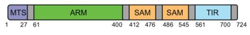 Fig. 1 Schematic representation of protein domains and motifs found in SARM1.1
Key structural properties of SARM1:
- Multi-domain self-suppression structure: It includes ARM, SAM and TIR domains
- NAD+ hydrolysis active center: Located within the TIR domain
- Allosteric regulation mechanism: remove axonal injury signal suppression, activate the enzyme activity
Functions of SARM1
The core function of the SARM1 gene is to perform programmed axonal degeneration, which is an active clearance mechanism after nerve injury. At the same time, it is also involved in the immune regulation and metabolic stress response of the nervous system.
| Function | Description |
| Axonal degeneration is performed in the core | When nerve injury or lesion occurs, its TIR domain is activated, actively triggering the self-degradation of axons by rapidly consuming NAD+ within the axons. |
| Neuroimmune regulation | Involved in regulating nerve inflammation, its activation associated molecular patterns of damage can be released, affect the function of microglial cells and other immune cells. |
| Metabolic stress sensor | As a key sensor of neuronal energy status (especially NAD+ levels), it initiates protective or destructive pathways when metabolic imbalance occurs. |
| Common pathways of neurodegenerative diseases | In various disease models such as Alzheimer's disease, Parkinson's disease, and amyotrophic lateral sclerosis, its activation has been confirmed to be a key downstream event of axonal injury. |
| Potential neuroprotective targets | Inhibiting its NAD+ hydrolytic activity has been proven to effectively delay or prevent axonal degeneration in various animal models and has therapeutic potential. |
The activation of SARM1 has a "all or none" switching characteristic, and its NAD+ hydrolysis activity shows sharp positive feedback above the threshold, which contrasts sharply with the gentle oxygen-binding curve of myoglobin, reflecting its role as a decisive executive molecule in injury response.
Applications of SARM1 and SARM1 Antibody in Literature
- Liu, Huitao, et al. "SARM1 promotes neuroinflammation and inhibits neural regeneration after spinal cord injury through NF-κB signaling." Theranostics 11.9 (2021): 4187. https://doi.org/10.7150/thno.49054
The article indicates that after spinal cord injury, SARM1 is upregulated in neurons and astrocytes, mediating axonal degeneration and neuroinflammation. Inhibiting the NF-κB pathway and upregulating HSP70 can promote nerve regeneration and functional recovery. Targeting SARM1 is a potential therapeutic strategy.
- Miao, Xuemeng, et al. "SARM1 promotes neurodegeneration and memory impairment in mouse models of Alzheimer's disease." Aging and disease 15.1 (2024): 390. https://doi.org/10.14336/AD.2023.0516-1
The article indicates that in Alzheimer's disease, conditional knockout of SARM1 in the central nervous system can delay cognitive decline, reduce beta-amyloid protein deposition and inflammatory infiltration, and the mechanism may be related to the down-regulation of the TNF-α signaling pathway.
- Oliveira, Samuel dos Santos, et al. "SARM1: a key multifaceted component in immunoregulation, inflammation and neurodegeneration." Frontiers in Immunology 16 (2025): 1521364. https://doi.org/10.3389/fimmu.2025.1521364
The article indicates that SARM1 plays a unique role in the TLR pathway. It inhibits inflammatory signals, mediates mitochondrial damage and cell death by degrading NAD+ to regulate the immune system and prevent overreactions, making it a potential target for treating neurodegenerative and inflammatory diseases.
- Wang, Lina, et al. "SARM1 senses dsDNA to promote NAD+ degradation and cell death." Cell (2025). https://doi.org/10.1016/j.cell.2025.09.026
The article indicates that the DNA perception mechanism remains unclear. SARM1 is a key axonic executive protein with NADase activity. Its unique octamer cyclic activated state structure suggests that linear molecules such as double-stranded DNA may directly activate SARM1, providing a new perspective for understanding the related disease mechanisms.
- McGuinness, Helen Y., et al. "SARM1-Dependent axon degeneration: nucleotide signaling, neurodegenerative disorders, toxicity, and therapeutic opportunities." The Neuroscientist 30.4 (2024): 473-492. https://doi.org/10.1177/10738584231162508
The article indicates that SARM1 is a key NAD+ hydrolase mediating axonal degeneration, and its activation is regulated by the NMN/NAD+ ratio, playing a core role in various neurodegenerative diseases. A thorough understanding of its structure and regulatory mechanism provides a new target for the development of axonal protection therapy.
Creative Biolabs: SARM1 Antibodies for Research
Creative Biolabs specializes in the production of high-quality SARM1 antibodies for research and industrial applications. Our portfolio includes monoclonal antibodies tailored for ELISA, Flow Cytometry, Western blot, immunohistochemistry, and other diagnostic methodologies.
- Custom SARM1 Antibody Development: Tailor-made solutions to meet specific research requirements.
- Bulk Production: Large-scale antibody manufacturing for industry partners.
- Technical Support: Expert consultation for protocol optimization and troubleshooting.
- Aliquoting Services: Conveniently sized aliquots for long-term storage and consistent experimental outcomes.
For more details on our SARM1 antibodies, custom preparations, or technical support, contact us at email.
Reference
- McGuinness, Helen Y., et al. "SARM1-Dependent axon degeneration: nucleotide signaling, neurodegenerative disorders, toxicity, and therapeutic opportunities." The Neuroscientist 30.4 (2024): 473-492. https://doi.org/10.1177/10738584231162508
Anti-SARM1 antibodies
 Loading...


Hot products 
-
Mouse Anti-14-3-3 Pan Recombinant Antibody (V2-9272) (CBMAB-1181-LY)

-
Mouse Anti-AKR1C3 Recombinant Antibody (V2-12560) (CBMAB-1050-CN)

-
Rat Anti-4-1BB Recombinant Antibody (V2-1558) (CBMAB-0953-LY)

-
Mouse Anti-ABL2 Recombinant Antibody (V2-179121) (CBMAB-A0364-YC)

-
Mouse Anti-BMI1 Recombinant Antibody (CBYC-P026) (CBMAB-P0108-YC)

-
Mouse Anti-DLL4 Recombinant Antibody (D1090) (CBMAB-D1090-YC)

-
Mouse Anti-dsDNA Recombinant Antibody (22) (CBMAB-AP1954LY)

-
Mouse Anti-ALPL Antibody (B4-78) (CBMAB-1009CQ)

-
Mouse Anti-ACTG1 Recombinant Antibody (V2-179597) (CBMAB-A0916-YC)

-
Mouse Anti-BIRC7 Recombinant Antibody (88C570) (CBMAB-L0261-YJ)

-
Mouse Anti-ENO2 Recombinant Antibody (H14) (CBMAB-E1341-FY)

-
Mouse Anti-dsRNA Recombinant Antibody (2) (CBMAB-D1807-YC)

-
Mouse Anti-ABIN2 Recombinant Antibody (V2-179106) (CBMAB-A0349-YC)

-
Mouse Anti-EMP3 Recombinant Antibody (CBFYE-0100) (CBMAB-E0207-FY)

-
Mouse Anti-FAS2 Monoclonal Antibody (1D4) (CBMAB-0071-CN)

-
Rat Anti-CD63 Recombinant Antibody (7G4.2E8) (CBMAB-C8725-LY)

-
Mouse Anti-FLT1 Recombinant Antibody (11) (CBMAB-V0154-LY)

-
Mouse Anti-BLK Recombinant Antibody (CBYY-0618) (CBMAB-0621-YY)

-
Mouse Anti-CFL1 (Phospho-Ser3) Recombinant Antibody (CBFYC-1770) (CBMAB-C1832-FY)

-
Mouse Anti-BAX Recombinant Antibody (CBYY-0216) (CBMAB-0217-YY)

- AActivation
- AGAgonist
- APApoptosis
- BBlocking
- BABioassay
- BIBioimaging
- CImmunohistochemistry-Frozen Sections
- CIChromatin Immunoprecipitation
- CTCytotoxicity
- CSCostimulation
- DDepletion
- DBDot Blot
- EELISA
- ECELISA(Cap)
- EDELISA(Det)
- ESELISpot
- EMElectron Microscopy
- FFlow Cytometry
- FNFunction Assay
- GSGel Supershift
- IInhibition
- IAEnzyme Immunoassay
- ICImmunocytochemistry
- IDImmunodiffusion
- IEImmunoelectrophoresis
- IFImmunofluorescence
- IGImmunochromatography
- IHImmunohistochemistry
- IMImmunomicroscopy
- IOImmunoassay
- IPImmunoprecipitation
- ISIntracellular Staining for Flow Cytometry
- LALuminex Assay
- LFLateral Flow Immunoassay
- MMicroarray
- MCMass Cytometry/CyTOF
- MDMeDIP
- MSElectrophoretic Mobility Shift Assay
- NNeutralization
- PImmunohistologyp-Paraffin Sections
- PAPeptide Array
- PEPeptide ELISA
- PLProximity Ligation Assay
- RRadioimmunoassay
- SStimulation
- SESandwich ELISA
- SHIn situ hybridization
- TCTissue Culture
- WBWestern Blot
