SIAE Antibodies
Background
SIAE encodes a protein called "sialic acid acetylesterase", which is mainly distributed in the human immune system and digestive system. This enzyme participates in regulating the glycosylation modification process on the cell surface by catalyzing the hydrolysis of acetyl groups from N-acetylneuraminic acid, and plays a key role in maintaining immune system homeostasis and preventing autoimmune responses. In 2001, the Japanese scientist team led by Yamaguchi first identified that mutations in this gene are closely related to the occurrence of various autoimmune diseases, especially to the pathogenesis of rheumatoid arthritis and systemic lupus erythematosus. Its unique enzyme active site structure and precise substrate recognition mechanism provide important targets for the development of new immunomodulatory drugs, promoting research progress in the fields of glycobiology and immunotherapy.
Structure of SIAE
Myoglobin is a relatively small protein with a molecular weight of approximately 16.7 kDa. This weight may slightly vary between species due to minor differences in amino acid sequence.
| Species | Human | Mouse | Ganges Monkey | Rabbit |
| Molecular Weight (kDa) | 48.2 | 47.8 | 49.1 | 48.5 |
| Primary Structural Differences | Contains specific glycosylation sites | Highly conserved structure of catalytic domain | The homology with human was 93% | There is variation in the substrate binding region |
SIAE is composed of 421 amino acids and has a typical α/β hydrolase folding structure. Its active center contains a catalytic triplet composed of Ser146, Asp248 and His278, which is responsible for hydrolyzing the 9-O-acetyl group of sialic acid. This enzyme functions through two key domains: the transmembrane domain at the N-terminal (amino acids at positions 1-28) and the catalytic domain at the C-terminal (amino acids at positions 29-421). Its tertiary structure contains three pairs of conserved disulfide bonds (Cys54-Cys67, Cys123-Cys139, Cys201-Cys215), which are crucial for maintaining enzyme activity. The unique substrate-binding pocket of SIAE is long and narrow and hydrophobic, which can specifically recognize acetylated sialic acid. This precise molecular recognition mechanism enables it to play a key role in immune regulation.
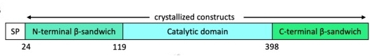 Fig. 1 Domain organization of SIAE. 1
Key structural properties of SIAE:
- Typical α/β hydrolase folding structure
- Composed of Ser - Asp - His triplets catalytic center
- Highly conservative substrate bonding pocket
- Three pairs of disulfide bonds maintain structural stability
- N-terminal transmembrane domain
Functions of SIAE
The main function of the SIAE gene is to regulate glycosylation modifications in the immune system, but it is also involved in a variety of physiological and pathological processes, including autoimmune regulation and control of inflammatory responses.
| Function | Description |
| Sialic acid deacetylation | Hydrolyze the 9-O-acetyl group of sialic acid, regulate the sugar chain structure on the cell surface, and affect the recognition and signal transduction of immune cells. |
| Maintenance of B-cell tolerance | By modifying B-cell receptor (BCR) -related sugar chains, excessive activation of autoimmune responses can be prevented. |
| Inflammatory regulation | Toll-like receptor (TLR) signaling pathway, adjust intensity of innate immune response. |
| Association with autoimmune diseases | Defects in SIAE function are closely related to autoimmune diseases such as rheumatoid arthritis and systemic lupus erythematosus. |
| Regulation of the tumor microenvironment | By altering the glycosylation pattern on the surface of tumor cells, it affects the potential for immune escape and metastasis. |
The enzymatic activity of SIAE shows a strict PH-dependent (optimal pH 6.5-7.5), and its substrate-specific curve exhibits typical Mie kinetic characteristics, indicating its high selectivity for 9-O-acetylated sialic acid. Unlike other sialic acid-modifying enzymes, SIAE plays an irreplaceable role in maintaining immune homeostasis by precisely regulating the hydrolysis of specific acetyl groups.
Applications of SIAE and SIAE Antibody in Literature
1. Sevdali, Eirini, et al. "SIAE rare variants in juvenile idiopathic arthritis and primary antibody deficiencies." Journal of Immunology Research 2017.1 (2017): 1514294. https://doi.org/10.1155/2017/1514294
Research has found that rare variations in the SIAE gene (such as p.Q343P, p.Y495X, etc.) are only present in patients with autoimmune juvenile idiopathic arthritis (aJIA) and their healthy relatives, but not in the population with primary antibody deficiency (PAD). The results indicated that a single SIAE deficiency was not a direct pathogenic factor of autoimmunity.
2. Chellappa, Vasant, et al. "M89V Sialic acid Acetyl Esterase (SIAE) and all other non-synonymous common variants of this gene are catalytically normal." PLoS One 8.1 (2013): e53453. https://doi.org/10.1371/journal.pone.0053453
Research has found that SIAE-catalytically deficient rare variations (uncommon variations) are associated with autoimmunity, but normal lymphocytes do not express SIAE. Reanalysis confirmed that such variations are enriched in patients with autoimmune diseases, suggesting that they may increase disease susceptibility. Further research is needed.
3. Kumari, Romika, et al. "Prognostic significance of esterase gene expression in multiple myeloma." British Journal of Cancer 124.8 (2021): 1428-1436. https://doi.org/10.1038/s41416-020-01237-1
Research has found that in multiple myeloma (MM), abnormal expression of esterase genes such as SIAE is associated with disease progression. High expression of SIAE is an adverse prognostic marker for new-onset and relapsed/refractory multiple myeloma, suggesting that it may be involved in the biological process of multiple myeloma.
4. Zhang, Kun, et al. "Identifying diagnostic markers and constructing a prognostic model for small-cell lung cancer based on blood exosome-related genes and machine-learning methods." Frontiers in Oncology 12 (2022): 1077118. https://doi.org/10.3389/fonc.2022.1077118
This study screened out 13 diagnostic markers for small cell lung cancer (SCLC) through exosome RNA analysis and constructed a prognostic model including 7 genes such as SIAE. This model can effectively distinguish between high-risk and low-risk patients. The AUC values in 1/3/5 years reach 0.820-0.989, providing a new strategy for the diagnosis and treatment of SCLC.
5. Zhang, Hui, et al. "A three-genes signature predicting colorectal cancer relapse reveals Lemd1 promoting crc cells migration by Rhoa/Rock1 signaling pathway." Frontiers in Oncology 12 (2022): 823696. https://doi.org/10.3389/fonc.2022.823696
This study constructed a colorectal cancer recurrence prediction model including LEMD1, SERPINE1 and SIAE through multi-database analysis. Experimental verification has found that LEMD1 promotes the migration and invasion of CRC cells through the RhoA/ROCK1 pathway, indicating that genes such as SIAE may serve as potential markers and therapeutic targets for postoperative recurrence.
Creative Biolabs: SIAE Antibodies for Research
Creative Biolabs specializes in the production of high-quality SIAE antibodies for research and industrial applications. Our portfolio includes monoclonal antibodies tailored for ELISA, Flow Cytometry, Western blot, immunohistochemistry, and other diagnostic methodologies.
- Custom SIAE Antibody Development: Tailor-made solutions to meet specific research requirements.
- Bulk Production: Large-scale antibody manufacturing for industry partners.
- Technical Support: Expert consultation for protocol optimization and troubleshooting.
- Aliquoting Services: Conveniently sized aliquots for long-term storage and consistent experimental outcomes.
For more details on our SIAE antibodies, custom preparations, or technical support, contact us at email.
Reference
- Ide, Danilo, et al. "Structural analysis of mammalian sialic acid esterase." Journal of Molecular Biology 436.22 (2024): 168801. https://doi.org/10.1016/j.jmb.2024.168801
Anti-SIAE antibodies
 Loading...


Hot products 
-
Mouse Anti-CD33 Recombinant Antibody (P67.6) (CBMAB-C10189-LY)

-
Mouse Anti-BIRC3 Recombinant Antibody (16E63) (CBMAB-C3367-LY)

-
Mouse Anti-ANXA7 Recombinant Antibody (A-1) (CBMAB-A2941-YC)

-
Mouse Anti-ALB Recombinant Antibody (V2-55272) (CBMAB-H0819-FY)

-
Human Anti-SARS-CoV-2 S1 Monoclonal Antibody (CBFYR-0120) (CBMAB-R0120-FY)

-
Rat Anti-AChR Recombinant Antibody (V2-12500) (CBMAB-0990-CN)

-
Mouse Anti-CDK7 Recombinant Antibody (CBYY-C1783) (CBMAB-C3221-YY)

-
Mouse Anti-FOXA3 Recombinant Antibody (2A9) (CBMAB-0377-YC)

-
Rat Anti-ADGRE4 Recombinant Antibody (V2-160163) (CBMAB-F0011-CQ)

-
Mouse Anti-BPGM Recombinant Antibody (CBYY-1806) (CBMAB-2155-YY)

-
Mouse Anti-CTNND1 Recombinant Antibody (CBFYC-2414) (CBMAB-C2487-FY)

-
Mouse Anti-GLP1R Recombinant Antibody (4F3) (CBMAB-G0521-LY)

-
Mouse Anti-ATP1A2 Recombinant Antibody (M7-PB-E9) (CBMAB-A4013-YC)

-
Mouse Anti-BIRC3 Recombinant Antibody (315304) (CBMAB-1214-CN)

-
Mouse Anti-FN1 Monoclonal Antibody (D6) (CBMAB-1240CQ)

-
Mouse Anti-FYN Recombinant Antibody (10) (CBMAB-S6332-CQ)

-
Mouse Anti-AMH Recombinant Antibody (5/6) (CBMAB-A2527-YC)

-
Mouse Anti-DLC1 Recombinant Antibody (D1009) (CBMAB-D1009-YC)

-
Mouse Anti-ELAVL4 Recombinant Antibody (6B9) (CBMAB-1132-YC)

-
Mouse Anti-BANF1 Recombinant Antibody (3F10-4G12) (CBMAB-A0707-LY)

- AActivation
- AGAgonist
- APApoptosis
- BBlocking
- BABioassay
- BIBioimaging
- CImmunohistochemistry-Frozen Sections
- CIChromatin Immunoprecipitation
- CTCytotoxicity
- CSCostimulation
- DDepletion
- DBDot Blot
- EELISA
- ECELISA(Cap)
- EDELISA(Det)
- ESELISpot
- EMElectron Microscopy
- FFlow Cytometry
- FNFunction Assay
- GSGel Supershift
- IInhibition
- IAEnzyme Immunoassay
- ICImmunocytochemistry
- IDImmunodiffusion
- IEImmunoelectrophoresis
- IFImmunofluorescence
- IGImmunochromatography
- IHImmunohistochemistry
- IMImmunomicroscopy
- IOImmunoassay
- IPImmunoprecipitation
- ISIntracellular Staining for Flow Cytometry
- LALuminex Assay
- LFLateral Flow Immunoassay
- MMicroarray
- MCMass Cytometry/CyTOF
- MDMeDIP
- MSElectrophoretic Mobility Shift Assay
- NNeutralization
- PImmunohistologyp-Paraffin Sections
- PAPeptide Array
- PEPeptide ELISA
- PLProximity Ligation Assay
- RRadioimmunoassay
- SStimulation
- SESandwich ELISA
- SHIn situ hybridization
- TCTissue Culture
- WBWestern Blot
