STMN2 Antibodies
Background
The STMN2 gene encodes a phosphoprotein called stathmin-2 or SCG10-like protein, which is mainly highly expressed in neurons. This protein plays a crucial role in regulating the dynamic balance of microtubules in neurons by binding to microtubule dimer and promoting microtubule depolymerization. It is essential for axon growth, regeneration, and intracellular transport. Studies have shown that STMN2 is highly active during the development of the nervous system, and changes in its expression level are closely related to neural plasticity and injury repair. It is notable that STMN2 is one of the first genes discovered to be significantly upregulated after nerve injury. This characteristic makes it an important molecular marker for studying the mechanisms of neural regeneration. In neurodegenerative diseases such as amyotrophic lateral sclerosis, abnormal regulation of STMN2 is believed to be related to axonal degeneration and motor neuron dysfunction, providing a new perspective for understanding the pathogenesis of such diseases and developing therapeutic targets.
Structure of STMN2
The protein encoded by the STMN2 gene has a molecular weight of approximately 20 kDa, and there are slight differences among different species. This protein is highly conserved in the nervous system, and its structural and functional characteristics show certain variations among different species.
| Species | Human | Mouse | Rat | Chicken | Zebrafish |
| Molecular Weight (kDa) | 20.2 | 20.1 | 20.1 | 19.8 | 19.5 |
| Primary Structural Differences | Contains typical SLD domain | High homology with humans | Highly similar to mice | Strong sequence conservation | Early vertebrate model |
The STMN2 protein is composed of approximately 179 amino acids and its three-dimensional structure presents a typical globular conformation consisting of α-helix and β-sheet combinations. The N-terminal of this protein contains a highly conserved stathmin domain, which is the core functional region for regulating the dynamic instability of microtubules. The protein sequence is dotted with multiple phosphorylation sites, including serine residues Ser16 and Ser63, which can be recognized and modified by various protein kinases. Through changes in the phosphorylation state, STMN2 can precisely regulate its binding activity to microtubule protein dimers. In the C-terminal region, the α-helix structure forms a specific hydrophobic interface, responsible for the interaction with microtubule proteins. The entire protein molecule achieves fine regulation of its biological functions through conformational changes.
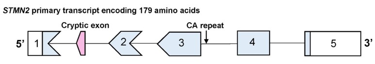 Fig. 1 Schematic of the STMN2 primary transcript and the location of the CA repeat.1
Key structural properties of STMN2:
- A highly conserved stathmin domain
- The coiled-coil region mediates protein interactions
- Multiple phosphorylation sites regulate microtubule binding activity
- The Ser16 and Ser63 sites respond to kinase signals and regulate functions
Functions of STMN2
The main function of STMN2 is to regulate the dynamic balance of microtubules in neurons and axon growth. In addition, it is involved in various physiological processes such as nervous system development and injury repair.
| Function | Description |
| Microtubule dynamic regulation | The STMN2 protein binds to free tubulin dimers and promotes tubulin depolymerization, maintaining the dynamic balance of the cytoskeleton. |
| Axonal growth support | Regulates axonal extension and branching formation during neuronal differentiation, playing a crucial role in the construction of neural circuits. |
| Neural injury response | Expresses at an elevated level after nerve damage, participating in the regulation of axonal regeneration and repair processes. |
| Intracellular transport regulation | Indirectly regulates axoplasmic transport of vesicles and organelles by influencing microtubule stability. |
| Neurodegenerative association | Abnormal expression is closely related to axonal degeneration processes in diseases such as ALS. |
The expression regulation of STMN2 in neurons shows dynamic characteristics, corresponding to the periodic fluctuations of microtubule polymerization. Its activity is mainly regulated rapidly by phosphorylation modifications rather than through allosteric effects.
Applications of STMN2 and STMN2 Antibody in Literature
1. Theunissen, Frances, et al. "Novel STMN2 variant linked to amyotrophic lateral sclerosis risk and clinical phenotype." Frontiers in Aging Neuroscience 13 (2021): 658226. https://doi.org/10.3389/fnagi.2021.658226
The study found that the CA repeat polymorphism of the STMN2 gene is associated with sporadic ALS: the long CA genotype significantly increases the risk of disease onset, and carriers have an earlier age of onset and shorter survival periods for patients with bulbar onset. Moreover, the expression of STMN2 is reduced, which can serve as a potential disease marker.
2. Wang, Xinyue, et al. "Rescue RM/CS-AKI by blocking strategy with one-dose anti-myoglobin RabMAb." Nature Communications 16.1 (2025): 1044. https://doi.org/10.1186/s40478-025-01977-2
The study found that the poly-PR protein produced by the C9ORF72 repeat amplification disrupted the RNA-binding protein SRSF7, leading to a reduction in STMN2 and subsequently inhibiting axonal regeneration. This reveals the association between the DPR toxic effect and the loss of STMN2 function in the pathogenesis of ALS.
3. Krus, Kelsey L., et al. "Reduced STMN2 and pathogenic TDP-43, two hallmarks of ALS, synergize to accelerate motor decline in mice." bioRxiv (2024). https://doi.org/10.1101/2024.03.19.585052
The study found that partial deletion of STMN2 would exacerbate the TDP-43-related phenotypes: the double mutant mice exhibited early-onset progressive movement disorders, accompanied by abnormal mitochondrial morphology in the distal axons. This indicates that restoring STMN2 may improve the TDP-43-related neurodegenerative disorders.
4. Pickles, Sarah, et al. "CRISPR interference to evaluate modifiers of C9ORF72-mediated toxicity in FTD." Frontiers in Cell and Developmental Biology 11 (2023): 1251551. https://doi.org/10.3389/fcell.2023.1251551
The study found that the STMN2 protein in FTD patients was significantly reduced. By using the new CRISPRi mice to knock down Stmn2 and combine it with C9ORF72 repeat amplification, the C9 pathology can be simulated, and the absence of Stmn2 reduces the phosphorylated Tdp-43 inclusions, providing new evidence for the role of STMN2 in FTD/ALS.
5. Liu, Yunqing, et al. "Stathmin 2 is a potential treatment target for TDP-43 proteinopathy in amyotrophic lateral sclerosis." Translational Neurodegeneration 13.1 (2024): 20. https://doi.org/10.1186/s40035-024-00413-0
The research has found that the TDP-43 pathology leads to abnormal splicing of STMN2, resulting in a significant reduction of STMN2 in ALS patients. STMN2 regulates microtubule dynamics and axonal regeneration, and its absence causes progressive motor nerve disease. Restoring STMN2 is expected to become a new strategy for the treatment of ALS.
Creative Biolabs: STMN2 Antibodies for Research
Creative Biolabs specializes in the production of high-quality STMN2 antibodies for research and industrial applications. Our portfolio includes monoclonal and polyclonal antibodies tailored for ELISA, Flow Cytometry, Western blot, immunohistochemistry, and other diagnostic methodologies.
- Custom STMN2 Antibody Development: Tailor-made solutions to meet specific research requirements.
- Bulk Production: Large-scale antibody manufacturing for industry partners.
- Technical Support: Expert consultation for protocol optimization and troubleshooting.
- Aliquoting Services: Conveniently sized aliquots for long-term storage and consistent experimental outcomes.
For more details on our STMN2 antibodies, custom preparations, or technical support, contact us at email.
Reference
- Theunissen, Frances, et al. "Novel STMN2 variant linked to amyotrophic lateral sclerosis risk and clinical phenotype." Frontiers in Aging Neuroscience 13 (2021): 658226. Distributed under Open Access license CC BY 4.0, and cropped from the original figure. https://doi.org/10.3389/fnagi.2021.658226
Anti-STMN2 antibodies
 Loading...


Hot products 
-
Mouse Anti-C1QC Recombinant Antibody (CBFYC-0600) (CBMAB-C0654-FY)

-
Mouse Anti-AZGP1 Recombinant Antibody (CBWJZ-007) (CBMAB-Z0012-WJ)

-
Mouse Anti-CSPG4 Recombinant Antibody (CBFYM-1050) (CBMAB-M1203-FY)

-
Mouse Anti-AGK Recombinant Antibody (V2-258056) (CBMAB-M0989-FY)

-
Mouse Anti-HTLV-1 gp46 Recombinant Antibody (CBMW-H1006) (CBMAB-V208-1154-FY)

-
Mouse Anti-ARID1B Recombinant Antibody (KMN1) (CBMAB-A3546-YC)

-
Mouse Anti-CGAS Recombinant Antibody (CBFYM-0995) (CBMAB-M1146-FY)

-
Mouse Anti-ABL2 Recombinant Antibody (V2-179121) (CBMAB-A0364-YC)

-
Rabbit Anti-ADRA1A Recombinant Antibody (V2-12532) (CBMAB-1022-CN)

-
Mouse Anti-APP Recombinant Antibody (5C2A1) (CBMAB-A3314-YC)

-
Armenian hamster Anti-CD40 Recombinant Antibody (HM40-3) (CBMAB-C10365-LY)

-
Mouse Anti-BRD3 Recombinant Antibody (CBYY-0801) (CBMAB-0804-YY)

-
Rat Anti-ADGRE4 Recombinant Antibody (V2-160163) (CBMAB-F0011-CQ)

-
Human Anti-SARS-CoV-2 Spike Recombinant Antibody (CBC05) (CBMAB-CR005LY)

-
Mouse Anti-BIRC7 Recombinant Antibody (88C570) (CBMAB-L0261-YJ)

-
Mouse Anti-EPO Recombinant Antibody (CBFYR0196) (CBMAB-R0196-FY)

-
Mouse Anti-CD33 Recombinant Antibody (P67.6) (CBMAB-C10189-LY)

-
Mouse Anti-FOXA3 Recombinant Antibody (2A9) (CBMAB-0377-YC)

-
Mouse Anti-CASP8 Recombinant Antibody (CBYY-C0987) (CBMAB-C2424-YY)

-
Mouse Anti-COL1A2 Recombinant Antibody (CF108) (V2LY-1206-LY626)

- AActivation
- AGAgonist
- APApoptosis
- BBlocking
- BABioassay
- BIBioimaging
- CImmunohistochemistry-Frozen Sections
- CIChromatin Immunoprecipitation
- CTCytotoxicity
- CSCostimulation
- DDepletion
- DBDot Blot
- EELISA
- ECELISA(Cap)
- EDELISA(Det)
- ESELISpot
- EMElectron Microscopy
- FFlow Cytometry
- FNFunction Assay
- GSGel Supershift
- IInhibition
- IAEnzyme Immunoassay
- ICImmunocytochemistry
- IDImmunodiffusion
- IEImmunoelectrophoresis
- IFImmunofluorescence
- IGImmunochromatography
- IHImmunohistochemistry
- IMImmunomicroscopy
- IOImmunoassay
- IPImmunoprecipitation
- ISIntracellular Staining for Flow Cytometry
- LALuminex Assay
- LFLateral Flow Immunoassay
- MMicroarray
- MCMass Cytometry/CyTOF
- MDMeDIP
- MSElectrophoretic Mobility Shift Assay
- NNeutralization
- PImmunohistologyp-Paraffin Sections
- PAPeptide Array
- PEPeptide ELISA
- PLProximity Ligation Assay
- RRadioimmunoassay
- SStimulation
- SESandwich ELISA
- SHIn situ hybridization
- TCTissue Culture
- WBWestern Blot
