TBK1 Antibodies
Background
The TBK1 gene encodes a serine/threonine protein kinase called Tank-binding kinase 1, which functions as a key regulatory factor in the innate immune signaling pathway. It activates type I interferon production by phosphorylating transcription factors such as IRF3 and NF-κB, thereby playing a core role in the antiviral immune response. The abnormal expression of this gene is closely related to autoimmune diseases and neurodegenerative disorders. A study in the journal Nature in 2017 confirmed that its mutation can lead to normal pressure hydrocephalus. Due to its unique signal transduction function, TBK1 has become an important target for the study of inflammatory pathways and drug development, providing a key molecular basis for understanding the immune balance mechanism.
Structure of TBK1
TBK1 is a serine/threonine kinase with a molecular weight of approximately 84 kDa. Its precise molecular weight varies slightly among different species due to differences in amino acid sequences.
| Species | Human | Mouse | Rat | Bovine |
| Molecular Weight (kDa) | 84.3 | 84.1 | 83.9 | 84.2 |
| Primary Structural Differences | Contains the kinase domain and the C-terminal coiled-coil region | Highly conservative kinase domain structure | Approximately 95% homology to human TBK1 | The functional domains are highly similar |
This protein is composed of 729 amino acids and forms a typical kinase folding structure. Its N-terminal is the kinase catalytic domain, which forms the active center through an α -helix and β -folding. The C-terminal contains a coiled helical region that mediates protein interactions. The phosphorylation of the crucial serine residue at position 172 plays a core regulatory role in kinase activity, while the dimerization interface maintains its functional conformation, jointly fulfilling the signal transduction function.
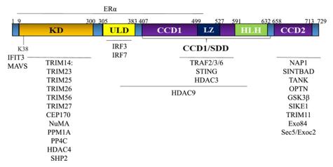 Fig. 1 TBK1 protein structure and interaction partners.1
Key structural properties of TBK1:
- The functional dimer is composed of the kinase domain and the C-terminal coil region
- Kinase domain structure forming inside the conservative ATP combined with pockets and substrate interface
- Phosphorylation site Ser172 is located in the activation link
- Two polymerization interface by hydrophobic interaction between spiral maintain signal complex assembly
Functions of TBK1
The core function of the TBK1 gene is to regulate the innate immune response and autophagy of cells. Meanwhile, it is also widely involved in the signal transduction of various pathophysiological processes.
| Function | Description |
| Activation of immune response | By phosphorylating transcription factors such as IRF3 and NF-κB, the expression of type I interferons and inflammatory factors is initiated. |
| Autophagy regulation | As a adaptor protein of the selective autophagy receptor OPTN/NDP52, it promotes the clearance of damaged organelles. |
| Regulation of cell survival | Balance the survival and death decisions of cells through the mTOR pathway under energy stress conditions. |
| Virus defense | After recognizing the viral nucleic acid, signal bodies are formed, activating the antiviral immune response program. |
| Maintenance of inflammatory balance | Negative feedback regulates the activity of signaling molecules such as STING to prevent excessive immune damage. |
The signal activation of TBK1 exhibits rapid transient characteristics, and its activity is strictly regulated by multiple phosphorylation modifications. This dynamic equilibrium feature makes it a core molecular switch of innate immunity.
Applications of TBK1 and TBK1 Antibody in Literature
1. Zhang, Min, et al. "Inhibitory targeting cGAS-STING-TBK1 axis: Emerging strategies for autoimmune diseases therapy." Frontiers in Immunology 13 (2022): 954129. https://doi.org/10.3389/fimmu.2022.954129
Research has found that abnormal activation of the cGAS-STING-TBK1 signal drives autoimmune diseases. A review of its key pathways, related diseases and the research and development progress of targeted inhibitors indicates that inhibiting this axis is a feasible therapeutic strategy.
2. Endo, Ryu, et al. "TBK1 adaptor AZI2/NAP1 regulates NDP52-driven mitochondrial autophagy." Journal of Biological Chemistry 300.10 (2024). https://doi.org/10.1016/j.jbc.2024.107775
Research reveals that in NDP52-driven mitochondrial autophagy, the adaptor protein AZI2 (rather than TBKBP1) and TBK1 work together to play a key role. AZI2 is recruited into damaged mitochondria and phosphorylated, a process that is crucial for mitochondrial degradation.
3. Vargas, Jose Norberto S., et al. "Spatiotemporal control of ULK1 activation by NDP52 and TBK1 during selective autophagy." Molecular cell 74.2 (2019): 347-362. https://doi.org/10.1016/j.molcel.2019.02.010
Research has revealed that the autophagy receptor NDP52, with the assistance of TBK1 kinase, can precisely recruit the ULK1 complex to damaged organelles, thereby initiating selective autophagy. This provides a key model for the core mechanism.
4. Wang, Banglu, et al. "TBK1 is paradoxical in tumor development: a focus on the pathway mediating IFN-I expression." Frontiers in Immunology 15 (2024): 1433321. https://doi.org/10.3389/fimmu.2024.1433321
Research reveals that TBK1 plays a "double-edged sword" role in tumor development. It can not only promote tumor survival but also mediate anti-tumor immunity by activating type I interferon (IFN). The bidirectness of the downstream IFN signal determines the complex function of TBK1.
5. Runde, Austin P., et al. "The role of TBK1 in cancer pathogenesis and anticancer immunity." Journal of Experimental & Clinical Cancer Research 41.1 (2022): 135. https://doi.org/10.1186/s13046-022-02352-y
The article indicates that TBK1 is a key kinase regulating innate immunity and cellular activities, and its abnormal activation can drive the occurrence of cancer. Studies have shown that inhibiting TBK1 can directly suppress cancer cells and activate T-cell immunity, making it a highly potential anti-cancer target.
Creative Biolabs: TBK1 Antibodies for Research
Creative Biolabs specializes in the production of high-quality TBK1 antibodies for research and industrial applications. Our portfolio includes monoclonal antibodies tailored for ELISA, Flow Cytometry, Western blot, immunohistochemistry, and other diagnostic methodologies.
- Custom TBK1 Antibody Development: Tailor-made solutions to meet specific research requirements.
- Bulk Production: Large-scale antibody manufacturing for industry partners.
- Technical Support: Expert consultation for protocol optimization and troubleshooting.
- Aliquoting Services: Conveniently sized aliquots for long-term storage and consistent experimental outcomes.
For more details on our TBK1 antibodies, custom preparations, or technical support, contact us at email.
Reference
- Runde, Austin P., et al. "The role of TBK1 in cancer pathogenesis and anticancer immunity." Journal of Experimental & Clinical Cancer Research 41.1 (2022): 135. https://doi.org/10.1186/s13046-022-02352-y
Anti-TBK1 antibodies
 Loading...


Hot products 
-
Mouse Anti-C4B Recombinant Antibody (CBYY-C2996) (CBMAB-C4439-YY)

-
Mouse Anti-COL12A1 Recombinant Antibody (CBYY-C3117) (CBMAB-C4560-YY)

-
Mouse Anti-GFAP Recombinant Antibody (24) (CBMAB-G2927-LY)

-
Mouse Anti-Acetyl SMC3 (K105/K106) Recombinant Antibody (V2-634053) (CBMAB-AP052LY)

-
Mouse Anti-2C TCR Recombinant Antibody (V2-1556) (CBMAB-0951-LY)

-
Mouse Anti-AZGP1 Recombinant Antibody (CBWJZ-007) (CBMAB-Z0012-WJ)

-
Mouse Anti-HTLV-1 gp46 Recombinant Antibody (CBMW-H1006) (CBMAB-V208-1154-FY)

-
Mouse Anti-AGO2 Recombinant Antibody (V2-634169) (CBMAB-AP203LY)

-
Mouse Anti-ADGRE5 Recombinant Antibody (V2-360335) (CBMAB-C2088-CQ)

-
Mouse Anti-BHMT Recombinant Antibody (CBYY-0547) (CBMAB-0550-YY)

-
Mouse Anti-AAV8 Recombinant Antibody (V2-634028) (CBMAB-AP022LY)

-
Mouse Anti-ARIH1 Recombinant Antibody (C-7) (CBMAB-A3563-YC)

-
Rabbit Anti-ADRA1A Recombinant Antibody (V2-12532) (CBMAB-1022-CN)

-
Mouse Anti-CECR2 Recombinant Antibody (CBWJC-2465) (CBMAB-C3533WJ)

-
Mouse Anti-ENPP1 Recombinant Antibody (CBFYE-0159) (CBMAB-E0375-FY)

-
Mouse Anti-CORO1A Recombinant Antibody (4G10) (V2LY-1206-LY806)

-
Mouse Anti-AHCYL1 Recombinant Antibody (V2-180270) (CBMAB-A1703-YC)

-
Rabbit Anti-Acetyl-Histone H4 (Lys16) Recombinant Antibody (V2-623415) (CBMAB-CP1021-LY)

-
Mouse Anti-DMPK Recombinant Antibody (CBYCD-324) (CBMAB-D1200-YC)

-
Mouse Anti-CTNND1 Recombinant Antibody (CBFYC-2414) (CBMAB-C2487-FY)

- AActivation
- AGAgonist
- APApoptosis
- BBlocking
- BABioassay
- BIBioimaging
- CImmunohistochemistry-Frozen Sections
- CIChromatin Immunoprecipitation
- CTCytotoxicity
- CSCostimulation
- DDepletion
- DBDot Blot
- EELISA
- ECELISA(Cap)
- EDELISA(Det)
- ESELISpot
- EMElectron Microscopy
- FFlow Cytometry
- FNFunction Assay
- GSGel Supershift
- IInhibition
- IAEnzyme Immunoassay
- ICImmunocytochemistry
- IDImmunodiffusion
- IEImmunoelectrophoresis
- IFImmunofluorescence
- IGImmunochromatography
- IHImmunohistochemistry
- IMImmunomicroscopy
- IOImmunoassay
- IPImmunoprecipitation
- ISIntracellular Staining for Flow Cytometry
- LALuminex Assay
- LFLateral Flow Immunoassay
- MMicroarray
- MCMass Cytometry/CyTOF
- MDMeDIP
- MSElectrophoretic Mobility Shift Assay
- NNeutralization
- PImmunohistologyp-Paraffin Sections
- PAPeptide Array
- PEPeptide ELISA
- PLProximity Ligation Assay
- RRadioimmunoassay
- SStimulation
- SESandwich ELISA
- SHIn situ hybridization
- TCTissue Culture
- WBWestern Blot
