CD44 Antibodies
Background
CD44 is a transmembrane glycoprotein widely distributed on the cell surface, mainly involved in cell-cell and cell-matrix interactions. As the main receptor of hyaluronic acid (HA), CD44 plays a key role in physiological processes such as cell migration, proliferation and signal transduction. In 1991, scientists successfully cloned the CD44 gene for the first time. The discovery of multiple subtypes (such as CD44v6) produced by its alternative splicing opened up a new direction for the study of tumor metastasis. Due to its unique structural features and significant biological functions, CD44 has become a classic example in the research of tumor stem cell markers, providing new ideas for targeted cancer therapy.
Structure of CD44
CD44 is a transmembrane glycoprotein with a molecular weight of approximately 80-95kDa, and its molecular weight varies depending on the degree of glycosylation and alternative splicing subtypes.
| Species | Human | Mice | Rats |
| Molecular Weight (kDa) | 80-95 | 85-100 | 82-97 |
| Primary Structural Differences | Containing variable exons (such as CD44v6) | Higher conservation, but different glycosylation patterns | There is strong homology with human CD44 |
The core structure of CD44 includes the N-terminal hyaluronic acid binding domain, transmembrane region and cytoplasmic tail region. Its extracellular segment contains multiple glycosylation sites, which affect the ligand binding ability. CD44v subtypes (such as CD44v6) form additional domains through the insertion of variable exons, enhancing the potential for tumor metastasis. Conformational changes in proteins can regulate their interactions with ligands such as hyaluronic acid and growth factors, playing a key role in the tumor microenvironment.
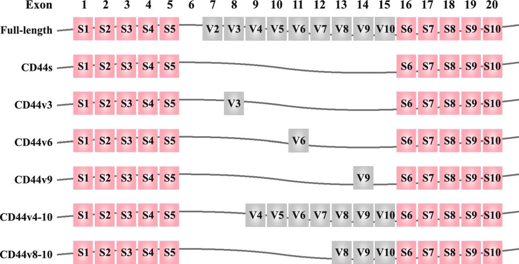 Fig. 1 Structure of CD44 gene. 1
Key structural properties of CD44:
- Highly glycosylated extracellular domains (containing hyaluronic acid binding sites)
- Maintain protein anchoring in the transmembrane region
- Cytoplasmic tail region involved in signal transduction
- Alternative splicing exons (such as the v6 region) determine functional diversity
- Sugar chain modification regulates ligand binding specificity
Functions of CD44
The main function of CD44 is to mediate the interactions between cells and between cells and matrix, and it also plays a key role in tumor development.
| Function | Description |
| Hyaluronic acid binding | As the main receptor of hyaluronic acid, it participates in cell migration and tissue remodeling. |
| Signal transduction | Activate pathways such as EGFR and RAS to regulate cell proliferation and survival. |
| Tumor metastasis | Promote epithelial-mesenchymal transition (EMT) and enhance the invasiveness of cancer cells. |
| Stem cell maintenance | As a biomarker of tumor stem cells, it maintains the characteristics of stem cells. |
| Immune regulation | Participate in the activation of lymphocytes and the regulation of inflammatory responses. |
The affinity of CD44 for ligand binding is affected by the degree of glycosylation and splicing variants. Its multivalent binding property enables it to participate in multiple signaling pathways simultaneously. In the tumor microenvironment, the CD44v6 subtype works in synergy with growth factors, significantly enhancing the metastatic potential.
Applications of CD44 and CD44 Antibody in Literature
1. Thapa, Ranjeeta, and George D. Wilson. "The importance of CD44 as a stem cell biomarker and therapeutic target in cancer." Stem cells international 2016.1 (2016): 2087204. https://doi.org/10.1155/2016/2087204
The article indicates that CD44 is a cell surface glycoprotein bound to hyaluronic acid (HA), which is widely overexpressed in epithelial-derived tumors and serves as a biomarker for cancer stem cells in various solid tumors. The HA-CD44 interaction promotes tumor growth, metastasis and chemotherapy resistance by activating the EGFR pathway. Monoclonal antibodies targeting the CD44 variant subtype in combination with radiotherapy and chemotherapy are expected to become a new treatment strategy for advanced malignant tumors.
2. Jauw, Yvonne WS, et al. "Assessment of target-mediated uptake with immuno-PET: analysis of a phase I clinical trial with an anti-CD44 antibody." EJNMMI research 8 (2018): 1-9. https://doi.org/10.1186/s13550-018-0358-8
In this study, immune PET imaging was performed using the zirconium-89-labeled anti-CD44 monoclonal antibody RG7356 to analyze its targeted uptake in normal tissues. The results showed that with the increase of antibody dose (1-450 mg), the specific uptake in tissues such as the spleen and liver decreased in a dose-dependent manner, confirming that immunoPET can non-invasively assess the targeted distribution characteristics of antibodies in normal tissues.
3. Chen, Han, et al. "The promotion of nanoparticle delivery to two populations of gastric cancer stem cells by CD133 and CD44 antibodies." Biomedicine & Pharmacotherapy 115 (2019): 108857. https://doi.org/10.1016/j.biopha.2019.108857
In this study, a dual antibody nanoparticle CD44/CD133-ATRA-PLPN targeting both CD44 and CD133 was developed for the delivery of all-trans retinoic acid to gastric cancer stem cells. Experiments have confirmed that this dual-targeted nanoparticle can specifically recognize the CD44+ and CD133+ subsets of gastric cancer stem cells. Compared with single-targeted nanoparticles, it shows a stronger growth inhibition effect, providing a new strategy for targeting multiphenotypic tumor stem cells.
4. Xu, Hanxiao, et al. "CD44 as a tumor biomarker and therapeutic target." Experimental hematology & oncology 9 (2020): 1-14. https://doi.org/10.1186/s40164-020-00192-0
This study indicates that CD44 is a polymorphic transmembrane glycoprotein, and its abnormal expression is closely related to tumorigenesis and development. As a biomarker of cancer stem cells, CD44 is involved in regulating tumor proliferation, metastasis and drug resistance, and can be used as an indicator of poor prognosis. Monoclonal antibodies targeting CD44 have entered the preclinical and clinical trial stages, providing new strategies for tumor treatment.
5. Maisel, Daniela, et al. "Targeting tumor cells with anti-CD44 antibody triggers macrophage-mediated immune modulatory effects in a cancer xenograft model." PloS one 11.7 (2016): e0159716.https://doi.org/10.1371/journal.pone.0159716
This study demonstrates that the anti-CD44 antibody RG7356 activates the immune system by blocking the CD44-HA interaction, promotes the secretion of macrophage chemokines, and induces antibody-dependent phagocytosis (ADCP), thereby inhibiting tumor growth. RNA sequencing confirmed that this antibody can reshape the tumor microenvironment and enhance the anti-tumor immune response.
Creative Biolabs: CD44 Antibodies for Research
Creative Biolabs specializes in the production of high-quality CD44 antibodies for research and industrial applications. Our portfolio includes monoclonal antibodies tailored for ELISA, Flow Cytometry, Western blot, immunohistochemistry, and other diagnostic methodologies.
- Custom CD44 Antibody Development: Tailor-made solutions to meet specific research requirements.
- Bulk Production: Large-scale antibody manufacturing for industry partners.
- Technical Support: Expert consultation for protocol optimization and troubleshooting.
- Aliquoting Services: Conveniently sized aliquots for long-term storage and consistent experimental outcomes.
For more details on our CD44 antibodies, custom preparations, or technical support, please contact us.
Reference
- Xu, Hanxiao, et al. "CD44 as a tumor biomarker and therapeutic target." Experimental hematology & oncology 9 (2020): 1-14. https://doi.org/10.1186/s40164-020-00192-0
Anti-CD44 antibodies
 Loading...


Hot products 
-
Mouse Anti-FYN Recombinant Antibody (10) (CBMAB-S6332-CQ)

-
Human Anti-SARS-CoV-2 S1 Monoclonal Antibody (CBFYR-0120) (CBMAB-R0120-FY)

-
Mouse Anti-CD24 Recombinant Antibody (ALB9) (CBMAB-0176CQ)

-
Mouse Anti-4-Hydroxynonenal Recombinant Antibody (V2-502280) (CBMAB-C1055-CN)

-
Mouse Anti-GIPC2 Recombinant Antibody (10) (CBMAB-G0476-LY)

-
Mouse Anti-ARSA Recombinant Antibody (CBYC-A799) (CBMAB-A3679-YC)

-
Mouse Anti-BMI1 Recombinant Antibody (CBYC-P026) (CBMAB-P0108-YC)

-
Mouse Anti-BIRC3 Recombinant Antibody (16E63) (CBMAB-C3367-LY)

-
Mouse Anti-ADGRE5 Recombinant Antibody (V2-360335) (CBMAB-C2088-CQ)

-
Mouse Anti-BSN Recombinant Antibody (219E1) (CBMAB-1228-CN)

-
Mouse Anti-FAS2 Monoclonal Antibody (1D4) (CBMAB-0071-CN)

-
Mouse Anti-BHMT Recombinant Antibody (CBYY-0547) (CBMAB-0550-YY)

-
Mouse Anti-BIRC7 Recombinant Antibody (88C570) (CBMAB-L0261-YJ)

-
Mouse Anti-CCDC6 Recombinant Antibody (CBXC-0106) (CBMAB-C5397-CQ)

-
Mouse Anti-ACE2 Recombinant Antibody (V2-179293) (CBMAB-A0566-YC)

-
Rat Anti-ADGRE4 Recombinant Antibody (V2-160163) (CBMAB-F0011-CQ)

-
Mouse Anti-BIRC5 Recombinant Antibody (6E4) (CBMAB-CP2646-LY)

-
Mouse Anti-ADAM29 Recombinant Antibody (V2-179787) (CBMAB-A1149-YC)

-
Rabbit Anti-CCL5 Recombinant Antibody (R0437) (CBMAB-R0437-CN)

-
Rabbit Anti-ALDOA Recombinant Antibody (D73H4) (CBMAB-A2314-YC)

- AActivation
- AGAgonist
- APApoptosis
- BBlocking
- BABioassay
- BIBioimaging
- CImmunohistochemistry-Frozen Sections
- CIChromatin Immunoprecipitation
- CTCytotoxicity
- CSCostimulation
- DDepletion
- DBDot Blot
- EELISA
- ECELISA(Cap)
- EDELISA(Det)
- ESELISpot
- EMElectron Microscopy
- FFlow Cytometry
- FNFunction Assay
- GSGel Supershift
- IInhibition
- IAEnzyme Immunoassay
- ICImmunocytochemistry
- IDImmunodiffusion
- IEImmunoelectrophoresis
- IFImmunofluorescence
- IGImmunochromatography
- IHImmunohistochemistry
- IMImmunomicroscopy
- IOImmunoassay
- IPImmunoprecipitation
- ISIntracellular Staining for Flow Cytometry
- LALuminex Assay
- LFLateral Flow Immunoassay
- MMicroarray
- MCMass Cytometry/CyTOF
- MDMeDIP
- MSElectrophoretic Mobility Shift Assay
- NNeutralization
- PImmunohistologyp-Paraffin Sections
- PAPeptide Array
- PEPeptide ELISA
- PLProximity Ligation Assay
- RRadioimmunoassay
- SStimulation
- SESandwich ELISA
- SHIn situ hybridization
- TCTissue Culture
- WBWestern Blot
