CDSN Antibodies
Background
The CDSN gene encodes the corneodesmosin protein, which is mainly present in the desmosin structure of the stratum corneum of vertebrate skin. This protein is involved in intercellular adhesion and promotes keratinocyte differentiation, which is crucial for maintaining the function of the epidermal barrier. Skin barrier-related diseases (such as psoriasis and atopic dermatitis) are often associated with variations in the CDSN gene, as they directly affect the process of keratinocyte desquamation. This gene was first identified in 1997 and was subsequently confirmed as a key component of the psoriasis susceptibility gene locus PSORS1. The core role of the protein encoded by it in the terminal differentiation of the epidermis has been deeply studied, providing an important perspective for understanding the formation of the skin barrier, the desquamation mechanism and the molecular basis of inflammatory skin diseases.
Structure of CDSN
The Corneodesmosin protein encoded by the CDSN gene is a secreted glycoprotein with a molecular weight of approximately 52-66 kDa. The size difference mainly stems from post-translational glycosylation modifications.
| Species | Human | Mouse | Bovine |
| Molecular Weight (kDa) | About 52-66 | About 48 | About 54 |
| Primary Structural Differences | Domains rich in glutamate and glutamine are easily cross-linked | Structurally homologous, but there are differences in glycosylation patterns | Core structure domain and human highly conservative |
This protein is composed of 690 amino acids, and its primary structure includes an N-terminal signaling peptide, a conserved "G" domain, and a repeating region rich in glutamic acid/glutamine. Its secondary structure is mainly β -folded and coiled helices, and these structures form stable cores through disulfide bonds. The hydrophobic core is responsible for maintaining structural integrity, while the hydrophilic glutamate-rich region binds to keratinized envelope proteins through transglutaminase-mediated covalent cross-linking in the extracellular matrix, thereby forming strong intercellular bonding bonds between keratinocytes, which is the key molecular basis of the skin's physical barrier.
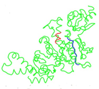 Fig. 1 Structural Localization of CdsL Binding Domains on the CdsN Monomer.1
Key structural properties of CDSN:
- Rich in β-fold and coiled helix modular structure
- Crosslinking function of hydrophilic glutamic acid rich regions
- Calcium ion-dependent regulatory domain of cell adhesion
Functions of CDSN
The main function of the keratinized desmosin encoded by the CDSN gene is to construct and maintain the intercellular adhesion and barrier integrity of the stratum corneum of the skin. Its specific functions include:
| Function | Description |
| The stratum corneum adheres | As a core component of the desmosomal structure, it binds to keratinized envelope proteins through covalent cross-linking to form a firm intercellular connection. |
| Barrier formation | The physical crosslinking network structure skin barrier to prevent moisture loss and external pathogens and allergens. |
| Desquamation regulation | Orderly degradation of the upper stratum corneum by specific proteases (such as KLK) enables normal exfoliation (desquamation) of keratinocytes. |
| Inflammatory regulation | Degradation products can act as innate immune signals and participate in the local regulation of skin inflammation and repair responses. |
| Disease association | The loss of function or abnormal degradation directly leads to desquamation disorder, which is closely related to psoriasis, atopic dermatitis and some hereditary ichthyosis. |
The functional realization of this protein depends on its fine spatiotemporal regulation from synthesis, secretion to orderly degradation. Once the dynamic balance between its expression and degradation is disrupted, it will disrupt the homeostasis of the skin barrier, which is one of the key pathological bases for many common inflammatory and desquamation skin diseases.
Applications of CDSN and CDSN Antibody in Literature
1. van der Velden, Jaap JAJ, et al. "Mutations in the CDSN gene cause peeling skin disease and hypotrichosis simplex of the scalp." The Journal of dermatology 47.1 (2020): 3-7. https://doi.org/10.1111/1346-8138.15136
This article reports a rare skin disease caused by compound heterozygous mutations in the CDSN gene. The patient presented with generalized skin abruption. His mother carried a nonsense mutation of the same gene (c.598C>T), and the clinical manifestation was early hair thinning. This study confirmed the association between CDSN gene mutations and exfoliative skin diseases as well as simple oligotrichosis of the scalp.
2. Zingkou, Eleni, et al. "Deletion of the Epidermal Protease KLK5 Aggravates the Symptoms of Congenital Ichthyosis CDSN-nEDD." International Journal of Molecular Sciences 26.17 (2025): 8605. https://doi.org/10.3390/ijms26178605
This study explores the treatment strategies for ichthyosis caused by CDSN gene mutations. By constructing equivalent models of mouse and human skin, the study found that inhibiting the KLK5 serine protease not only failed to alleviate excessive desquamation but also exacerbated the disease phenotype. This indicates that a single targeted elimination of proteolysis cannot effectively treat this disease, and more precise regulation of enzyme activity is required.
3. Stone, Chris B., et al. "Chlamydia pneumoniae CdsL regulates CdsN ATPase activity, and disruption with a peptide mimetic prevents bacterial invasion." Frontiers in microbiology 2 (2011): 21. https://doi.org/10.3389/fmicb.2011.00021
This article found that the secretory system protein CdsL of Chlamydia pneumoniae type III can inhibit the activity of ATPase CdsN, and two key binding domains were identified through epitope scanning. The treatment with CdsN peptide mimics can effectively inhibit chlamydia from invading cells, providing a basis for the development of new anti-virulence therapies.
4. Wiśniewski, Andrzej, et al. "HLA-C* 06: 02-independent, gender-related association of PSORS1C3 and PSORS1C1/CDSN single-nucleotide polymorphisms with risk and severity of psoriasis." Molecular Genetics and Genomics 293.4 (2018): 957-966. https://doi.org/10.1007/s00438-018-1435-4
This study found in 461 patients with psoriasis that multiple SNPS in the PSORS1 region were significantly associated with disease risk. The results revealed for the first time that variations in the PSORS1C3 and CDSN genes have male-specific effects on the risk and severity of psoriasis, and are independent of the HLA-C*06:02 allele.
5. Fan, Xing, et al. "Fine mapping of the psoriasis susceptibility locus PSORS1 supports HLA-C as the susceptibility gene in the Han Chinese population." PLoS genetics 4.3 (2008): e1000038. https://doi.org/10.1371/journal.pgen.1000038
In this study, through family analysis of the Chinese population, the PSORS1 psoriasis susceptibility gene was located in the key regions near HLA-C. Analysis shows that HLA-Cw6 is the main risk allele, while the CDSN*TTC allele itself does not independently increase the risk of disease in the absence of HLA-Cw6, and does not support its status as the primary gene of PSORS1.
Creative Biolabs: CDSN Antibodies for Research
Creative Biolabs specializes in the production of high-quality CDSN antibodies for research and industrial applications. Our portfolio includes monoclonal antibodies tailored for ELISA, Flow Cytometry, Western blot, immunohistochemistry, and other diagnostic methodologies.
- Custom CDSN Antibody Development: Tailor-made solutions to meet specific research requirements.
- Bulk Production: Large-scale antibody manufacturing for industry partners.
- Technical Support: Expert consultation for protocol optimization and troubleshooting.
- Aliquoting Services: Conveniently sized aliquots for long-term storage and consistent experimental outcomes.
For more details on our CDSN antibodies, custom preparations, or technical support, contact us at email.
Reference
- Stone, Chris B., et al. "Chlamydia pneumoniae CdsL regulates CdsN ATPase activity, and disruption with a peptide mimetic prevents bacterial invasion." Frontiers in microbiology 2 (2011): 21. https://doi.org/10.3389/fmicb.2011.00021
Anti-CDSN antibodies
 Loading...


Hot products 
-
Rat Anti-AChR Recombinant Antibody (V2-12500) (CBMAB-0990-CN)

-
Mouse Anti-GFP Recombinant Antibody (28) (CBMAB-G3038-LY)

-
Mouse Anti-FLI1 Recombinant Antibody (CBXF-0733) (CBMAB-F0435-CQ)

-
Mouse Anti-ADIPOR2 Recombinant Antibody (V2-179983) (CBMAB-A1369-YC)

-
Mouse Anti-C5b-9 Recombinant Antibody (aE11) (CBMAB-AO138LY)

-
Mouse Anti-ADGRE2 Recombinant Antibody (V2-261270) (CBMAB-C0813-LY)

-
Mouse Anti-CCNH Recombinant Antibody (CBFYC-1054) (CBMAB-C1111-FY)

-
Mouse Anti-ADGRL2 Recombinant Antibody (V2-58519) (CBMAB-L0166-YJ)

-
Mouse Anti-ACTN4 Recombinant Antibody (V2-6075) (CBMAB-0020CQ)

-
Mouse Anti-CEMIP Recombinant Antibody (3C12) (CBMAB-K0296-LY)

-
Mouse Anti-AKT1 Recombinant Antibody (V2-180546) (CBMAB-A2070-YC)

-
Mouse Anti-CASP8 Recombinant Antibody (CBYY-C0987) (CBMAB-C2424-YY)

-
Mouse Anti-ACVR1C Recombinant Antibody (V2-179685) (CBMAB-A1041-YC)

-
Mouse Anti-DDC Recombinant Antibody (8E8) (CBMAB-0992-YC)

-
Mouse Anti-BIRC5 Recombinant Antibody (6E4) (CBMAB-CP2646-LY)

-
Mouse Anti-ALPL Antibody (B4-78) (CBMAB-1009CQ)

-
Mouse Anti-DHFR Recombinant Antibody (D0821) (CBMAB-D0821-YC)

-
Mouse Anti-CECR2 Recombinant Antibody (CBWJC-2465) (CBMAB-C3533WJ)

-
Mouse Anti-ARHGAP5 Recombinant Antibody (54/P190-B) (CBMAB-P0070-YC)

-
Mouse Anti-AK4 Recombinant Antibody (V2-180419) (CBMAB-A1891-YC)

- AActivation
- AGAgonist
- APApoptosis
- BBlocking
- BABioassay
- BIBioimaging
- CImmunohistochemistry-Frozen Sections
- CIChromatin Immunoprecipitation
- CTCytotoxicity
- CSCostimulation
- DDepletion
- DBDot Blot
- EELISA
- ECELISA(Cap)
- EDELISA(Det)
- ESELISpot
- EMElectron Microscopy
- FFlow Cytometry
- FNFunction Assay
- GSGel Supershift
- IInhibition
- IAEnzyme Immunoassay
- ICImmunocytochemistry
- IDImmunodiffusion
- IEImmunoelectrophoresis
- IFImmunofluorescence
- IGImmunochromatography
- IHImmunohistochemistry
- IMImmunomicroscopy
- IOImmunoassay
- IPImmunoprecipitation
- ISIntracellular Staining for Flow Cytometry
- LALuminex Assay
- LFLateral Flow Immunoassay
- MMicroarray
- MCMass Cytometry/CyTOF
- MDMeDIP
- MSElectrophoretic Mobility Shift Assay
- NNeutralization
- PImmunohistologyp-Paraffin Sections
- PAPeptide Array
- PEPeptide ELISA
- PLProximity Ligation Assay
- RRadioimmunoassay
- SStimulation
- SESandwich ELISA
- SHIn situ hybridization
- TCTissue Culture
- WBWestern Blot
