CIDEA Antibodies
Background
CIDEA is a cell apoptosis-inducing factor that is mainly expressed in the adipose tissue and immune cells of mammals. The protein encoded by this gene is located in the mitochondria and participates in energy balance regulation by regulating energy metabolism, lipid droplet formation, and the heat production process. Its core functions include promoting fatty acid oxidation, inhibiting lipid storage, and mediating mitochondrial uncoupling, thereby influencing the transformation of white fat to brown fat. When researchers first cloned the CIDEA gene in 1997, they found that it showed significant expression changes during adipocyte differentiation. Subsequent studies further revealed its crucial role in metabolic diseases such as obesity and diabetes. As a core regulatory factor of fat metabolism, CIDEA, due to its unique dual regulatory mechanism - being able to induce cell apoptosis as well as regulate lipid metabolic homeostasis - has become an important molecular model in the field of metabolism research. The related achievements continue to promote a deeper understanding of the molecular mechanisms of energy balance and metabolic diseases.
Structure of CIDEA
CIDEA is a protein with a molecular weight of approximately 25 kDa and shows significant differences among different species. This protein consists of about 220 amino acids and has a unique dimeric domain structure: the N-terminal domain is rich in α-helices and participates in protein interactions and cell localization; the C-terminal domain forms a hydrophobic surface, mediating its binding to lipid droplets.
| Species | Human | Mouse | Pig |
| Molecular Weight (kDa) | 24.8 | 25.1 | 25.3 |
| Primary Structural Differences | The C-terminal lipid droplet binding domain regulates lipolysis and thermogenesis | The N-terminal helical structure is highly conserved and interacts with UCP1 | The sequence has a high similarity to that of humans and its function is conserved |
The three-dimensional structure of CIDEA enables it to anchor onto the surface of lipid droplets and interact with key metabolic proteins such as mitochondrial uncoupling protein 1 (UCP1) through its helical domain. Thus, it plays a central regulatory role in the energy dissipation and production processes of adipocytes. Its dysfunction is closely related to metabolic diseases such as obesity and insulin resistance.
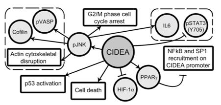 Fig. 1 Proposed mechanism of regulation of CIDEA and its role in glioma cell survival.1
Key structural properties of CIDEA:
- Double-domain configuration
- C-terminal hydrophobic region
- N-terminal regulatory motif
Functions of CIDEA
The core function of the CIDEA protein is to regulate the energy metabolism and heat production processes of adipocytes. Its specific physiological functions are as shown in the table below:
| Function | Description |
| Lipid Droplet Formation Regulation | By binding to the surface of lipid droplets through its C-terminal domain, it regulates the size, fusion, and lipid storage of lipid droplets. |
| Heat Production Activation | In brown adipose tissue, it interacts with the mitochondrial UCP1 protein, promoting uncoupled respiration and converting energy into heat. |
| Energy Metabolism Conversion | Promotes fatty acid oxidation, inhibits lipid synthesis, and drives the transformation of white fat to beige/brown fat-like phenotypes. |
| Cell Apoptosis Induction | Under specific metabolic stress, it can be transferred to the mitochondria and induce changes in mitochondrial membrane permeability, thereby initiating the apoptotic process. |
| Insulin Sensitivity Regulation | By improving the metabolic state and inflammatory levels of adipose tissue, it indirectly enhances insulin sensitivity throughout the body. |
Unlike single-function storage proteins, the regulatory mechanism of CIDEA exhibits "metabolic switch" characteristics: it promotes lipid storage and buffering when energy is abundant, and rapidly switches to the thermogenic and catabolic mode in response to cold or β-adrenergic stimulation. This bidirectional regulatory ability makes it a key molecular hub for the plasticity of adipose tissue and the overall energy balance of the body.
Applications of CIDEA and CIDEA Antibody in Literature
1. Chatterjee, A., et al. "PPARγ regulated CIDEA affects pro-apoptotic responses in glioblastoma." Cell death discovery 1.1 (2015): 1-9. https://doi.org/10.1038/cddiscovery.2015.38
This study found that PPARγ was upregulated and CIDEA was downregulated in glioblastoma. Inhibition of PPARγ could increase CIDEA, which then induced apoptosis, cell cycle arrest and inflammatory responses through JNK-dependent and -independent pathways. This study for the first time revealed the crucial role of the PPARγ-CIDEA regulatory axis in the survival of glioblastoma.
2. Tang, Jinhong, et al. "METTL16-mediated translation of CIDEA promotes non-alcoholic fatty liver disease progression via m6A-dependent manner."PeerJ 10 (2022): e14379. https://doi.org/10.7717/peerj.14379
In this study, a CIDEA dual-reporter gene mouse model was constructed. The fluorescence and bioluminescence signals from this model can accurately reflect the expression of CIDEA in adipose tissue. This system successfully monitored the thermogenic activation induced by cold exposure and drug administration, providing a non-invasive in vivo assessment tool for screening candidate molecules promoting fat thermogenesis.
3. He, Qi, et al. "SREBP1c mediates the effect of acetaldehyde on Cidea expression in Alcoholic fatty liver Mice." Scientific reports 8.1 (2018): 1200. https://doi.org/10.1038/s41598-018-19466-z
This study reveals that the expression of CIDEA is significantly upregulated in alcoholic fatty liver. The experiments show that the alcohol metabolite acetaldehyde specifically induces the expression of CIDEA in liver cells by activating the transcription factor SREBP1c, thereby promoting lipid droplet formation. This suggests that CIDEA plays a crucial role in the occurrence of alcoholic liver disease.
4. Ren, Rumeng, et al. "Hu-lu-su-pian ameliorates hepatic steatosis by regulating CIDEA expression in AKT-driven MASLD mice."Frontiers in Pharmacology 15 (2025): 1503247. https://doi.org/10.3389/fphar.2024.1503247
This study reveals that the traditional Chinese medicine Huashiluansi Tablets (HLSP) can effectively improve metabolic-related fatty liver diseases. The mechanism is by inhibiting the expression of the CIDEA gene in the liver, thereby reducing the synthesis of new fatty acids and the formation of lipid droplets, providing a new strategy for the treatment of this disease.
5. Zorzan, Eleonora, et al. "Hypermethylation-mediated silencing of CIDEA, MAL and PCDH17 tumour suppressor genes in canine DLBCL: From multi-omics analyses to mechanistic studies."International Journal of Molecular Sciences 23.7 (2022): 4021. https://doi.org/10.3390/ijms23074021
This study, through multi-omics analysis, discovered that in canine diffuse large B-cell lymphoma, genes such as CIDEA are silenced due to high DNA methylation. Demethylating drugs can restore their expression, suggesting that CIDEA is a potential tumor suppressor gene regulated by epigenetics.
Creative Biolabs: CIDEA Antibodies for Research
Creative Biolabs specializes in the production of high-quality CIDEA antibodies for research and industrial applications. Our portfolio includes monoclonal antibodies tailored for ELISA, Flow Cytometry, Western blot, immunohistochemistry, and other diagnostic methodologies.
- Custom CIDEA Antibody Development: Tailor-made solutions to meet specific research requirements.
- Bulk Production: Large-scale antibody manufacturing for industry partners.
- Technical Support: Expert consultation for protocol optimization and troubleshooting.
- Aliquoting Services: Conveniently sized aliquots for long-term storage and consistent experimental outcomes.
For more details on our CIDEA antibodies, custom preparations, or technical support, contact us at email.
Reference
- Chatterjee, A., et al. "PPARγ regulated CIDEA affects pro-apoptotic responses in glioblastoma." Cell death discovery 1.1 (2015): 1-9. Distributed under the same Creative Commons license CC BY 4.0 as the original. Cropped from the original figure.https://doi.org/10.1038/cddiscovery.2015.38
Anti-CIDEA antibodies
 Loading...


Hot products 
-
Mouse Anti-CARD11 Recombinant Antibody (CBFYC-0811) (CBMAB-C0866-FY)

-
Mouse Anti-ASTN1 Recombinant Antibody (H-9) (CBMAB-1154-CN)

-
Mouse Anti-BIRC5 Recombinant Antibody (6E4) (CBMAB-CP2646-LY)

-
Mouse Anti-ARID3A Antibody (A4) (CBMAB-0128-YC)

-
Mouse Anti-GFAP Recombinant Antibody (24) (CBMAB-G2927-LY)

-
Mouse Anti-BCL2L1 Recombinant Antibody (H5) (CBMAB-1025CQ)

-
Rabbit Anti-B2M Recombinant Antibody (CBYY-0059) (CBMAB-0059-YY)

-
Mouse Anti-CD1C Recombinant Antibody (L161) (CBMAB-C2173-CQ)

-
Mouse Anti-CRYAB Recombinant Antibody (A4345) (CBMAB-A4345-YC)

-
Rabbit Anti-BAD (Phospho-Ser136) Recombinant Antibody (CAP219) (CBMAB-AP536LY)

-
Mouse Anti-CD46 Recombinant Antibody (CBFYC-0076) (CBMAB-C0085-FY)

-
Mouse Anti-BIRC7 Recombinant Antibody (88C570) (CBMAB-L0261-YJ)

-
Mouse Anti-ALB Recombinant Antibody (V2-180650) (CBMAB-A2186-YC)

-
Mouse Anti-CD8 Recombinant Antibody (C1083) (CBMAB-C1083-LY)

-
Mouse Anti-CDK7 Recombinant Antibody (CBYY-C1783) (CBMAB-C3221-YY)

-
Mouse Anti-ABCA3 Recombinant Antibody (V2-178911) (CBMAB-A0145-YC)

-
Mouse Anti-CD33 Recombinant Antibody (P67.6) (CBMAB-C10189-LY)

-
Mouse Anti-BMI1 Recombinant Antibody (CBYC-P026) (CBMAB-P0108-YC)

-
Mouse Anti-AKT1 (Phosphorylated S473) Recombinant Antibody (V2-505430) (PTM-CBMAB-0067LY)

-
Mouse Anti-CCN2 Recombinant Antibody (CBFYC-2383) (CBMAB-C2456-FY)

- AActivation
- AGAgonist
- APApoptosis
- BBlocking
- BABioassay
- BIBioimaging
- CImmunohistochemistry-Frozen Sections
- CIChromatin Immunoprecipitation
- CTCytotoxicity
- CSCostimulation
- DDepletion
- DBDot Blot
- EELISA
- ECELISA(Cap)
- EDELISA(Det)
- ESELISpot
- EMElectron Microscopy
- FFlow Cytometry
- FNFunction Assay
- GSGel Supershift
- IInhibition
- IAEnzyme Immunoassay
- ICImmunocytochemistry
- IDImmunodiffusion
- IEImmunoelectrophoresis
- IFImmunofluorescence
- IGImmunochromatography
- IHImmunohistochemistry
- IMImmunomicroscopy
- IOImmunoassay
- IPImmunoprecipitation
- ISIntracellular Staining for Flow Cytometry
- LALuminex Assay
- LFLateral Flow Immunoassay
- MMicroarray
- MCMass Cytometry/CyTOF
- MDMeDIP
- MSElectrophoretic Mobility Shift Assay
- NNeutralization
- PImmunohistologyp-Paraffin Sections
- PAPeptide Array
- PEPeptide ELISA
- PLProximity Ligation Assay
- RRadioimmunoassay
- SStimulation
- SESandwich ELISA
- SHIn situ hybridization
- TCTissue Culture
- WBWestern Blot
