COL3A1 Antibodies
Background
The COL3A1 gene encodes the precursor peptide chain of type III collagen, which is a fibrous collagen abundant in the extracellular matrix and mainly distributed in elastic tissues such as skin, blood vessel walls and internal organs. It provides mechanical support and elastic recovery force for various soft tissues by forming a three-helix structure, and participates in processes such as cell migration and tissue repair. Mutations in this gene can disrupt the normal assembly of the collagen triple helix, leading to type IV Ellers-Danlos syndrome, which is clinically characterized by increased vascular rupture and organ fragility. Since its discovery in the 1970s, researchers have gradually revealed its genetic regulatory mechanism through gene sequencing and mutation analysis. In-depth studies on COL3A1 have not only enhanced people's understanding of the molecular mechanisms of connective tissue diseases, but also promoted the development of diagnosis and treatment strategies for collagen-related rare diseases.
Structure of COL3A1
The molecular weight of the type III collagen monomer encoded by the COL3A1 gene is approximately 138 kDa. This value varies to some extent among different mammals, mainly due to interspecific variations in the amino acid sequence of the gene coding region.
| Species | Human | Mouse | Bovine |
| Molecular Weight (kDa) | 138 | 136 | 139 |
| Primary Structural Differences | Typical Gly-X-Y repeats | Collagen structure domain highly conservative | The triple helix region is highly homologous to humans |
This protein, after processing its precursor peptide, forms a triple helix structure composed of three original α1 chains intertwined with each other, which is its characteristic higher conformation. Each peptide chain contains a continuous sequence of glycine-proline-hydroxyproline (Gly-X-Y) repeats, which is the molecular basis for the formation of a stable triple helix. This structure maintains stability through a large number of hydrogen bond networks and hydrophobic interactions between peptide chains, providing the necessary mechanical strength and elasticity for connective tissue.
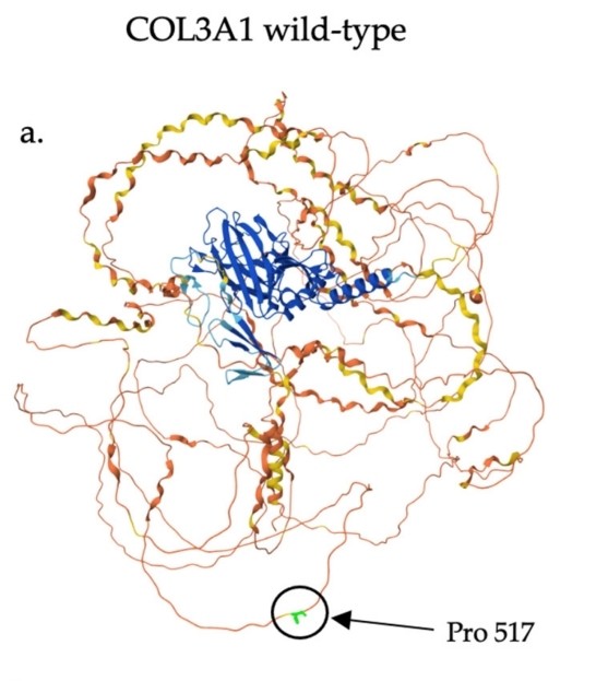 Fig. 1 Predicted structure of wild-type COL3A1, with proline 517 labeled.1
Key structural properties of COL3A1:
- Typical three-stranded helix conformation
- Glycine-proline-hydroxyproline repeats form the structural basis
- Three original alpha 1 chain through hydrogen bonding and hydrophobic action coordination stability
Functions of COL3A1
The main function of type III collagen encoded by the COL3A1 gene is to form the extracellular matrix framework and maintain tissue integrity. Its specific functions include:
| Function | Description |
| Structural support | Co-distributed with type I collagen, it forms an elastic fiber network, providing mechanical strength and toughness for soft tissues such as the skin and blood vessels. |
| Tissue development and repair | In the process of embryonic development, wound healing, guide cell migration and proliferation, promote tissue morphogenesis repair and regeneration. |
| Cell signal regulation | Cell-matrix interactions are mediated through receptors such as integrins, which affect cell adhesion, differentiation and survival. |
| Maintenance of vascular integrity | Blood vessels to form matrix core framework and ensure the vascular elasticity and stability, mutations can lead to blood vessel brittleness increased. |
Unlike some matrix proteins that only provide passive support, type III collagen plays a dual role in mechanical support and dynamic physiological regulation through its unique reticular fiber structure and cell interactions.
Applications of COL3A1 and COL3A1 Antibody in Literature
1. Shen, Yuchen, et al. "COL3A1: Potential prognostic predictor for head and neck cancer based on immune‐microenvironment alternative splicing." Cancer medicine 12.4 (2023): 4882-4894. https://doi.org/10.1002/cam4.5170
Based on tumor immune-related alternative splicing analysis, this study found that COL3A1 was significantly increased in head and neck squamous cell carcinoma and was independently associated with the degree of differentiation, T stage and prognosis of patients. Compared with risk models, COL3A1 can more effectively assess the infiltration, activity and expression of immune checkpoints of tumor immune cells.
2. Du, Haoyuan, et al. "An integrated analysis of bulk and single-cell sequencing data reveals that EMP1+/COL3A1+ fibroblasts contribute to the bone metastasis process in breast, prostate, and renal cancers." Frontiers in Immunology 14 (2023): 1313536. https://doi.org/10.3389/fimmu.2023.1313536
This study, by integrating single-cell and transcriptome data, for the first time reveals from a pan-cancer perspective the existence of a class of EMP1+ fibroblasts rich in COL3A1 in the bone metastasis microenvironment. This cell communicates with cancer cells through the COL3A1-ADGRG1 pathway and may drive the bone metastasis process of multiple cancer types such as breast cancer and prostate cancer, providing a new perspective for understanding the metastasis mechanism.
3. Niu, Ke, Xu Chen, and Yongxian Lu. "COL3A1 rs1800255 polymorphism is associated with pelvic organ prolapse susceptibility in Caucasian individuals: Evidence from a meta-analysis." PLoS One 16.4 (2021): e0250943. https://doi.org/10.1371/journal.pone.0250943
This study, through meta-analysis, found that the rs1800255 polymorphism of the COL3A1 gene is associated with the risk of pelvic organ prolapse in women. In the Caucasian population, the A allele and AA genotype significantly increase the risk of disease, indicating that COL3A1 is a candidate susceptibility gene for this population.
4. Tang, Min, et al. "COL3A1 and its related molecules as potential biomarkers in the development of human Ewing's sarcoma." BioMed Research International 2021.1 (2021): 7453500. https://doi.org/10.1155/2021/7453500
This study, through bioinformatics analysis, found that high expression of COL3A1 was significantly associated with distant metastasis, surgical margin status and poor prognosis of Ewing's sarcoma. Multivariate analysis confirmed that COL3A1 is an independent risk factor for patients, suggesting that it can serve as a potential diagnostic and prognostic biomarker for this tumor.
5. Hu, Xuantao, et al. "Deconvolution of synovial myeloid cell subsets across pathotypes and role of COL3A1+ macrophages in rheumatoid arthritis remission." Frontiers in Immunology 15 (2024): 1307748. https://doi.org/10.3389/fimmu.2024.1307748
In this study, a special type of COL3A1+ macrophage was discovered in the synovium of rheumatoid arthritis. This cell subtype is enriched in the myeloid pathological type, and its abundance is negatively correlated with disease activity. Studies have shown that COL3A1+ macrophages maintain immune homeostasis through mechanisms such as expressing anti-inflammatory factors and enhancing cell adhesion, and are expected to become new targets for inducing disease remission.
Creative Biolabs: COL3A1 Antibodies for Research
Creative Biolabs specializes in the production of high-quality COL3A1 antibodies for research and industrial applications. Our portfolio includes monoclonal antibodies tailored for ELISA, Flow Cytometry, Western blot, immunohistochemistry, and other diagnostic methodologies.
- Custom COL3A1 Antibody Development: Tailor-made solutions to meet specific research requirements.
- Bulk Production: Large-scale antibody manufacturing for industry partners.
- Technical Support: Expert consultation for protocol optimization and troubleshooting.
- Aliquoting Services: Conveniently sized aliquots for long-term storage and consistent experimental outcomes.
For more details on our COL3A1 antibodies, custom preparations, or technical support, contact us at email.
Reference
- Norgan Radler, Charlene, et al. "The Novel Association of a Single Nucleotide Variant in the COL3A1 Gene with Diffuse Coronary Aneurysms." Current Issues in Molecular Biology 47.2 (2025): 82. https://doi.org/10.3390/cimb47020082
Anti-COL3A1 antibodies
Products List
 Loading...


Hot products 
-
Mouse Anti-ENPP1 Recombinant Antibody (CBFYE-0159) (CBMAB-E0375-FY)

-
Mouse Anti-CTNND1 Recombinant Antibody (CBFYC-2414) (CBMAB-C2487-FY)

-
Rabbit Anti-DLK1 Recombinant Antibody (9D8) (CBMAB-D1061-YC)

-
Mouse Anti-BIRC5 Recombinant Antibody (6E4) (CBMAB-CP2646-LY)

-
Mouse Anti-C5b-9 Recombinant Antibody (aE11) (CBMAB-AO138LY)

-
Mouse Anti-ATP1A2 Recombinant Antibody (M7-PB-E9) (CBMAB-A4013-YC)

-
Rabbit Anti-BRCA2 Recombinant Antibody (D9S6V) (CBMAB-CP0017-LY)

-
Mouse Anti-AHCYL1 Recombinant Antibody (V2-180270) (CBMAB-A1703-YC)

-
Rat Anti-FABP3 Recombinant Antibody (CBXF-2299) (CBMAB-F1612-CQ)

-
Mouse Anti-ADV Recombinant Antibody (V2-503423) (CBMAB-V208-1364-FY)

-
Mouse Anti-APOE Recombinant Antibody (A1) (CBMAB-0078CQ)

-
Mouse Anti-ARSA Recombinant Antibody (CBYC-A799) (CBMAB-A3679-YC)

-
Mouse Anti-AGK Recombinant Antibody (V2-258056) (CBMAB-M0989-FY)

-
Mouse Anti-BLK Recombinant Antibody (CBYY-0618) (CBMAB-0621-YY)

-
Mouse Anti-ADGRE5 Recombinant Antibody (V2-360335) (CBMAB-C2088-CQ)

-
Human Anti-SARS-CoV-2 Spike Recombinant Antibody (CBC05) (CBMAB-CR005LY)

-
Mouse Anti-dsDNA Recombinant Antibody (22) (CBMAB-AP1954LY)

-
Mouse Anti-CORO1A Recombinant Antibody (4G10) (V2LY-1206-LY806)

-
Mouse Anti-GGT1 Recombinant Antibody (1F9) (CBMAB-G3273-LY)

-
Rat Anti-4-1BB Recombinant Antibody (V2-1558) (CBMAB-0953-LY)

- AActivation
- AGAgonist
- APApoptosis
- BBlocking
- BABioassay
- BIBioimaging
- CImmunohistochemistry-Frozen Sections
- CIChromatin Immunoprecipitation
- CTCytotoxicity
- CSCostimulation
- DDepletion
- DBDot Blot
- EELISA
- ECELISA(Cap)
- EDELISA(Det)
- ESELISpot
- EMElectron Microscopy
- FFlow Cytometry
- FNFunction Assay
- GSGel Supershift
- IInhibition
- IAEnzyme Immunoassay
- ICImmunocytochemistry
- IDImmunodiffusion
- IEImmunoelectrophoresis
- IFImmunofluorescence
- IGImmunochromatography
- IHImmunohistochemistry
- IMImmunomicroscopy
- IOImmunoassay
- IPImmunoprecipitation
- ISIntracellular Staining for Flow Cytometry
- LALuminex Assay
- LFLateral Flow Immunoassay
- MMicroarray
- MCMass Cytometry/CyTOF
- MDMeDIP
- MSElectrophoretic Mobility Shift Assay
- NNeutralization
- PImmunohistologyp-Paraffin Sections
- PAPeptide Array
- PEPeptide ELISA
- PLProximity Ligation Assay
- RRadioimmunoassay
- SStimulation
- SESandwich ELISA
- SHIn situ hybridization
- TCTissue Culture
- WBWestern Blot
