CYCS Antibodies
Background
The CYCS encoded by the CYCS gene is a small mitochondrial heme protein that exists in the intermembrane space of mitochondria in eukaryotic cells. This protein serves as a key component of the electron transport chain, transferring electrons between mitochondrial complex III and IV through its heme group. It also plays a central regulatory role in the mitochondrial-mediated apoptosis pathway - when cells are stimulated for apoptosis, the CYCS released by mitochondria enters the cytoplasm and binds with Apaf-1 to form an apoptosome, thereby initiating the caspase cascade reaction. As early as 1925, CYCS was discovered and named. It was the first cytochrome protein to be purified and sequenced, and its three-dimensional structure was resolved through X-ray crystallography in the 1970s. As one of the most intensively studied heme proteins, the relationship between the structure and function of CYCS has been widely elucidated, providing a classic model for understanding electron transfer, protein-protein interactions, and the regulatory mechanisms of apoptosis. Its highly conserved evolutionary characteristics also make it an important marker for molecular evolution research.
Structure of CYCS
The CYCS encoded by the CYCS gene is a small heme protein with a molecular weight of approximately 12 kDa. This protein shows high sequence conservation among different species, with only slight differences in molecular weight but overall stability.
| Species | Human | Mouse | Rat | Cattle |
| Molecular Weight (kDa) | 11.6 | 11.6 | 11.6 | 12.0 |
| Primary Structural Differences | 104 amino acids, highly conserved | 95% homology with humans | Only a few residues differ from mice | Additional C-terminal extension |
The CYCS protein consists of 104 amino acids and has a compact spherical spatial structure. It is formed by multiple α helices folding around the heme cofactor. The protein structure center contains a heme cofactor (a ferrous porphyrin ring), which is covalently linked to the protein part through a bond. This is the basis of its electron transfer function. The red color of CYCS comes from the heme group. Its secondary structure is mainly composed of multiple α helices, forming a special hydrophobic environment to stabilize the heme cofactor. The iron atom of the heme forms an axial coordination bond with His18, and Met80 provides another coordination bond. This six-coordinate structure distinguishes it from oxygenated proteins and is suitable for reversible electron transfer.
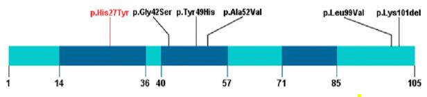 Fig. 1 Schematic presentation of linear CYCS protein with all variants.1
Key structural properties of CYCS:
- Compact α-helical folding structure
- Hemoglobin covalently bound to Cys14 and Cys17
- Iron-containing porphyrin ring facilitating electron transfer
- Met80 and His18 coordinate to stabilize the iron of hemoglobin
Functions of CYCS
The main function of CYCS is mitochondrial electron transfer. In addition, it is involved in the regulation of cell apoptosis, the clearance of reactive oxygen species, and the mitochondrial stress response.
| Function | Description |
| Electron transfer | Transfers a single electron between mitochondrial complex III and IV, driving oxidative phosphorylation to generate ATP. |
| Apoptosis initiation | Released from the mitochondria into the cytoplasm when the cell is damaged, it combines with Apaf-1 to form an apoptosome and activates the caspase cascade reaction. |
| Oxygen radicals regulation | Participates in the regulation of electron leakage through its heme group, affecting the mitochondrial oxygen radical level. |
| Cell death signal integration | Acts as the hub of the mitochondrial-dependent apoptosis pathway, receiving regulation from the Bcl-2 family proteins. |
| Tissue hypoxia response | Regulates the balance between cell survival and death through its release mechanism under hypoxic conditions. |
The redox potential characteristics of CYCS present a typical single-electron transfer curve, which is different from the cooperative binding curve of hemoglobin. This indicates the high efficiency of it as an electron carrier and its fixed position in the mitochondrial respiratory chain.
Applications of CYCS and CYCS Antibody in Literature
1. Che, Fengyu, et al. "A novel heterozygous pathogenic variation in CYCS gene cause autosomal dominant non-syndromic thrombocytopenia 4 in a large Chinese family." Frontiers in Genetics 12 (2022): 783455. https://doi.org/10.3389/fgene.2021.783455
An 8-member family was found to have thrombocytopenia. Genetic testing revealed a heterozygous missense variation of c.79C>T in the CYCS gene, confirming the diagnosis of autosomal dominant hereditary thrombocytopenia type 4. This variation did not cause abnormal bleeding or mitochondrial disorders, enriching the mutation spectrum of this disease.
2. Kan, Jingbao, et al. "Declined expressions of vast mitochondria-related genes represented by CYCS and transcription factor ESRRA in skeletal muscle aging." Bioengineered 12.1 (2021): 3485-3502. https://doi.org/10.1080/21655979.2021.1948951
The research has found that the aging of skeletal muscles is related to the downregulation of mitochondrial function-related gene expression. CYCS and ESRRA are key molecules whose expression significantly decreases with age, and they can serve as potential biomarkers for age-related sarcopenia.
3. Li, Yan, et al. "[Retracted] The Identification and Clinical Value Evaluation of CYCS Related to Asthma through Bioinformatics Analysis and Functional Experiments." Disease Markers 2023.1 (2023): 5746940. https://doi.org/10.1155/2023/5746940
The research has found that the CYCS gene is upregulated in asthma and is related to immune cells, which can promote the proliferation of asthma cells. CYCS may serve as a new diagnostic marker and therapeutic target for asthma.
4. Zhan, Xiaoshu, et al. "MiR-29b inhibits COC expansion and oocyte in vitro maturation via induction of ROS by targeting CYCS." Animal Reproduction Science 270 (2024): 107598. https://doi.org/10.1016/j.anireprosci.2024.107598
Studies have shown that miR-29b targets and inhibits the CYCS gene, leading to the accumulation of reactive oxygen species, thereby inhibiting the expansion of the cumulus-oocyte complex and the maturation of oocytes in pigs.
5. Ong, Lily, Kirstin O. McDonald, and Elizabeth C. Ledgerwood. "Differentiation and cell density upregulate CYCS levels in megakaryoblastic cell lines: Implications for analysis of CYCS-associated thrombocytopenia." Plos one 12.12 (2017): e0190433. https://doi.org/10.1371/journal.pone.0190433
The study found that mutations in the CYCS gene cause autosomal dominant hereditary thrombocytopenia. The endogenous expression of CYCS increases along with the differentiation of megakaryocytes and the increase in cell density, suggesting that this gene plays an important role in cell proliferation and differentiation.
Creative Biolabs: CYCS Antibodies for Research
Creative Biolabs specializes in the production of high-quality CYCS antibodies for research and industrial applications. Our portfolio includes monoclonal and polyclonal antibodies tailored for ELISA, Flow Cytometry, Western blot, immunohistochemistry, and other diagnostic methodologies.
- Custom CYCS Antibody Development: Tailor-made solutions to meet specific research requirements.
- Bulk Production: Large-scale antibody manufacturing for industry partners.
- Technical Support: Expert consultation for protocol optimization and troubleshooting.
- Aliquoting Services: Conveniently sized aliquots for long-term storage and consistent experimental outcomes.
For more details on our CYCS antibodies, custom preparations, or technical support, contact us at email.
Reference
- Che, Fengyu, et al. "A novel heterozygous pathogenic variation in CYCS gene cause autosomal dominant non-syndromic thrombocytopenia 4 in a large Chinese family." Frontiers in Genetics 12 (2022): 783455. Distributed under Open Access license CC BY 4.0, cropped from the original figure. https://doi.org/10.3389/fgene.2021.783455
Anti-CYCS antibodies
 Loading...


Hot products 
-
Mouse Anti-2C TCR Recombinant Antibody (V2-1556) (CBMAB-0951-LY)

-
Mouse Anti-ASH1L Monoclonal Antibody (ASH5H03) (CBMAB-1372-YC)

-
Rat Anti-ADAM10 Recombinant Antibody (V2-179741) (CBMAB-A1103-YC)

-
Mouse Anti-AMOT Recombinant Antibody (CBYC-A564) (CBMAB-A2552-YC)

-
Mouse Anti-C5b-9 Recombinant Antibody (aE11) (CBMAB-AO138LY)

-
Mouse Anti-CAT Recombinant Antibody (724810) (CBMAB-C8431-LY)

-
Mouse Anti-AK4 Recombinant Antibody (V2-180419) (CBMAB-A1891-YC)

-
Mouse Anti-CD59 Recombinant Antibody (CBXC-2097) (CBMAB-C4421-CQ)

-
Mouse Anti-DLL4 Recombinant Antibody (D1090) (CBMAB-D1090-YC)

-
Mouse Anti-APP Recombinant Antibody (DE2B4) (CBMAB-1122-CN)

-
Mouse Anti-CHRNA9 Recombinant Antibody (8E4) (CBMAB-C9161-LY)

-
Rabbit Anti-ABL1 (Phosphorylated Y185) Recombinant Antibody (V2-443434) (PTM-CBMAB-0001YC)

-
Mouse Anti-BACE1 Recombinant Antibody (61-3E7) (CBMAB-1183-CN)

-
Mouse Anti-GFP Recombinant Antibody (28) (CBMAB-G3038-LY)

-
Rat Anti-AChR Recombinant Antibody (V2-12500) (CBMAB-0990-CN)

-
Mouse Anti-BRCA2 Recombinant Antibody (CBYY-1728) (CBMAB-2077-YY)

-
Mouse Anti-CD2AP Recombinant Antibody (BR083) (CBMAB-BR083LY)

-
Rat Anti-FABP3 Recombinant Antibody (CBXF-2299) (CBMAB-F1612-CQ)

-
Mouse Anti-BrdU Recombinant Antibody (IIB5) (CBMAB-1038CQ)

-
Mouse Anti-8-oxoguanine Recombinant Antibody (V2-7719) (CBMAB-1898CQ)

- AActivation
- AGAgonist
- APApoptosis
- BBlocking
- BABioassay
- BIBioimaging
- CImmunohistochemistry-Frozen Sections
- CIChromatin Immunoprecipitation
- CTCytotoxicity
- CSCostimulation
- DDepletion
- DBDot Blot
- EELISA
- ECELISA(Cap)
- EDELISA(Det)
- ESELISpot
- EMElectron Microscopy
- FFlow Cytometry
- FNFunction Assay
- GSGel Supershift
- IInhibition
- IAEnzyme Immunoassay
- ICImmunocytochemistry
- IDImmunodiffusion
- IEImmunoelectrophoresis
- IFImmunofluorescence
- IGImmunochromatography
- IHImmunohistochemistry
- IMImmunomicroscopy
- IOImmunoassay
- IPImmunoprecipitation
- ISIntracellular Staining for Flow Cytometry
- LALuminex Assay
- LFLateral Flow Immunoassay
- MMicroarray
- MCMass Cytometry/CyTOF
- MDMeDIP
- MSElectrophoretic Mobility Shift Assay
- NNeutralization
- PImmunohistologyp-Paraffin Sections
- PAPeptide Array
- PEPeptide ELISA
- PLProximity Ligation Assay
- RRadioimmunoassay
- SStimulation
- SESandwich ELISA
- SHIn situ hybridization
- TCTissue Culture
- WBWestern Blot
