
DLX5 Antibodies
Background
The DLX5 gene belongs to the DLX gene family and is a homologous box transcription factor that plays a key role in the development of vertebrates. This gene mainly participates in the development process of craniofacial structures, limb bones and the central nervous system (especially GABAergic neurons) by regulating the expression of downstream target genes. DLX5 is actively expressed in the early stage of embryonic development, and its functional abnormalities are closely related to human congenital malformations such as craniosynostosis, hearing impairment and limb development defects. Since its discovery in the early 1990s, DLX5 has become an important model molecule in developmental biology and genetic disease research, greatly promoting people's understanding of transcriptional regulatory mechanisms, embryonic morphogenesis and evolutionary conservation.
Structure of DLX5
The molecular weight of the protein encoded by the DLX5 gene is approximately 31.9 kDa, and its precise value varies slightly among different species. This difference mainly stems from specific variations in the amino acid sequences of each species.
| Species | Human | Mouse | Rat | Zebrafish | Chicken |
| Molecular Weight (kDa) | 31.9 | 32.1 | 32.0 | 31.7 | 31.8 |
| Primary Structural Differences | Field containing homologous structure, sequence is highly conserved | High homology with human, similar function | Core structure domain highly consistent | There are specific amino acid substitutions | Typical of the DLX family |
The DLX5 protein is a transcription factor composed of 289 amino acids. Its three-dimensional structure forms a characteristic homologous domain, which is made up of three α -helixes connected by a circular region. The core of the protein is its highly conserved DNA-binding motif, especially the third α -helix which can directly embed into the large DNA groove, recognize and bind to specific target sequences. This combination is mediated by multiple arginine and histidine residues, which form crucial hydrogen bonds with DNA bases, thereby precisely regulating the transcriptional activity of downstream genes.
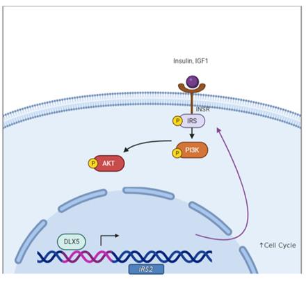 Fig. 1 DLX5 upregulates IRS2 expression to enhance AKT signaling.1
Key structural properties of DLX5:
- Homologous domains constitute the core of DNA binding
- Helix-loop-helix structures mediate protein dimerization
- Highly conserved DNA recognition helices
Functions of DLX5
The main function of the DLX5 gene is to regulate gene transcription during embryonic development. However, it is also involved in a variety of physiological and pathological processes, including osteogenic differentiation and neurogenesis.
| Function | Description |
| Regulation of bone development | Dominating the morphogenesis of craniofacial and limb bones is achieved by activating the expression of osteoblast-specific genes. |
| Neural pattern formation | In the central nervous system development decide the fate of the GABA neurons can affect the normal of the cerebral cortex. |
| Development of the auditory system | Regulation of the inner ear ear bone formation and development of the cochlea, loss of its function will lead to hearing impairment. |
| Participation in damage repair | In adulthood, it participates in the process of bone tissue injury repair and nerve regeneration, and is reactivated to promote tissue reconstruction. |
| Association of disease occurrence | The abnormal expression and craniosynostosis, mental retardation, and closely related to the occurrence of certain types of cancer development. |
As a transcription factor, DLX5 exhibits DNA binding characteristics in a co-heterodimer form, which contrasts with other family members in the homodimer form, demonstrating higher precision and functional specificity in target gene selection in developmental regulation.
Applications of DLX5 and DLX5 Antibody in Literature
1. Aouci, Rym, et al. "Dlx5/6 expression levels in mouse GABAergic neurons regulate adult parvalbumin neuronal density and anxiety/compulsive behaviours." Cells 11.11 (2022): 1739. https://doi.org/10.3390/cells11111739
The article indicates that in adult mice, a reduction in the dose of the Dlx5/6 gene in GABAergic neurons leads to a linear decrease in the density of Parvalbumin-positive inhibitory interneurons in the prefrontal cortex and hippocampus, and aggravates anxiety and obsessive-like behaviors. This indicates that the expression level of Dlx5/6 is associated with the risk of mental illness.
2. Ambrosetti, Irene, et al. "Split hand-foot and deafness in a patient with 7q21. 13-q21. 3 deletion not including the Dlx5/6 genes." Genes 14.8 (2023): 1526. https://doi.org/10.3390/genes14081526
Research has found that the deletion of two exons acting as enhancers in the DYNC1I1 gene on chromosome 7q21 will down-regulate the expression of the distal DLX5/6 gene, thereby leading to acromegaly (SHFM1) and inner ear abnormalities. This reveals that enhancer destruction is the pathogenic mechanism of congenital malformations.
3. Mullen, Rachel D., et al. "Distal-less homeobox genes Dlx5/6 regulate Müllerian duct regression." Frontiers in Endocrinology 13 (2022): 916173. https://doi.org/10.3389/fendo.2022.916173
Studies have shown that in male mouse embryos, the Dlx5 and Dlx6 genes are induced to express anti-Mullerian hormone (AMH), with functional redundancy. Single-gene homozygous mutations of Dlx5 or Dlx6 can lead to incomplete degeneration of the Mullerian duct, while double-gene mutations result in more severe degeneration defects, confirming their crucial role in the normal development of the male reproductive tract.
4. Lu, Ye, et al. "Anti‐Dlx5 retards the progression of osteoarthritis through inhibiting chondrocyte hypertrophy and apoptosis." Evidence‐Based Complementary and Alternative Medicine 2022.1 (2022): 5019920. https://doi.org/10.1155/2022/5019920
Studies have shown that in osteoarthritis models, the use of anti-DLX5 treatment can delay disease progression. The mechanism is to inhibit the excessive hypertrophy of chondrocytes by down-regulating factors such as BMP-2 and SP7, and reduce the expression of caspase-3 to decrease cell apoptosis, thereby improving the pathological damage of cartilage.
5. MacKenzie, Rachel K., Parvathy Ravi Sankar, and Andrew J. Bendall. "Dlx5 and Dlx6 can antagonize cell division at the G1/S checkpoint." BMC Molecular and Cell Biology 20.1 (2019): 8. https://doi.org/10.1186/s12860-019-0191-6
Studies have shown that the transcription factors Dlx5 and Dlx6 promote the differentiation of various progenitor cells by inhibiting cell proliferation, reducing the proportion of S-phase cells and prolonging the cell cycle. This function of inhibiting the cell cycle is an important mechanism independent of the cell lineage that drives differentiation.
Creative Biolabs: DLX5 Antibodies for Research
Creative Biolabs specializes in the production of high-quality DLX5 antibodies for research and industrial applications. Our portfolio includes monoclonal antibodies tailored for ELISA, Flow Cytometry, Western blot, immunohistochemistry, and other diagnostic methodologies.
- Custom DLX5 Antibody Development: Tailor-made solutions to meet specific research requirements.
- Bulk Production: Large-scale antibody manufacturing for industry partners.
- Technical Support: Expert consultation for protocol optimization and troubleshooting.
- Aliquoting Services: Conveniently sized aliquots for long-term storage and consistent experimental outcomes.
For more details on our DLX5 antibodies, custom preparations, or technical support, contact us at email.
Reference
- Tan, Yinfei, and Joseph R. Testa. "DLX genes: roles in development and cancer." Cancers 13.12 (2021): 3005. https://doi.org/10.3390/cancers13123005
Anti-DLX5 antibodies
 Loading...
 Compare




Hot products 
-
Mouse Anti-AP4E1 Recombinant Antibody (32) (CBMAB-A2996-YC)

-
Mouse Anti-F11R Recombinant Antibody (402) (CBMAB-0026-WJ)

-
Mouse Anti-CD24 Recombinant Antibody (ALB9) (CBMAB-0176CQ)

-
Mouse Anti-ATP1B1 Recombinant Antibody (E4) (CBMAB-0463-LY)

-
Mouse Anti-ATP1B3 Recombinant Antibody (1E9) (CBMAB-A4021-YC)

-
Mouse Anti-DES Monoclonal Antibody (440) (CBMAB-AP1857LY)

-
Rat Anti-AChR Recombinant Antibody (V2-12500) (CBMAB-0990-CN)

-
Rat Anti-EMCN Recombinant Antibody (28) (CBMAB-E0280-FY)

-
Mouse Anti-BLK Recombinant Antibody (CBYY-0618) (CBMAB-0621-YY)

-
Mouse Anti-CDKL5 Recombinant Antibody (CBFYC-1629) (CBMAB-C1689-FY)

-
Mouse Anti-ABL2 Recombinant Antibody (V2-179121) (CBMAB-A0364-YC)

-
Mouse Anti-APP Recombinant Antibody (5C2A1) (CBMAB-A3314-YC)

-
Rat Anti-C5AR1 Recombinant Antibody (8D6) (CBMAB-C9139-LY)

-
Mouse Anti-CRYAB Recombinant Antibody (A4345) (CBMAB-A4345-YC)

-
Mouse Anti-BIRC3 Recombinant Antibody (315304) (CBMAB-1214-CN)

-
Mouse Anti-BrdU Recombinant Antibody (IIB5) (CBMAB-1038CQ)

-
Mouse Anti-AAV-5 Recombinant Antibody (V2-503417) (CBMAB-V208-1369-FY)

-
Mouse Anti-AZGP1 Recombinant Antibody (CBWJZ-007) (CBMAB-Z0012-WJ)

-
Mouse Anti-BZLF1 Recombinant Antibody (BZ.1) (CBMAB-AP705LY)

-
Mouse Anti-BCL6 Recombinant Antibody (CBYY-0442) (CBMAB-0445-YY)

- AActivation
- AGAgonist
- APApoptosis
- BBlocking
- BABioassay
- BIBioimaging
- CImmunohistochemistry-Frozen Sections
- CIChromatin Immunoprecipitation
- CTCytotoxicity
- CSCostimulation
- DDepletion
- DBDot Blot
- EELISA
- ECELISA(Cap)
- EDELISA(Det)
- ESELISpot
- EMElectron Microscopy
- FFlow Cytometry
- FNFunction Assay
- GSGel Supershift
- IInhibition
- IAEnzyme Immunoassay
- ICImmunocytochemistry
- IDImmunodiffusion
- IEImmunoelectrophoresis
- IFImmunofluorescence
- IGImmunochromatography
- IHImmunohistochemistry
- IMImmunomicroscopy
- IOImmunoassay
- IPImmunoprecipitation
- ISIntracellular Staining for Flow Cytometry
- LALuminex Assay
- LFLateral Flow Immunoassay
- MMicroarray
- MCMass Cytometry/CyTOF
- MDMeDIP
- MSElectrophoretic Mobility Shift Assay
- NNeutralization
- PImmunohistologyp-Paraffin Sections
- PAPeptide Array
- PEPeptide ELISA
- PLProximity Ligation Assay
- RRadioimmunoassay
- SStimulation
- SESandwich ELISA
- SHIn situ hybridization
- TCTissue Culture
- WBWestern Blot
