EEF1A1 Antibodies
Background
The EEF1A1 gene encodes eukaryotic elongation factor 1α1, which is a highly conserved and widely expressed GTP-binding protein. This protein is mainly responsible for transporting aminoacyl-tRNA to the ribosome during protein synthesis, ensuring the accuracy and efficiency of the translation process. As a key regulatory factor in cellular metabolism and proliferation, EEF1A1 also participates in various non-classical functions such as cytoskeleton regulation, apoptosis, and signal transduction. This gene was first identified in the study of protein synthesis mechanisms in the 1970s, and its structure and function have been systematically analyzed through biochemical and structural biology methods, becoming an important model for understanding the translation mechanism and cellular homeostasis. In-depth research on EEF1A1 not only reveals the core mechanism of translation elongation but also provides molecular-level insights into pathological processes such as tumors and neurodegenerative diseases.
Structure of EEF1A1
EEF1A1 is a highly conserved GTP-binding protein with a molecular weight of approximately 50.1 kDa. There are slight differences in its molecular weight among different species, mainly due to minor variations in the amino acid sequence.
| Species | Human | Mouse | Zebrafish | Yeast |
| Molecular Weight (kDa) | 50.1 | 50.0 | 49.8 | 50.5 |
| Primary Structural Differences | Containing 462 amino acids and multiple post-translational modification sites | Conservative sequence homology is extremely high, function | The core structural domain is highly similar | Homologous genes TEF1/2 perform similar functions |
This protein is composed of approximately 462 amino acids and its three-dimensional structure forms a typical globular domain. Its primary structure contains three highly conserved GTP-binding motifs (G1-G3), which determine its GTPase activity. The secondary structure is mainly composed of alternating α-helices and β-sheets, forming the classic guanine nucleotide-binding protein folding structure. The Switch I and Switch II regions undergo conformational changes when GTP and GDP are bound, and this allosteric mechanism is the core for its function as a translation elongation factor, responsible for accurately delivering aminoacyl-tRNA to the A site of the ribosome.
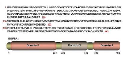 Fig. 1 EEF1A1: Protein Sequence, Truncated Variants (Nucleotide Sequences & Schematics).1
Key structural properties of EEF1A1:
- Conservative guanine nucleotide-binding protein (G protein) folding domain
- Three core GTP-binding motifs (G1-G3) constitute the active center
- The Switch I/II region, which is capable of conformational change, regulates its GTPase activity
Functions of EEF1A1
The main function of EEF1A1 is to act as a eukaryotic translation elongation factor, transporting aminoacyl-tRNA during protein synthesis. Additionally, it is involved in various cellular physiological processes, including cytoskeleton regulation, apoptosis, and viral infection responses.
| Function | Description |
| Protein Synthesis | During the extension phase of translation, the correct aminoacyl-tRNA is delivered to the A-site of the ribosome in a GTP-dependent manner to ensure the accurate elongation of the polypeptide chain. |
| Cytoskeleton Regulation | By binding and stabilizing actin microfilaments, it participates in processes such as cell shape maintenance, movement, and cytoplasmic division. |
| Apoptosis Regulation | Under specific cellular stress conditions, it can be cleaved or modified, thereby participating in the signaling pathways that induce cell apoptosis. |
| Virus Replication Support | Various viruses (such as poliovirus) utilize the EEF1A1 protein of the host cell to facilitate the replication and translation of their own genetic material. |
| Quality Control | Participates in directing misfolded or damaged proteins to the degradation pathway, maintaining protein homeostasis within the cell. |
The translation activity of EEF1A1 strictly depends on the conformational change driven by GTP hydrolysis, which is different from the oxygenation dissociation mechanism of myoglobin. The diversity of its function stems from its high abundance, conserved structure, and the ability to adapt to different cellular environments by interacting with various different ligands.
Applications of EEF1A1 and EEF1A1 Antibody in Literature
1. Xie, Baoping, et al. "Disruption of the eEF1A1/ARID3A/PKC‐δ Complex by Neferine Inhibits Macrophage Glycolytic Reprogramming in Atherosclerosis." Advanced Science 12.15 (2025): 2416158. https://doi.org/10.1002/advs.202416158
The article indicates that the natural small molecule Neferine is found to target eEF1A1, inhibit its formation with the ARID3A/PKC-δ complex, and prevent ARID3A from entering the nucleus to regulate the transcription of the key glycolytic enzyme ENO2, thereby blocking the glycolytic reprogramming and M1-type polarization of macrophages, providing a new strategy for the treatment of atherosclerosis.
2. Zhao, Jie, et al. "UCHL3 promotes hepatocellular carcinoma progression by stabilizing EEF1A1 through deubiquitination." Biology Direct 19.1 (2024): 53. https://doi.org/10.1186/s13062-024-00495-w
This study reveals that UCHL3 binds to and stabilizes EEF1A1 through deubiquitination, forming the UCHL3/EEF1A1 axis, which promotes the migration, stem cell characteristics and drug resistance of liver cancer cells, providing a new target for targeted therapy of liver cancer.
3. Fan, A‐hui, et al. "eEF1A1 promotes colorectal cancer progression and predicts poor prognosis of patients." Cancer Medicine 12.1 (2023): 513-524. https://doi.org/10.1002/cam4.4848
This study found that eEF1A1 is upregulated in colorectal cancer, and its high expression is associated with a poor prognosis for patients. Mechanistically, eEF1A1 promotes tumor proliferation by activating the MAPK signaling pathway (p38/JNK/ERK), suggesting that it can serve as a potential therapeutic target and prognostic marker.
4. Li, Mingli, et al. "Epigenetic Control of Translation Checkpoint and Tumor Progression via RUVBL1‐EEF1A1 Axis." Advanced Science 10.17 (2023): 2206584. https://doi.org/10.1002/advs.202206584
Research has found that RUVBL1, the core ATpase subunit of the histone acetyltransferase complex NuA4 in Ewing's sarcoma, promotes the expression and protein translation of its downstream target gene EEF1A1 by regulating the binding of MYC to chromatin, thereby driving tumor progression. Inhibiting RUVBL1 and MYC has a synergistic anti-tumor effect.
5. Wang, Xun, et al. "CDCA5-EEF1A1 interaction promotes progression of clear cell renal cell carcinoma by regulating mTOR signaling." Cancer Cell International 24.1 (2024): 147. https://doi.org/10.1186/s12935-024-03330-4
Research has found that CDCA5 is highly expressed in clear cell renal cell carcinoma and has a poor prognosis. It activates the mTOR signaling pathway through interaction with EEF1A1, promoting tumor proliferation, migration and sunitinib resistance, suggesting that CDCA5 is a potential therapeutic target.
Creative Biolabs: EEF1A1 Antibodies for Research
Creative Biolabs specializes in the production of high-quality EEF1A1 antibodies for research and industrial applications. Our portfolio includes monoclonal antibodies tailored for ELISA, Flow Cytometry, Western blot, immunohistochemistry, and other diagnostic methodologies.
- Custom EEF1A1 Antibody Development: Tailor-made solutions to meet specific research requirements.
- Bulk Production: Large-scale antibody manufacturing for industry partners.
- Technical Support: Expert consultation for protocol optimization and troubleshooting.
- Aliquoting Services: Conveniently sized aliquots for long-term storage and consistent experimental outcomes.
For more details on our EEF1A1 antibodies, custom preparations, or technical support, contact us at email.
Reference
- Zhao, Jie, et al. "UCHL3 promotes hepatocellular carcinoma progression by stabilizing EEF1A1 through deubiquitination." Biology Direct 19.1 (2024): 53. https://doi.org/10.1186/s13062-024-00495-w
Anti-EEF1A1 antibodies
 Loading...


Hot products 
-
Mouse Anti-ARID3A Antibody (A4) (CBMAB-0128-YC)

-
Mouse Anti-DHFR Recombinant Antibody (D0821) (CBMAB-D0821-YC)

-
Mouse Anti-GFAP Recombinant Antibody (20) (CBMAB-G2914-LY)

-
Mouse Anti-BAX Recombinant Antibody (CBYY-0216) (CBMAB-0217-YY)

-
Mouse Anti-AQP2 Recombinant Antibody (G-3) (CBMAB-A3359-YC)

-
Mouse Anti-DES Monoclonal Antibody (440) (CBMAB-AP1857LY)

-
Rabbit Anti-ABL1 (Phosphorylated Y185) Recombinant Antibody (V2-443434) (PTM-CBMAB-0001YC)

-
Mouse Anti-ATP1B3 Recombinant Antibody (1E9) (CBMAB-A4021-YC)

-
Mouse Anti-CCDC25 Recombinant Antibody (CBLC132-LY) (CBMAB-C9786-LY)

-
Rabbit Anti-AP2M1 (Phosphorylated T156) Recombinant Antibody (D4F3) (PTM-CBMAB-0610LY)

-
Mouse Anti-CD24 Recombinant Antibody (SN3) (CBMAB-C1037-CQ)

-
Mouse Anti-ATM Recombinant Antibody (2C1) (CBMAB-A3970-YC)

-
Mouse Anti-BACE1 Recombinant Antibody (61-3E7) (CBMAB-1183-CN)

-
Rabbit Anti-AKT3 Recombinant Antibody (V2-12567) (CBMAB-1057-CN)

-
Mouse Anti-FPR2 Recombinant Antibody (1D6) (CBMAB-F2628-CQ)

-
Mouse Anti-DDC Recombinant Antibody (8E8) (CBMAB-0992-YC)

-
Mouse Anti-dsRNA Recombinant Antibody (2) (CBMAB-D1807-YC)

-
Mouse Anti-BANF1 Recombinant Antibody (3F10-4G12) (CBMAB-A0707-LY)

-
Mouse Anti-14-3-3 Pan Recombinant Antibody (V2-9272) (CBMAB-1181-LY)

-
Mouse Anti-CTNND1 Recombinant Antibody (CBFYC-2414) (CBMAB-C2487-FY)

- AActivation
- AGAgonist
- APApoptosis
- BBlocking
- BABioassay
- BIBioimaging
- CImmunohistochemistry-Frozen Sections
- CIChromatin Immunoprecipitation
- CTCytotoxicity
- CSCostimulation
- DDepletion
- DBDot Blot
- EELISA
- ECELISA(Cap)
- EDELISA(Det)
- ESELISpot
- EMElectron Microscopy
- FFlow Cytometry
- FNFunction Assay
- GSGel Supershift
- IInhibition
- IAEnzyme Immunoassay
- ICImmunocytochemistry
- IDImmunodiffusion
- IEImmunoelectrophoresis
- IFImmunofluorescence
- IGImmunochromatography
- IHImmunohistochemistry
- IMImmunomicroscopy
- IOImmunoassay
- IPImmunoprecipitation
- ISIntracellular Staining for Flow Cytometry
- LALuminex Assay
- LFLateral Flow Immunoassay
- MMicroarray
- MCMass Cytometry/CyTOF
- MDMeDIP
- MSElectrophoretic Mobility Shift Assay
- NNeutralization
- PImmunohistologyp-Paraffin Sections
- PAPeptide Array
- PEPeptide ELISA
- PLProximity Ligation Assay
- RRadioimmunoassay
- SStimulation
- SESandwich ELISA
- SHIn situ hybridization
- TCTissue Culture
- WBWestern Blot
