FLCN Antibodies
Background
FLCN gene encoding the follicle protein is a kind of highly conservative tumor suppressor, mainly engaged in cytoplasm and nuclei, by participating in regulation of mTOR signaling pathways and cell metabolic processes to maintain homeostasis. This gene is closely related to Birt-Hogg-Dube syndrome, and its functional inactivation can lead to the occurrence of diseases such as kidney tumors, pulmonary cysts and skin fibromas. Since its first localization and cloning in 2002, the multi-level functional mechanisms of the FLCN gene - including its interaction with AMPK, lysosomal association regulation, and energy perception pathway regulation - have gradually been revealed, providing a key molecular perspective for understanding metabolism-related diseases and tumorigenesis, and continuously driving the development of targeted therapeutic strategies.
Structure of FLCN
The follicular protein encoded by the FLCN gene is a relatively large protein with a molecular weight of approximately 64 kDa. The molecular weight of this protein is highly conserved across different species, which is closely related to its key structural functional domain.
| Species | Human | Mouse | Zebrafish | Fruit fly |
|---|---|---|---|---|
| Molecular Weight (kDa) | About 64 | About 64 | About 65 | About 68 |
| Primary Structural Differences | Contains the DENN domain, which is involved in gtpase activation | Highly homologous structure, used to construct the model of the disease | Conservative core function domain, for development research | Homologous genes exist and are involved in metabolic regulation |
This protein contains 579 amino acids, and its three-dimensional structure presents a unique folding pattern. The core feature of the FLCN protein is that its C-terminal contains a highly conserved DENN domain, which is crucial for its function as a GTPase activator protein (GAP) and can regulate the activity of Rab GTPases. Its secondary structure is mainly composed of alternating α -helix and β -fold, forming a specific binding interface for binding with interacting proteins (such as FNIP1/2), jointly constituting the FLCN complex, and thereby participating in the regulation of key cellular signaling pathways (such as mTOR and AMPK pathways) as well as cellular metabolic processes.
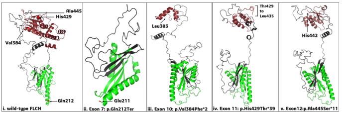 Fig. 1 FLCN Exonic Mutations: Structural and Protein Interaction Consequences.1
Key structural properties of FLCN:
- Contains a highly conserved DENN domain (acting on Rab GTP enzyme)
- Form a stable heterodimer complex with FNIP1/2 protein
- Through its the c-terminal domain structure involved in AMPK/mTOR signaling pathways control
- Have a sequence of check and ratify, can shuttle between the nucleus and cytoplasm
Functions of FLCN
The follicle-stimulating protein encoded by the FLCN gene mainly exerts tumor suppressive functions and participates in a wide range of cellular homeostasis maintenance processes by regulating multiple signaling pathways.
| Function | Description |
|---|---|
| mTOR Pathway Regulation | As a negative regulatory factor of the mTORC1 signal, it inhibits its excessive activation when nutrition is adequate, maintaining the metabolic balance of cells. |
| Cellular Metabolic regulation | It affects the energy perception, autophagy process and mitochondrial function of cells through pathways such as AMPK. |
| Cilia formation and Function | It is involved in regulating the formation and stability of primary cilia. The loss of its function will lead to abnormal cili-related signals. |
| Cell Proliferation and Survival | Under specific conditions (such as energy stress), it regulates the cell cycle process and apoptosis, and inhibits abnormal proliferation. |
| Cell-matrix Interaction | It affects cell adhesion, migration and the remodeling of extracellular matrix, and is related to the potential for tumor metastasis. |
As a key upstream regulatory factor, the inhibitory effect of FLCN on mTOR signals is more fundamental and persistent, which is different from many factors that directly act on the mTOR complex, highlighting its pivotal position in integrating nutritional signals and growth signals.
Applications of FLCN and FLCN Antibody in Literature
1. Wang, Guoyan, et al. "Role of FLCN Phosphorylation in Insulin‐Mediated mTORC1 Activation and Tumorigenesis." Advanced Science 10.17 (2023): 2206826. https://doi.org/10.1002/advs.202206826
The research has found that the Ser62 site of the FLCN protein can be phosphorylated by AKT1. This process relies on the mTORC2-AKT1 complex on the lysosome and activates mTORC1 by inhibiting the GTPase activity of RagC. This, in turn, regulates autophagy and tumor growth in various cancers and is positively correlated with the activity of mTORC1. It provides a new target for the treatment of related diseases.
2. Xia, Qin, et al. "Gal3‐CaN‐Smurf1 Complex Sequestrates FLCN‐FNIPs to Facilitate TFEB Activation in Response to Endomembrane Damage." Advanced Science 12.40 (2025): e13241. https://doi.org/10.1002/advs.202413241
The research reveals that when lysosomes are damaged, the Gal3-CaN-Smurf1 complex isolates and ubiquitinates FLCN-FNIPs, thereby relieving their inhibitory effect on RagC/D. This, in turn, blocks the phosphorylation of TFEB by mTORC1, ultimately activating TFEB to respond to stress. This provides a new approach for tumor treatment.
3. Glykofridis, Iris E., et al. "Phosphoproteomic analysis of FLCN inactivation highlights differential kinase pathways and regulatory TFEB phosphoserines." Molecular & Cellular Proteomics 21.9 (2022): 100263. https://doi.org/10.1016/j.mcpro.2022.100263
The research reveals that in BHD syndrome, the inactivation of FLCN drives the occurrence of renal cancer by enhancing the activity of the MAPK/ERK and PI3K pathways, and leads to the dephosphorylation of TFEB. Targeted inhibition of MAPK has significant toxicity on FLCN-deficient cells, providing a new target for the treatment of BHD-related renal tumors.
4. Zhao, Xuyang, et al. "FLCN regulates HIF2α nuclear import and proliferation of clear cell renal cell carcinoma." Frontiers in Molecular Biosciences 7 (2020): 121. https://doi.org/10.3389/fmolb.2020.00121
This study demonstrates that in renal clear cell carcinoma, the tumor suppressor gene FLCN binds to and degrades HIF2α, inhibiting the PI3K/mTORC2 signaling pathway, thereby down-regulating the expression of Cyclin D1 and MMP9, and inhibiting tumor proliferation and invasion, providing a new therapeutic target.
5. Hassan, Faisal A., et al. "Folliculin (FLCN) in Thyroid Tumors: Incidence, Significance, and Role as a Driver Gene and Secondary Alteration." Current Oncology 32.4 (2025): 224. https://doi.org/10.3390/curroncol32040224
This study demonstrates that in thyroid cancer, pathogenic mutations in FLCN are often associated with eosinophilic cell morphology and serve as an early driving factor; while gene homozygous deletion is more common in aggressive tumors such as anaplastic carcinoma. Incorporating FLCN testing in patients with BHD syndrome may enhance the screening and diagnostic efficacy of thyroid tumors.
Creative Biolabs: FLCN Antibodies for Research
Creative Biolabs specializes in the production of high-quality FLCN antibodies for research and industrial applications. Our portfolio includes monoclonal antibodies tailored for ELISA, Flow Cytometry, Western blot, immunohistochemistry, and other diagnostic methodologies.
- Custom FLCN Antibody Development: Tailor-made solutions to meet specific research requirements.
- Bulk Production: Large-scale antibody manufacturing for industry partners.
- Technical Support: Expert consultation for protocol optimization and troubleshooting.
- Aliquoting Services: Conveniently sized aliquots for long-term storage and consistent experimental outcomes.
For more details on our FLCN antibodies, custom preparations, or technical support, contact us at email.
Reference
- Ray, Anindita, et al. "Genetic insight into Birt–Hogg–Dubé syndrome in Indian patients reveals novel mutations at FLCN." Orphanet Journal of Rare Diseases 17.1 (2022): 176. https://doi.org/10.1186/s13023-022-02326-5
Anti-FLCN antibodies
 Loading...


Hot products 
-
Rat Anti-4-1BB Recombinant Antibody (V2-1558) (CBMAB-0953-LY)

-
Mouse Anti-CD2AP Recombinant Antibody (BR083) (CBMAB-BR083LY)

-
Mouse Anti-CFL1 Recombinant Antibody (CBFYC-1771) (CBMAB-C1833-FY)

-
Mouse Anti-DHFR Recombinant Antibody (D0821) (CBMAB-D0821-YC)

-
Mouse Anti-ADGRL2 Recombinant Antibody (V2-58519) (CBMAB-L0166-YJ)

-
Mouse Anti-ASTN1 Recombinant Antibody (H-9) (CBMAB-1154-CN)

-
Rat Anti-CCR2 Recombinant Antibody (475301) (CBMAB-C1338-LY)

-
Rat Anti-AChR Recombinant Antibody (V2-12500) (CBMAB-0990-CN)

-
Rabbit Anti-ALOX5AP Recombinant Antibody (CBXF-1219) (CBMAB-F0750-CQ)

-
Rabbit Anti-CAMK2A Recombinant Antibody (BA0032) (CBMAB-0137CQ)

-
Rat Anti-C5AR1 Recombinant Antibody (8D6) (CBMAB-C9139-LY)

-
Mouse Anti-ALOX5 Recombinant Antibody (33) (CBMAB-1890CQ)

-
Mouse Anti-ACLY Recombinant Antibody (V2-179314) (CBMAB-A0610-YC)

-
Mouse Anti-CECR2 Recombinant Antibody (CBWJC-2465) (CBMAB-C3533WJ)

-
Mouse Anti-AMACR Recombinant Antibody (CB34A) (CBMAB-CA034LY)

-
Mouse Anti-ACO2 Recombinant Antibody (V2-179329) (CBMAB-A0627-YC)

-
Mouse Anti-CASP8 Recombinant Antibody (CBYY-C0987) (CBMAB-C2424-YY)

-
Mouse Anti-AHCYL1 Recombinant Antibody (V2-180270) (CBMAB-A1703-YC)

-
Mouse Anti-GGT1 Recombinant Antibody (1F9) (CBMAB-G3273-LY)

-
Mouse Anti-ADAM12 Recombinant Antibody (V2-179752) (CBMAB-A1114-YC)

- AActivation
- AGAgonist
- APApoptosis
- BBlocking
- BABioassay
- BIBioimaging
- CImmunohistochemistry-Frozen Sections
- CIChromatin Immunoprecipitation
- CTCytotoxicity
- CSCostimulation
- DDepletion
- DBDot Blot
- EELISA
- ECELISA(Cap)
- EDELISA(Det)
- ESELISpot
- EMElectron Microscopy
- FFlow Cytometry
- FNFunction Assay
- GSGel Supershift
- IInhibition
- IAEnzyme Immunoassay
- ICImmunocytochemistry
- IDImmunodiffusion
- IEImmunoelectrophoresis
- IFImmunofluorescence
- IGImmunochromatography
- IHImmunohistochemistry
- IMImmunomicroscopy
- IOImmunoassay
- IPImmunoprecipitation
- ISIntracellular Staining for Flow Cytometry
- LALuminex Assay
- LFLateral Flow Immunoassay
- MMicroarray
- MCMass Cytometry/CyTOF
- MDMeDIP
- MSElectrophoretic Mobility Shift Assay
- NNeutralization
- PImmunohistologyp-Paraffin Sections
- PAPeptide Array
- PEPeptide ELISA
- PLProximity Ligation Assay
- RRadioimmunoassay
- SStimulation
- SESandwich ELISA
- SHIn situ hybridization
- TCTissue Culture
- WBWestern Blot
