HERC2 Antibodies
Background
HERC2 is a large E3 ubiquitin ligase that is widely present in various tissues of the human body, mainly located in the nucleus and cytoplasm. This protein modifies substrates through ubiquitination and participates in important biological processes such as DNA damage repair, cell cycle progression, and gene transcription. Mutations in the HERC2 gene can lead to the HERC2 Angel-like syndrome characterized by developmental delay and intellectual disability. It was first cloned and identified by the team of Chinese scientist Gong Xiaoyang in 2003. It is highly conserved in evolution with its homologous gene in fruit flies. HERC2 precisely regulates the stability and function of substrate proteins through the synergistic action of its RCC1-like domain and HECT catalytic domain, providing an important paradigm for understanding the molecular mechanism of ubiquitination modification in development and diseases.
Structure of HERC2
HERC2 is a large E3 ubiquitin ligase with a molecular weight of approximately 528 kDa. There are certain differences in the molecular weight and structure of this protein among different species.
| Species | Human | Mouse | Rat | Zebrafish | Fruit fly |
| Molecular Weight (kDa) | 528 | 528 | 528 | 509 | 306 |
| Primary Structural Differences | Contains the RCC1 domain and HECT catalytic domain | High homology with humans, similar functions | High homology with humans | Relatively simple structure | As a homologous gene, the structure is more primitive |
HERC2 mediates protein interactions through its multiple domains and participates in processes such as DNA damage repair, centrosome regulation, and ubiquitination modification.
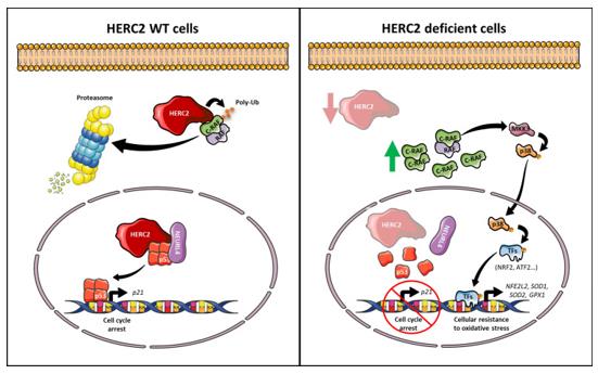 Fig. 1 Working model of HERC2 function in health and disease.1
Structural characteristics of HERC2 protein:
- Contains a RCC1-like domain and an HECT catalytic domain
- The RCC1 domain mediates protein interactions
- The HECT domain has ubiquitin ligase activity
- Multiple zinc finger structures are involved in substrate recognition
- Nuclear-cytoplasmic shuttling sequences regulate distribution
Functions of HERC2
HERC2, as an E3 ubiquitin ligase, participates in the regulation of various cellular processes. Its core function is to modify substrate proteins through ubiquitination, thereby influencing protein stability and function.
| Function | Description |
| DNA damage repair | Recruits repair proteins to the damage site to maintain genomic stability |
| Centrosome regulation | Participates in centrosome replication and maturation, ensuring the accuracy of cell division |
| Protein degradation | Mediates the degradation of target proteins through the ubiquitin-proteasome pathway |
| Transcription regulation | Interacts with transcription factors to affect gene expression |
| Neurodevelopment | Mutations in HERC2 lead to neurodevelopmental disorders related to the protein |
Applications of HERC2 and HERC2 Antibody in Literature
1. Sala-Gaston, Joan, et al. "HERC2 deficiency activates C-RAF/MKK3/p38 signalling pathway altering the cellular response to oxidative stress." Cellular and Molecular Life Sciences 79.11 (2022): 548. https://doi.org/10.1007/s00018-022-04586-7
The article indicates that mutations in the HERC2 gene lead to the activation of the C-RAF/MKK3/p38 signaling pathway, resulting in a stronger resistance of patient cells to oxidative stress. The study found that inhibiting the activity of RAF can block this pathway, providing a new idea for targeted treatment of the HERC2 angel-like syndrome.
2. Liu, Yunzhi, et al. "HERC2 promotes inflammation-driven cancer stemness and immune evasion in hepatocellular carcinoma by activating STAT3 pathway." Journal of Experimental & Clinical Cancer Research 42.1 (2023): 38. https://doi.org/10.1186/s13046-023-02609-0
The article indicates that high expression of HERC2 is associated with poor prognosis of liver cancer. It activates the JAK2/STAT3 pathway by binding to PTP1B, enhances the characteristics of tumor stem cells and the immune escape mediated by PD-L1, and promotes the occurrence of inflammation-related liver cancer.
3. Liu, Yunzhi, et al. "Hepatocyte‐Targeted Lipid Nanoparticle Delivery of HERC2 Plasmid Controls Drug‐Induced Hepatotoxicity by Limiting β‐Catenin‐Regulated CYP2E1 Expression." Advanced Science 11.46 (2024): 2401633. https://doi.org/10.1002/advs.202401633
The article indicates that HERC2 promotes the ubiquitination of β-catenin through interaction, regulates the expression of CYP2E1, and protects liver cells from drug metabolism damage. Hepatocyte-targeted delivery of HERC2 overexpression plasmids can alleviate acute liver injury induced by APAP.
4. Zhu, Mingzhang, et al. "HERC2 inactivation abrogates nucleolar localization of RecQ helicases BLM and WRN." Scientific Reports 11.1 (2021): 360. https://doi.org/10.1038/s41598-020-79715-y
The article indicates that HERC2 promotes the localization of DNA helicase BLM/WRN to the nucleolus, assisting in the unwinding of the G4 structure of rDNA. The loss of HERC2 function enhances the inhibitory effect of RNA polymerase I inhibitor CX-5461 on rRNA transcription, suggesting its potential value in cancer treatment.
5. Lai, Yongqiang, et al. "HERC2 regulates RPA2 by mediating ATR-induced Ser33 phosphorylation and ubiquitin-dependent degradation." Scientific reports 9.1 (2019): 14257. https://doi.org/10.1038/s41598-019-50812-x
The article indicates that HERC2 interacts with RPA through its HECT domain, regulating the ATR-mediated phosphorylation and ubiquitination degradation of RPA2, thereby maintaining the stability of RPA2 under low-level replication stress and participating in the inhibition of G4 DNA structure formation.
Creative Biolabs: HERC2 Antibodies for Research
Creative Biolabs specializes in the production of high-quality HERC2 antibodies for research and industrial applications. Our portfolio includes monoclonal and polyclonal antibodies tailored for ELISA, Flow Cytometry, Western blot, immunohistochemistry, and other diagnostic methodologies.
- Custom HERC2 Antibody Development: Tailor-made solutions to meet specific research requirements.
- Bulk Production: Large-scale antibody manufacturing for industry partners.
- Technical Support: Expert consultation for protocol optimization and troubleshooting.
- Aliquoting Services: Conveniently sized aliquots for long-term storage and consistent experimental outcomes.
For more details on our HERC2 antibodies, custom preparations, or technical support, contact us at email.
Reference
- Sala-Gaston, Joan, et al. "HERC2 deficiency activates C-RAF/MKK3/p38 signalling pathway altering the cellular response to oxidative stress." Cellular and Molecular Life Sciences 79.11 (2022): 548. Distributed under Open Access license CC BY 4.0, without modification. https://doi.org/10.1007/s00018-022-04586-7
Anti-HERC2 antibodies
 Loading...


Hot products 
-
Mouse Anti-FYN Recombinant Antibody (10) (CBMAB-S6332-CQ)

-
Mouse Anti-BIRC5 Recombinant Antibody (6E4) (CBMAB-CP2646-LY)

-
Mouse Anti-FAS2 Monoclonal Antibody (1D4) (CBMAB-0071-CN)

-
Mouse Anti-ACVR1C Recombinant Antibody (V2-179685) (CBMAB-A1041-YC)

-
Mouse Anti-CALR Recombinant Antibody (CBFYC-0763) (CBMAB-C0818-FY)

-
Mouse Anti-FLT1 Recombinant Antibody (11) (CBMAB-V0154-LY)

-
Rat Anti-ADGRE4 Recombinant Antibody (V2-160163) (CBMAB-F0011-CQ)

-
Mouse Anti-CA9 Recombinant Antibody (CBXC-2079) (CBMAB-C0131-CQ)

-
Mouse Anti-AZGP1 Recombinant Antibody (CBWJZ-007) (CBMAB-Z0012-WJ)

-
Mouse Anti-C5b-9 Recombinant Antibody (aE11) (CBMAB-AO138LY)

-
Mouse Anti-AKT1/AKT2/AKT3 (Phosphorylated T308, T309, T305) Recombinant Antibody (V2-443454) (PTM-CBMAB-0030YC)

-
Mouse Anti-CCT6A/B Recombinant Antibody (CBXC-0168) (CBMAB-C5570-CQ)

-
Mouse Anti-CD33 Recombinant Antibody (P67.6) (CBMAB-C10189-LY)

-
Mouse Anti-CDK7 Recombinant Antibody (CBYY-C1783) (CBMAB-C3221-YY)

-
Mouse Anti-AAV8 Recombinant Antibody (V2-634028) (CBMAB-AP022LY)

-
Mouse Anti-EMP3 Recombinant Antibody (CBFYE-0100) (CBMAB-E0207-FY)

-
Rat Anti-EPO Recombinant Antibody (16) (CBMAB-E1578-FY)

-
Mouse Anti-CRYAB Recombinant Antibody (A4345) (CBMAB-A4345-YC)

-
Mouse Anti-CD1C Recombinant Antibody (L161) (CBMAB-C2173-CQ)

-
Mouse Anti-CASP8 Recombinant Antibody (CBYY-C0987) (CBMAB-C2424-YY)

- AActivation
- AGAgonist
- APApoptosis
- BBlocking
- BABioassay
- BIBioimaging
- CImmunohistochemistry-Frozen Sections
- CIChromatin Immunoprecipitation
- CTCytotoxicity
- CSCostimulation
- DDepletion
- DBDot Blot
- EELISA
- ECELISA(Cap)
- EDELISA(Det)
- ESELISpot
- EMElectron Microscopy
- FFlow Cytometry
- FNFunction Assay
- GSGel Supershift
- IInhibition
- IAEnzyme Immunoassay
- ICImmunocytochemistry
- IDImmunodiffusion
- IEImmunoelectrophoresis
- IFImmunofluorescence
- IGImmunochromatography
- IHImmunohistochemistry
- IMImmunomicroscopy
- IOImmunoassay
- IPImmunoprecipitation
- ISIntracellular Staining for Flow Cytometry
- LALuminex Assay
- LFLateral Flow Immunoassay
- MMicroarray
- MCMass Cytometry/CyTOF
- MDMeDIP
- MSElectrophoretic Mobility Shift Assay
- NNeutralization
- PImmunohistologyp-Paraffin Sections
- PAPeptide Array
- PEPeptide ELISA
- PLProximity Ligation Assay
- RRadioimmunoassay
- SStimulation
- SESandwich ELISA
- SHIn situ hybridization
- TCTissue Culture
- WBWestern Blot
